
Method for enriching adherent monocyte populations

a technology of adherent monocytes and adherent monocytes, which is applied in the field of enriching adherent monocyte populations, can solve the problems of osmotically shocked buffy coat, no closed system elutriator has yet been developed for sterile monocyte isolation, and no method for cryopreservation of adherent monocytes has been reported

Active Publication Date: 2008-07-01
AMERICAN NAT RED CROSS
View PDF4 Cites 16 Cited by
  • Summary
  • Abstract
  • Description
  • Claims
  • Application Information

AI Technical Summary

Benefits of technology

"The patent is about a new method for isolating monocytes from blood using non-woven filters. This method is faster and more efficient than existing methods and can be performed in a closed system. The isolated monocytes are highly activated and can be differentiated into various lineages without the use of exogenous cytokines. The technical effect of this patent is to provide a more efficient and reliable method for isolating monocytes for clinical use."

Problems solved by technology

However, no closed system elutriator has yet been developed for sterile monocyte isolation.
However, the technique requires two gradients, discontinuous gradients can be difficult to prepare, and the procedure is not easily carried out in closed systems.
In addition, no method for cryopreservation of adherent monocytes from osmotically shocked buffy coat has been reported.
Another disadvantage is that non-filter methods are time consuming and often require more than two hours to complete the isolation procedures.
One disadvantage of such a method, however, is that many of these leukocyte reduction filters bind platelets, which are released upon backflushing the filter and thus contaminate the final product.
Platelets can interfere with the further culture of monocytes to dendritic cells, osteoclasts, and endothelial cells.
Additionally, filtration methods afford especially high quantities of activated monocytes.
Most, if not all, of the purification methods are difficult or impossible to carry out in a closed system, making maintenance of sterility a problem in producing clinical grade cell preparations.
Similarly, most purification procedures require more than two hours for completion, making the isolation process costly and time consuming.

Method used


Image

  • Method for enriching adherent monocyte populations
  • Method for enriching adherent monocyte populations
  • Method for enriching adherent monocyte populations

Examples


example 1

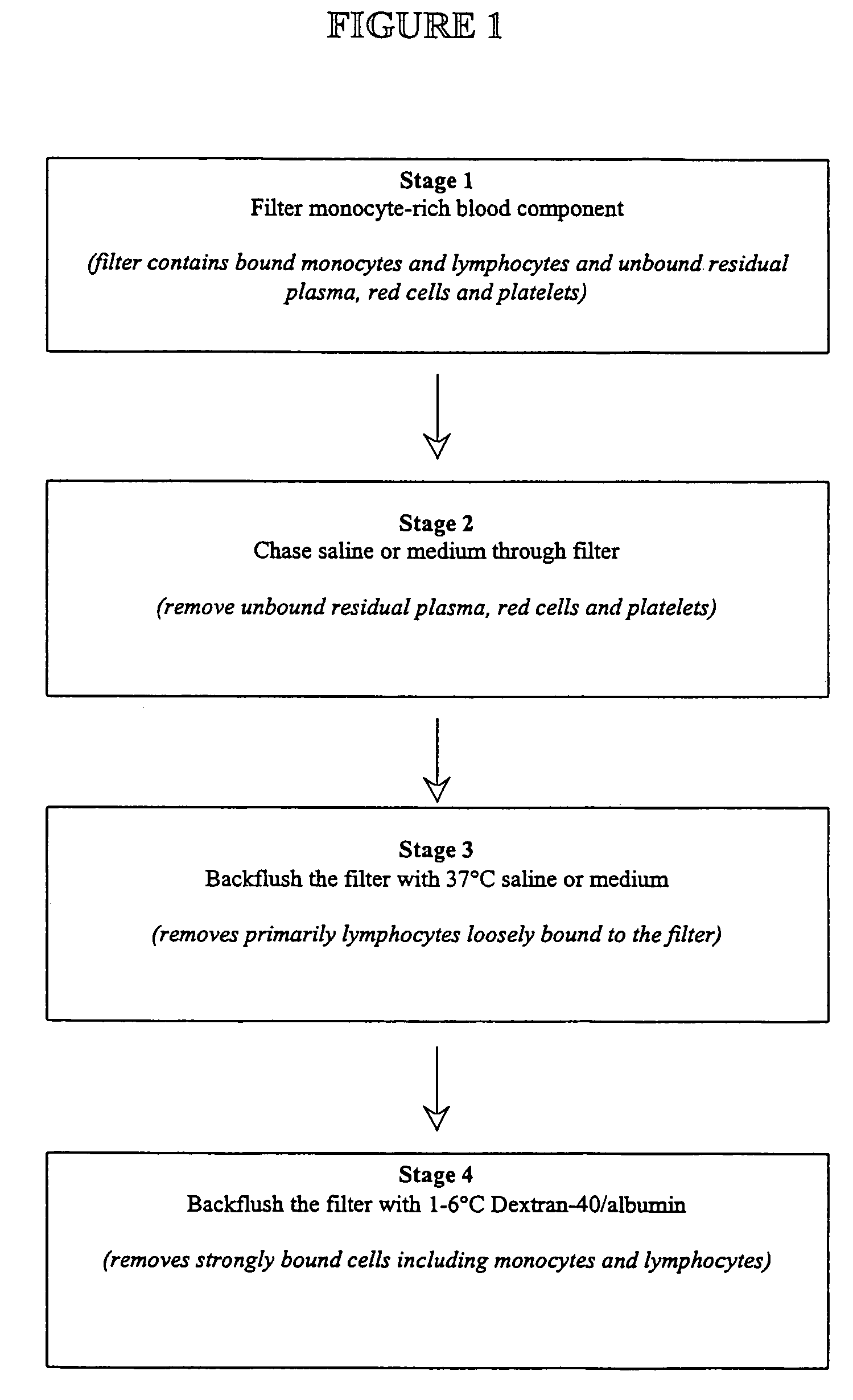
[0031]Monocytes were collected using a mononuclear apheresis procedure. Thirteen apheresis units were collected. Each unit contained 100 mL of approximately 5×10⁹ mononuclear cells and 1×10⁹ monocytes. Each unit was sterile docked to a cord blood collection filter (Asahi Medical Corp.) and the mononuclear suspension was allowed to pass through the filter. When 80-90% of the mononuclear suspension had passed through the filter, the filter was then gently backflushed with 50 mL of 37° C. saline twice. The filter was then strongly backflushed using 50 mL of 1-6° C. Dextran-40 / albumin twice. Isolated white cells from the 1-6° C. backflush fractions were characterized for monocyte purity, monocyte recovery, viability, and phagocytosis.

[0032]Cell viability was measured by standard microscopic methods (Nikon Labophot) using a 30 μM propidium iodide / 20 μM acridine orange stain in phosphate buffered saline.

[0033]Cell populations were determined by fluorescent microscopy using acridine orange...

example 2

[0037]Eight monocyte preparations from Example 1 were subsequently cryopreserved. A suspension of 2×10⁷ monocyte / mL was added to an equal volume of cold HES / DMSO / albumin to the monocyte / macrophage preparation according to standard methods (17). Final concentrations of the cyroprotectants were 6% hydroxyethyl starch (McGaw Inc, Irvine, Calif.), 5% dimethylsulfoxide (Cryoserve, Research Industries Corp., Salt Lake, Utah), 4% human serum albumin (American Red Cross, Hyland, Calif.). One-mL aliquots were aseptically transferred to cryovials and surrounded by 1 inch styrofoam insulation and frozen in a −80° C. mechanical freezer.

[0038]Following frozen storage, cryovials were rapidly thawed in a regulated heating block at 37±3° C. for 8±1 minutes. Thawed preparations were maintained on ice until use. Thawed preparations and prefreeze preparations were analyzed for viability, phagocytosis, and monocyte purity as described in Example 1.

[0039]

TABLE 2(% ± std dev)Pre-freeze1 month frozen stor...

example 3

[0040]Endothelial-like cells can be cultured from monocytes isolated from a cord blood filter. A monocyte preparation was isolated by filtration of an apheresis unit from Example 1. Cells were then processed according endothelial culture techniques as taught by Hebbel and colleagues (35). Cells were centrifuged and resuspended in EGM-2 medium (Clonetics) containing 5% human AB serum, and approximately 107 cells were plated into a well of a cell well plate coated with either collagen or fibronectin (Becton-Dickinson). Cells were incubated at 37° C. with 5% CO2. The following day, non-attached cells and medium was removed by aspiration and replaced with fresh medium. Medium was subsequently changed every one to two days.

[0041]Once cells were nearly confluent, they were passed into additional fibronectin or collagen coated wells or flasks by washing the cells with Hank's balanced salt solution twice and then incubating with 0.5× trypsin plus 1 mM EDTA for two minutes. The trypsin was n...


PUM

PropertyMeasurementUnit
timeaaaaaaaaaa
physiological solutionsaaaaaaaaaa
physiologically compatibleaaaaaaaaaa

Abstract

The present invention provides methods and apparatus for the rapid and efficient collection and purification of activated monocytes. The methods and apparatus of the present invention provide means for effecting the collection and purification in an aseptic environment. The method involves filtering a blood component mixture through a monocyte-adhering filter; washing the blood component mixture; backflushing the filter with a physiological solution; and backflushing the filter with Dextran-40 / serum albumin.

Description

CROSS-REFERENCE TO RELATED APPLICATIONS[0001]This application is a National Phase of International Application No. PCT / US03 / 031759, which designates the United States of America and was filed on Oct. 7, 2003, and claims priority to U.S. Provisional Application No. 60 / 416,527, filed on Oct. 8, 2002, the entire disclosures of which are hereby incorporated by reference.FIELD OF THE INVENTION[0002]The invention relates to methods and apparatus for isolating monocytes from blood using non-woven filters. The invention can be performed in a completely closed system and under aseptic conditions throughout. It is both rapid and efficient compared with existing methods. The resulting monocytes are highly activated, and demonstrate a high level of plasticity such that they can be differentiated into various lineages without the use of exogenous cytokines.BACKGROUND OF THE INVENTION[0003]Monocytes are an important leukocyte subset for potential clinical use as cell therapy agents. In culture, a...

Claims


Application Information

Patent Timeline
Patent Type & Authority Patents(United States)
IPC IPC(8): A01N1/02A61K35/14C12N5/0786
CPCA01N1/02C12N5/0645A01N1/0226A01N1/0205
Inventor WAGNER, STEPHEN J.MYRUP, ANDREWCELLUZZI, CHRISTINA
Owner AMERICAN NAT RED CROSS
Learn More