
Fast x-ray lenses and fabrication method therefor

a technology of x-ray lenses and fabrication methods, applied in the field of x-ray radiography, can solve the problems of loose strength, affecting the accuracy of x-ray rays, and limiting the width of features that can be fabricated, so as to improve the performance, reduce the cost, and improve the sensitivity of elemental analysis

Inactive Publication Date: 2008-04-29
CARL ZEISS X RAY MICROSCOPY
View PDF7 Cites 59 Cited by
  • Summary
  • Abstract
  • Description
  • Claims
  • Application Information

AI Technical Summary

Benefits of technology

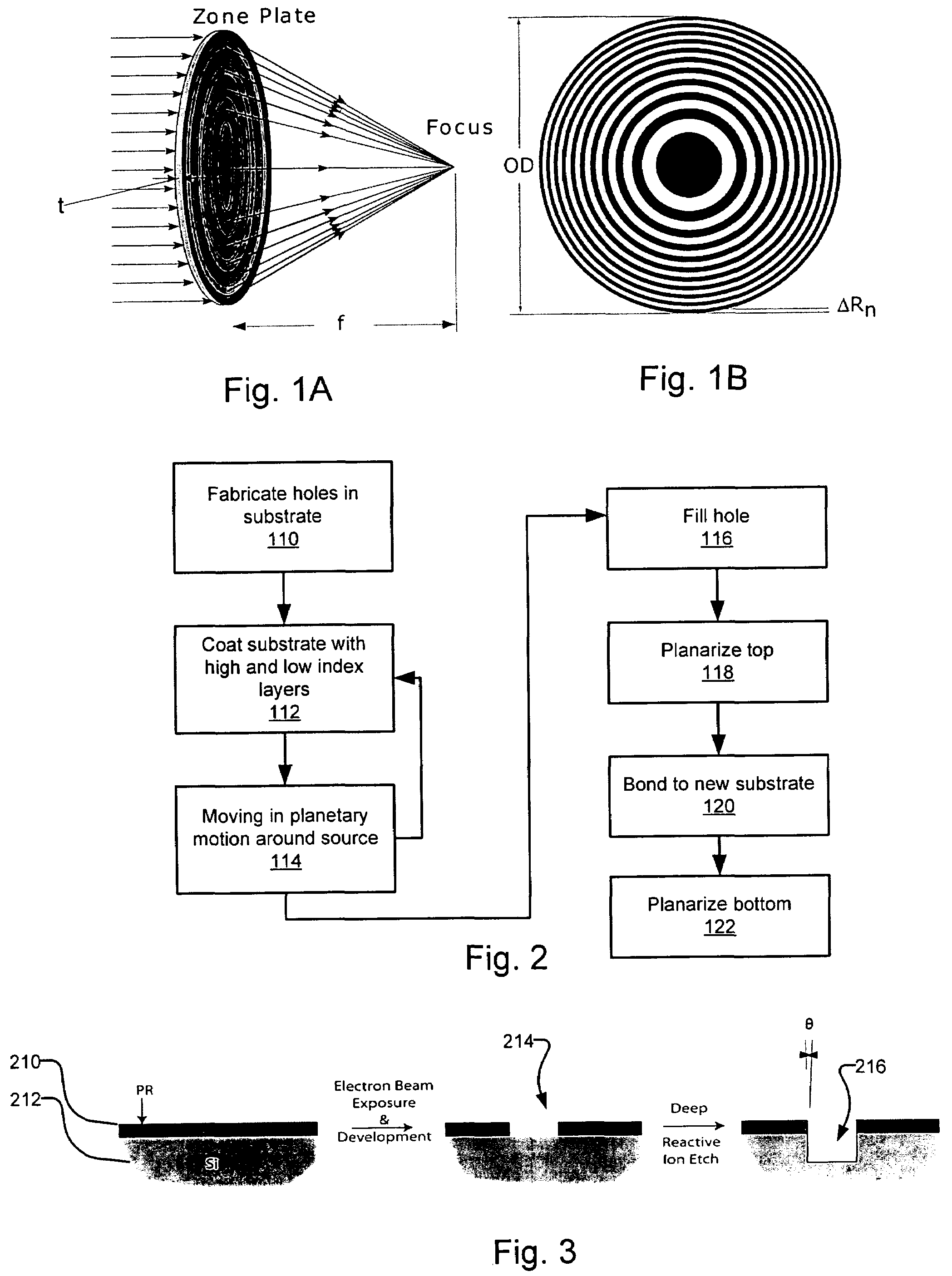
[0014]The approach can be used to develop a fast x-ray lens that will improve the performance of all the x-ray techniques described above, which are limited by the currently available x-ray lenses. One example is a diffractive zone plate lens with an outermost zone width of about 7 nm and with a thickness optimized for a desired x-ray energy working over a large range of energies. It can thus have a numerical aperture (NA) that is 5-10 times bigger than the NA of x-ray focusing optics currently available. Its figures of merit, such as resolution and throughput, are proportional to NA or NA2 respectively. It will enable the development of 3-D x-ray microscopy and x-ray elemental mapping with sub-10 nm resolution using synchrotron x-ray sources, and a compact laboratory source-based protein crystallography system with a throughput more than 10 times better than the best commercially available laboratory system at a significantly lower cost.
[0015]An x-ray microscope utilizing the disclosed lenses technology can have better than 20 nm spatial resolution for 2-D imaging and 60 nm for 3-D imaging. These values are significantly inferior to those of available electron microscopes but the proposed x-ray microscope is well compensated by its other unique capabilities, such as imaging single cells and relatively large tissue samples in their entirety without cross sectioning and in their natural state without staining, and substantially higher sensitivity for elemental analysis. Furthermore, its resolution may be sufficient for many important applications and it will offer significantly higher throughput in 3-D imaging of single cells and tissues compared to electron microscopy.

Problems solved by technology

While the progress in the fabrication of hard x-ray zone plates has significantly advanced within the last few years, the existing pattern transfer fabrication processes may reach a practical limit very soon.
As the polymer structures of the electroplating mold become smaller and smaller in width they loose strength and tend to collapse during the fabrication process.
Also the directionality of the reactive ion etch may impose practical limits to the achievable sidewall angle in the resist, limiting the achievable width of features that can be fabricated.
It has been found, however, that for these “Sputter-Sliced zone plates” imperfections in the wire and defects during deposition lead to accumulative errors in the fine outermost zones, which are deposited last.
Furthermore, it proved very difficult to slice the wire without deforming the delicate zone plate structure.
The resulting zone plates therefore fell short of expectations leading to the abandonment of this project.

Method used


Image

  • Fast x-ray lenses and fabrication method therefor
  • Fast x-ray lenses and fabrication method therefor
  • Fast x-ray lenses and fabrication method therefor

Examples


Embodiment Construction

[0029]The two most important parameters for a home-lab based protein crystallography system are the throughput and the quality of diffraction spots. Because a zone plate lens has the best focusing property among x-ray focusing elements as demonstrated by its achievement of sub-100 nm resolution and a large field of view, it provides high quality diffraction data sets with clean diffraction spots. The figure of merit of a zone plate lens in terms of throughput for a zone plate based home-lab protein crystallography system is discussed below.

[0030]In general, the flux F (in photons per second) incident on a protein crystal in a home-lab protein crystallography system using a focusing optic such as a zone plate or a reflection based focusing mirror is given by

F=ηBcL2Δθ2,  (1)

[0031]where Bc, L, and Δθ is the beam brightness, the linear dimension, and the divergence of the illumination beam at the position of the protein crystal, respectively; and η the focusing efficiency of the focusin...


PUM


Abstract

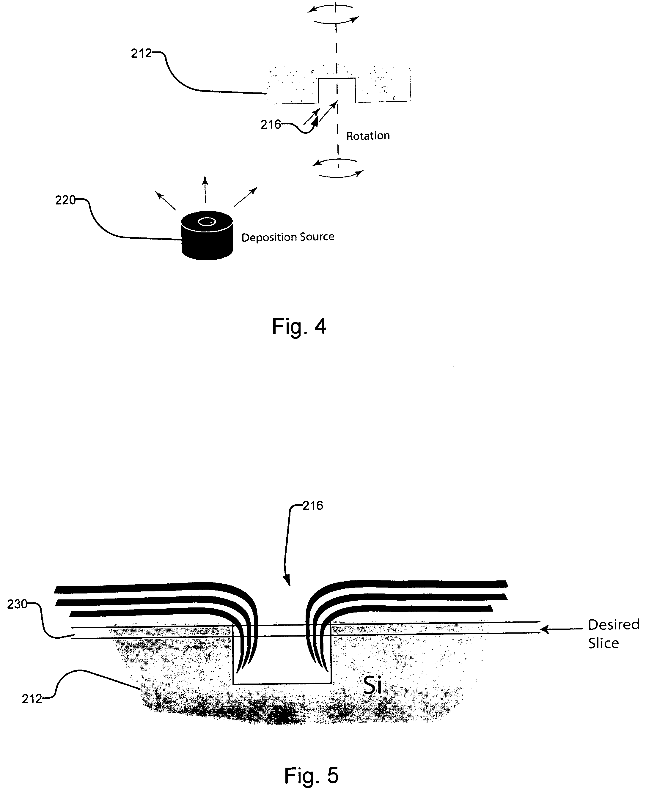
A fabrication process for zone plate lenses is based on controlled thin layer deposition for fabricating structures as small as 2 nanometers (nm) in width, and potentially smaller. The substrate for deposition will take the form of a precision hole, fabricated in a substrate, such as silicon by electron beam lithography and subsequent reactive ion etching. A controlled layer deposition is then used to form the required zone plate structure. A subsequent thinning process is used to section the hole and produce a zone plate with the required layer thicknesses.

Description

RELATED APPLICATIONS[0001]This application claims the benefit of U.S. Provisional Application No. 60 / 600,159 filed on Aug. 10, 2004 which is incorporated herein by reference in its entirety.BACKGROUND OF THE INVENTION[0002]X-ray imaging technology plays a critical and important role in healthcare and biomedical research. X-ray radiography is one of the most important and widely deployed medical diagnostic methods that can be traced back to the invention of x-rays more than 100 years ago. Recently, soft x-ray cryo-microscopy has been demonstrated to offer non-destructive high resolution three dimensional tomographic imaging of single biological cells with a resolution of about 25 nanometers (nm) in two dimensions (2-D) and 60 nm in three dimensions (3-D). Important trace elements in a biological specimen such as a cell or tissue can be mapped with sensitivity better than parts per billion at a spatial resolution of about 100 nm using a synchrotron based x-ray fluorescence microscope....

Claims


Application Information

Patent Timeline
IPC IPC(8): B02B3/08
CPCG21K1/06G02B5/1876G02B5/1847
Inventor YUN, WENBINGFESER, MICHAELLYON, ALAN
Owner CARL ZEISS X RAY MICROSCOPY
Learn More