
Microneedle device for extraction and sensing of bodily fluids

a micro-needle and bodily fluid technology, applied in the field of micro-needle devices for the extraction and sensing of biological fluids, can solve the problems of increasing the risk of disease transmission, inconvenient, expensive, undesirable, inconvenient and expensive, etc., and achieves painless and convenient manner, minimal or no damage, pain or irritation to the tissue, and minimal or no damage

Inactive Publication Date: 2008-03-18
GEORGIA TECH RES CORP +1
View PDF77 Cites 559 Cited by
  • Summary
  • Abstract
  • Description
  • Claims
  • Application Information

AI Technical Summary

Benefits of technology

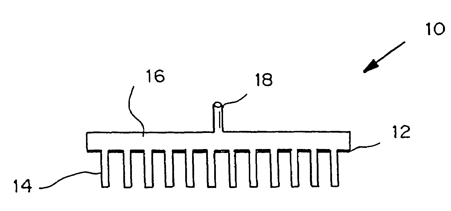
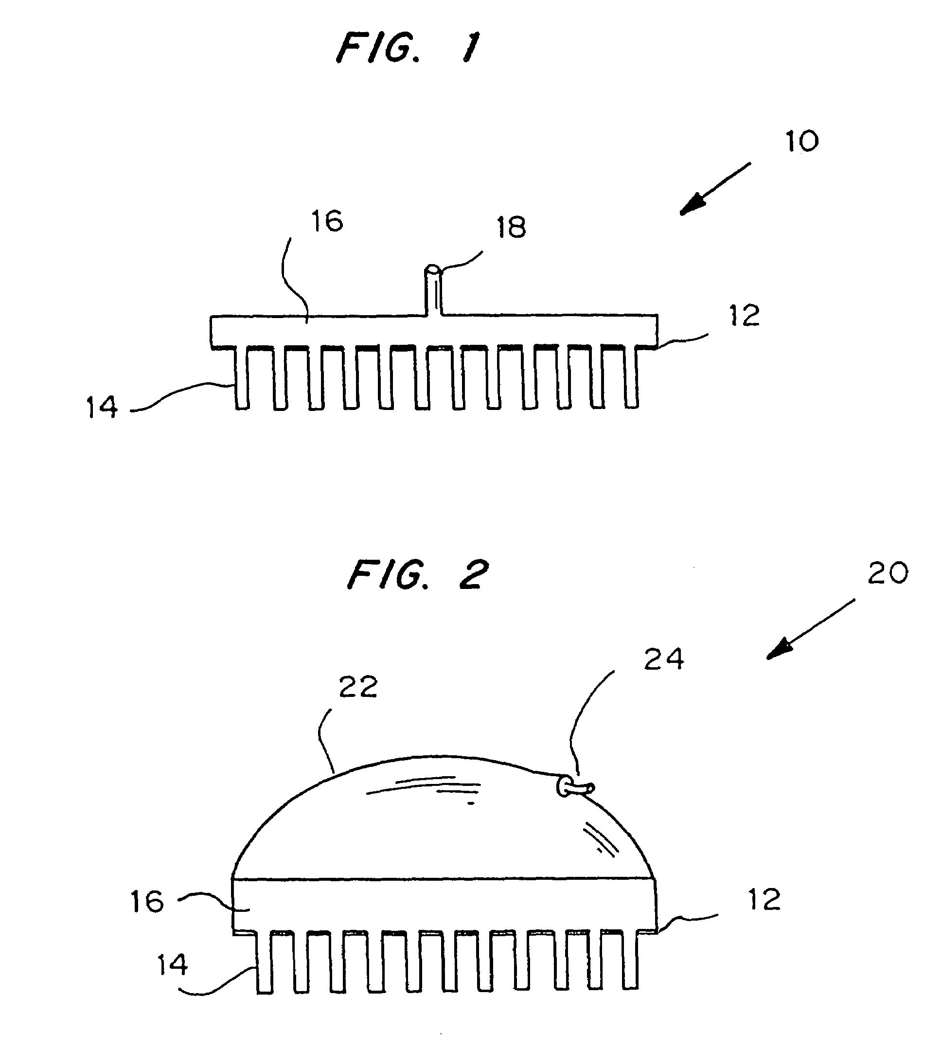
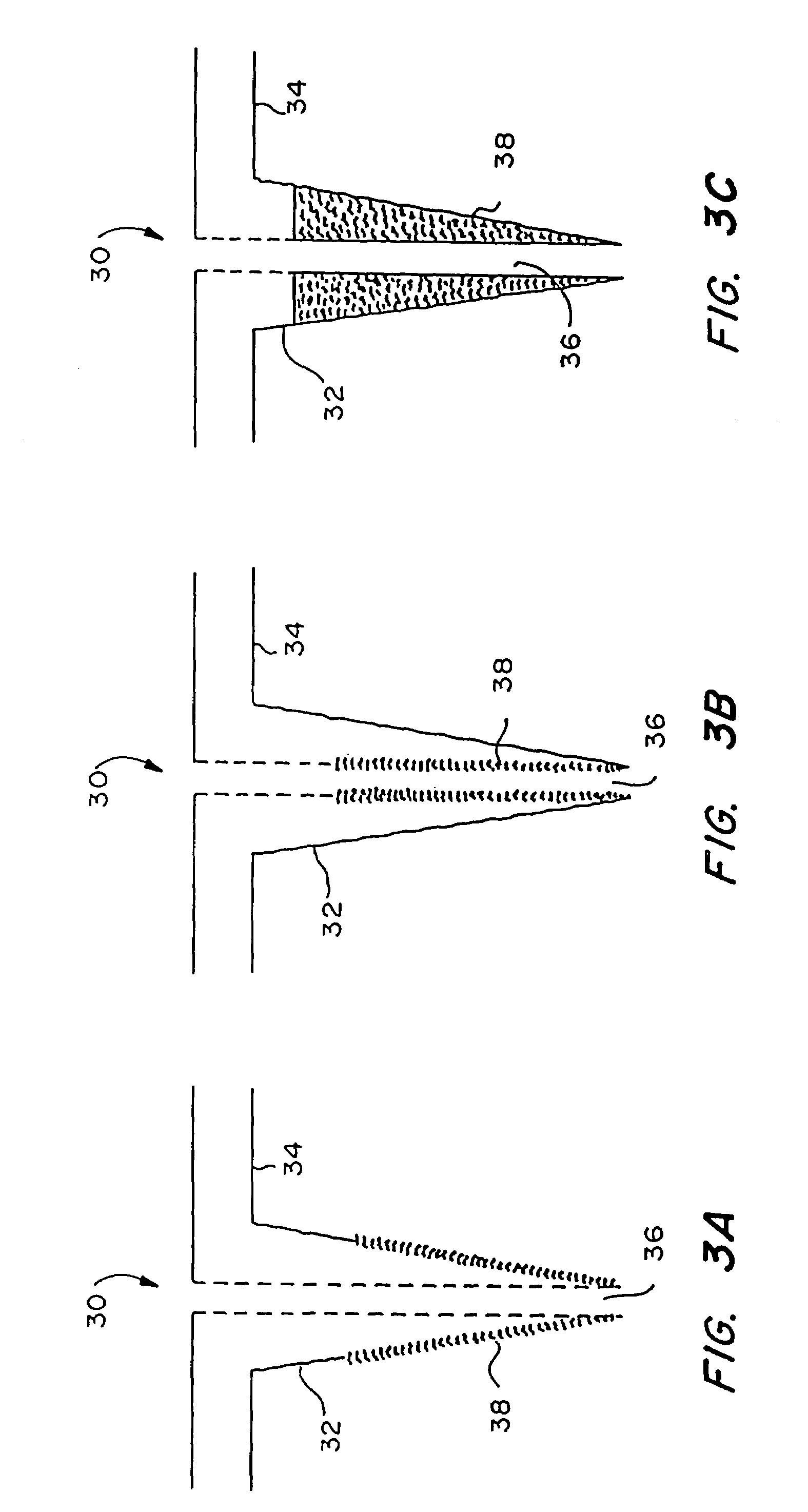
[0008]Microneedle devices for controlled sampling of biological fluids in a minimally-invasive, painless, and convenient manner, are provided. The microneedle devices permit in vivo sensing or withdrawal of biological fluids from the body, particularly from or through the skin or other tissue barriers, with minimal or no damage, pain, or irritation to the tissue.

Problems solved by technology

This method of glucose measurement has the major disadvantage that it is painful, so diabetics do not like to obtain a glucose measurement as often as is medically indicated.
While effective for this purpose, needles are invasive and generally cause pain; local damage to the skin at the site of insertion; and bleeding, which can increase the risk of disease transmission.
Needle techniques also generally require administration by one trained in its use, and are not preferred for frequent routine use due to the vascular damage caused by repeated punctures.
Some proposed alternatives to the needle require the use of (1) lasers or heat to create a hole in the skin, which is inconvenient, expensive, or undesirable for repeated use; (2) electric fields or ultrasound, which also is inconvenient and expensive; or (3) chemical or biological penetration enhancing agents, which can be irritating to the tissue and undesirable for repeated use.

Method used


Image

  • Microneedle device for extraction and sensing of bodily fluids
  • Microneedle device for extraction and sensing of bodily fluids
  • Microneedle device for extraction and sensing of bodily fluids

Examples


example 1

Transport of Molecules Through Microneedles Inserted into Skin

[0144]Studies were performed to demonstrate transport of molecules using either solid silicon microneedles or using hollow silicon microneedles. Transport was measured across human cadaver epidermis in vitro using Franz diffusion chambers at 37° C. using methods described in Henry, et al., “Microfabricated microneedles: A novel method to increase transdermal drug delivery”J. Pharm. Sci. 87:922-25 (1998).

[0145]Removal of calcein was measured. Removal refers to the ability to transport calcein from the viable epidermis side of the epidermis to the stratum corneum side. This is the direction of transport associated with removing from the body compounds found in the body, such as glucose. Other compounds were tested for delivery only.

[0146]In all cases shown in Table 1, transport of these compounds across skin occurred at levels below our detection limit when no needles were inserted into the skin. When solid microneedles wer...

example 2

Flow of Water Through Hollow Microneedles

[0148]To demonstrate that fluid can be forced through hollow microneedles at meaningful rates, the flow rate of water through a microneedle array was measures as a function of pressure. The array used contained 100 hollow silicon microneedles having an inner diameter of 50 μm and an outer diameter of 80 μm. The results, which are shown in Table 2, demonstrate that significant flow rates of water through microneedles can be achieved at modest pressures. The measured flow rates are comparable to flow rates through hypodermic needles attached to syringes.

[0149]

TABLE 2Flow Rate of Water Through Hollow Silicon MicroneedlesAs a Function of Applied PressurePressure (psi)Flow rate (ml / min)1.0161.5242.0312.5383.045

example 3

Fabrication of Solid Silicon Microneedles

[0150]A chromium masking material was deposited onto silicon wafers and patterned into dots having a diameter approximately equal to the base of the desired microneedles. The wafers were then loaded into a reactive ion etcher and subjected to a carefully controlled plasma based on fluorine / oxygen chemistries to etch very deep, high aspect ratio valleys into the silicon. Those regions protected by the metal mask remain and form the microneedles.

[0151]-oriented, prime grade, 450-550 μm thick, 10-15 Ω-cm silicon wafers (Nova Electronic Materials Inc., Richardson, Tex.) were used as the starting material. The wafers were cleaned in a solution of 5 parts by volume deionized water, 1 part 30% hydrogen peroxide, and 1 part 30% ammonium hydroxide (J. T. Baker, Phillipsburg, N.J.) at approximately 80° C. for 15 minutes, and then dried in an oven (Blue M Electric, Watertown, Wis.) at 150° C. for 10 minutes. Approximately 1000 Å of chromium (Mat-Vac Tec...


PUM


Abstract

Microneedle devices are provided for controlled sampling of biological fluids in a minimally-invasive, painless, and convenient manner. The microneedle devices permit in vivo sensing or withdrawal of biological fluids from the body, particularly from or through the skin or other tissue barriers, with minimal or no damage, pain, or irritation to the tissue. The microneedle device includes one or more microneedles, preferably in a three-dimensional array, a substrate to which the microneedles are connected, and at least one collection chamber and / or sensor in communication with the microneedles. Preferred embodiments further include a means for inducing biological fluid to be drawn through the microneedles and into the collection chamber for analysis. In a preferred embodiment, this induction is accomplished by use of a pressure gradient, which can be created for example by selectively increasing the interior volume of the collection chamber, which includes an elastic or movable portion engaged to a rigid base. Preferred biological fluids for withdrawal and / or sensing include blood, lymph, interstitial fluid, and intracellular fluid. Examples of analytes in the biological fluid to be measured include glucose, cholesterol, bilirubin, creatine, metabolic enzymes, hemoglobin, heparin, clotting factors, uric acid, carcinoembryonic antigen or other tumor antigens, reproductive hormones, oxygen, pH, alcohol, tobacco metabolites, and illegal drugs.

Description

CROSS-REFERENCE TO RELATED APPLICATIONS[0001]This application is a continuation-in-part of U.S. patent application Ser. No. 09 / 316,229, now U.S. Pat. No. 6,334,856, filed May 21, 1999, which is a continuation-in-part of U.S. patent application Ser. No. 09 / 095,221, now U.S. Pat. No. 6,503,231, filed Jun. 10, 1998. This application claims the benefit of the filing date of U.S. provisional application Ser. No. 60 / 146,200, filed Jul. 29, 1999.STATEMENT REGARDING FEDERALLY SPONSORED RESEARCH[0002]This invention was made with government support under Contract Number BES-9813321 awarded by the U.S. National Science Foundation. The government has certain rights in the invention.BACKGROUND OF THE INVENTION[0003]This invention is generally in the field of devices for the withdrawal and sensing of biological fluids, such as in the analysis of blood glucose levels.[0004]The extraction of analytes through the skin is critical to diabetic patients, who typically must measure blood glucose several...

Claims


Application Information

Patent Timeline
IPC IPC(8): A61B5/00
CPCA61B5/1411A61M37/0015A61B5/14532A61B5/418A61B5/150022A61B5/150221A61B5/150229A61B5/150282A61B5/150358A61B5/150984B33Y80/00
Inventor PRAUSNITZ, MARK R.ALLEN, MARK G.GUJRAL, INDER-JEET
Owner GEORGIA TECH RES CORP
Learn More