MR imaging providing tissue/blood contrast image
- Summary
- Abstract
- Description
- Claims
- Application Information
AI Technical Summary
Benefits of technology
Problems solved by technology
Method used
Image
Examples
first embodiment
[0075]A first embodiment of the present invention will now be described FIGS. 2 to 7.
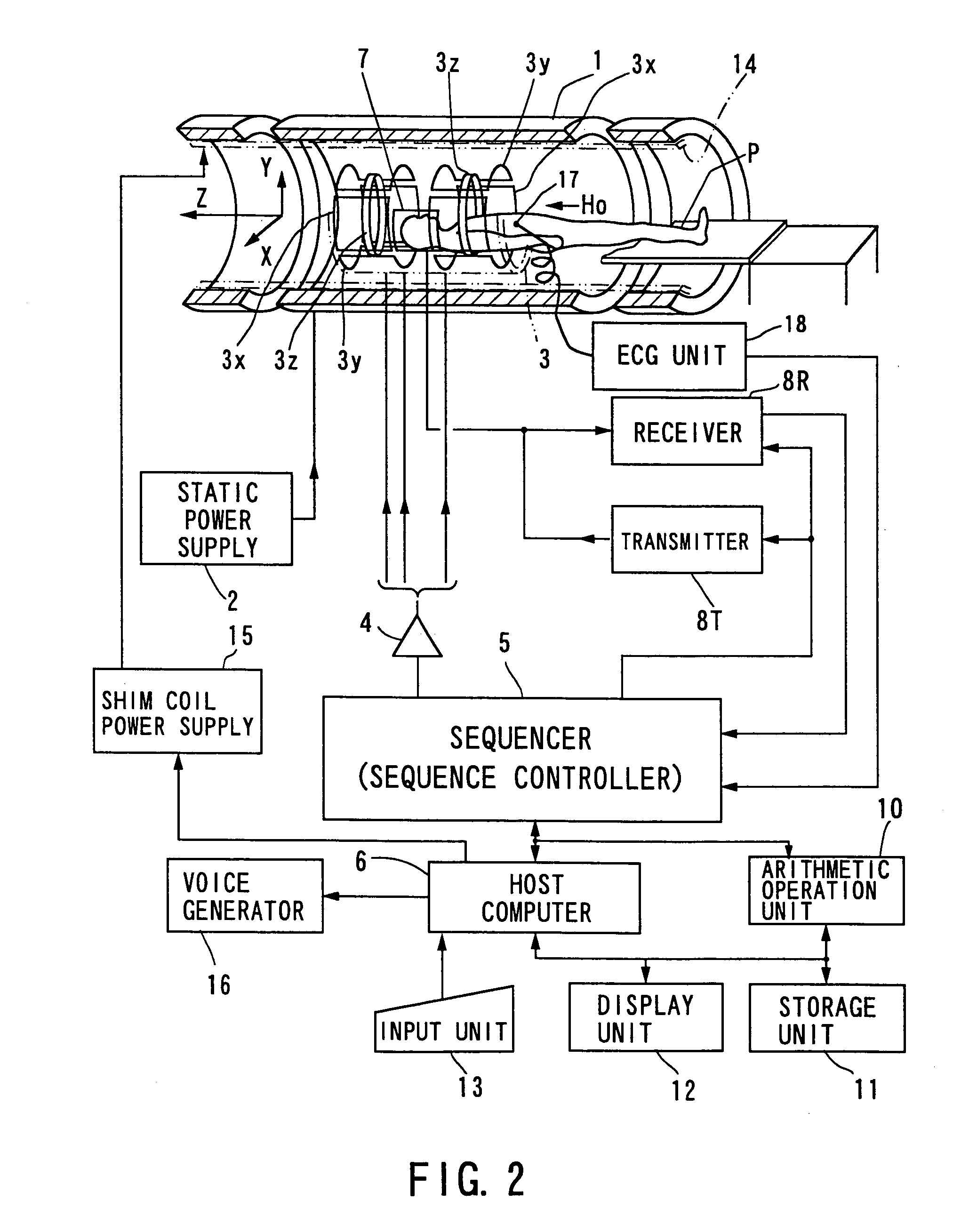
[0076]FIG. 2 shows the outlined configuration of a magnetic resonance imaging (MRI) system in accordance with the embodiment of the present invention, which will be described below.
[0077]The MRI system comprises a patient couch on which a patient P lies down, static magnetic field generating components for generating a static magnetic field, magnetic field gradient generating components for appending positional information to a static magnetic field, transmitting / receiving components for transmitting and receiving a radio-frequency (RF) signal, control and arithmetic operation components responsible for control of the whole system and for image reconstruction, electrocardiographing components for acquiring an ECG signal of a patient, which is a representative of signals indicative of cardiac temporal phases of the patient, and breath hold instructing components for instructing the patient to perform...
second embodiment
[0146]Referring to FIGS. 10 to 17, a second embodiment of the present invention will be described.
[0147]In this embodiment, using a plurality of divided MT pulses described above, the parenchyma of the lungs of a patient will be imaged.
[0148]For MR-imaging the lungs, three approaches have been known primarily, which are to use a hyper-polarized gas (e.g., xenon or helium), to perform a perfusion imaging using a contrast medium Gd-DTPA (refer to “Hatabu H, et al., MRM 36:503-508, 1996”), and to perform imaging with suction of oxygen using oxygen molecules (refer to “Edelman RR, et al., Nature Medicine 2, 11, 1236-1239, 1996).
[0149]Of these, the first approach is based on imaging at the MR frequency of, for example, a xenon gas (Xe) suctioned into the lungs. The second one is a technique to observe a perfused state of Gd-DTPA in blood. The third one utilizes a report that oxygen molecules, which are weakly paramagnetic, cause a signal from water to change sufficiently at the surface o...
third embodiment
[0195]Referring to FIGS. 18 to 20, a third embodiment of the present invention will be described.
[0196]The hardware construction of an MRI system of this embodiment is similar to those described in the foregoing embodiments.
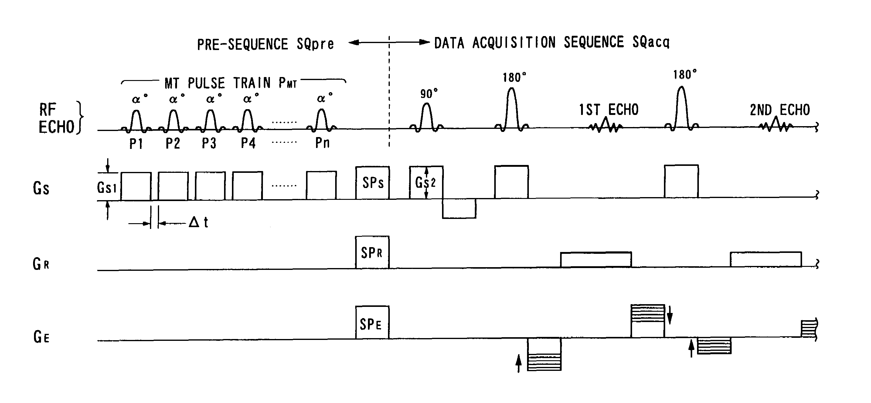
[0197]The basic feature of the MRI system according to this embodiment is to use pulse sequences based on a single-shot (RF excitation) fast SE (spin echo) method on three-dimensional or two-dimensional slab (thick slice) scanning under ECG gating, in which MT (magnetization transfer) pulses of which flip angles and of which number are changeable are applied. The MT pulses are applied to give images MT contrasts based on MT effects fitted to imaging objects, and functions as means for produce an effective contrast between tissue and blood. Such pulse sequences are most effective in depicting the cardiac blood vessel systems. For instance, abdomen organs such as the heart can be imaged to provide T2-weighted contrast images. These images provides a tissue contrast...
PUM
 Abstract
Description
Claims
Application Information
 - R&D
- Intellectual Property
- Life Sciences
- Materials
- Tech Scout
- Unparalleled Data Quality
- Higher Quality Content
- 60% Fewer Hallucinations
Browse by: Latest US Patents, China's latest patents, Technical Efficacy Thesaurus, Application Domain, Technology Topic, Popular Technical Reports.
© 2025 PatSnap. All rights reserved.Legal|Privacy policy|Modern Slavery Act Transparency Statement|Sitemap|About US| Contact US: help@patsnap.com


