
Movement-corrected multi-shot method for diffusion-weighted imaging in magnetic resonance tomography

a magnetic resonance tomography and motion correction technology, applied in the field of nuclear magnetic resonance tomography, can solve the problems of strong b0 field dependence of measurement signal, marked sensitivity, and strong image artifacts

Active Publication Date: 2007-04-17
SIEMENS HEALTHCARE GMBH
View PDF5 Cites 64 Cited by
  • Summary
  • Abstract
  • Description
  • Claims
  • Application Information

AI Technical Summary

Benefits of technology

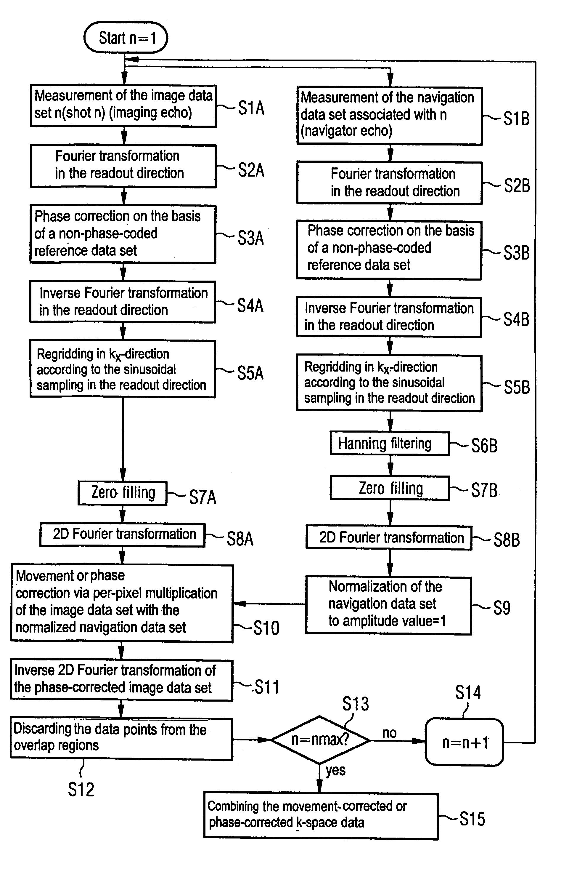
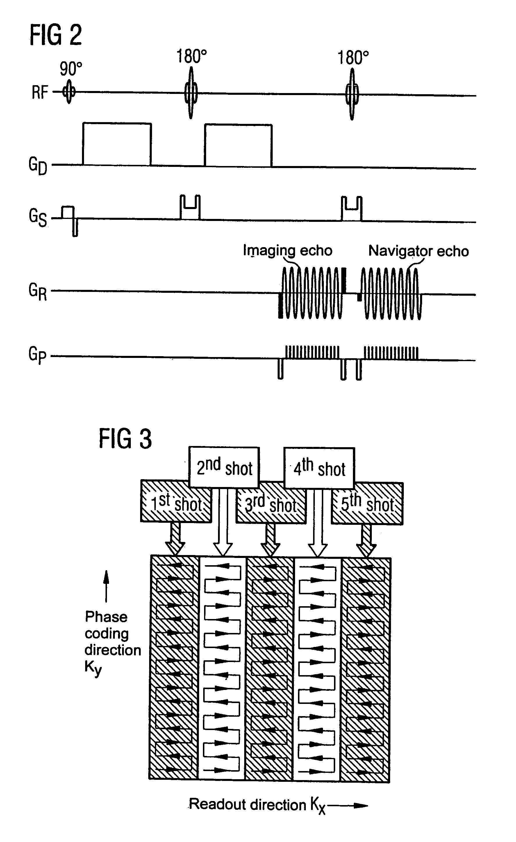
The present invention provides an improved method for movement-corrected diffusion-weighted imaging in magnetic resonance tomography. The method involves a multi-shot method for diffusion-weighted imaging, where scanning of the k-matrix ensues in segments in the readout direction. The imaging sequence is designed such that, simultaneously with the readout of a segment by means of an imaging echo, a navigation data set is acquired by readout of the central region of the k-matrix by means of a navigator echo. The image data sets and the navigation data sets are then combined using a pixel-by-pixel multiplication method. The readout advantageously ensues along serpentine path. The invention also includes optional standard phase correction, Hanning filtering, and zero-filling steps on each data set and corresponding navigation data set. The method allows for a serpentine shaped scan of the respective sub-range by activating short phase-coded gradient pulses during each zero point of the respective readout gradient pulse train.

Problems solved by technology

One problem of the diffusion-weighted imaging is, however, the marked sensitivity to non-diffusion-like movement types such as: heart movement, breathing movement, etc. and the movements associated with these such as, for example, brain pulsation (movement of the brain in cerebral fluid).
In particular in multi-shot sequences in MRT, such movements cause phase shifts in the nuclear magnetic resonance signal during the respective diffusion preparation, which leads to strong image artifacts.
A disadvantage of SSEPI, however, is that, due to the low bandwidth per pixel in the phase coding direction, a strong B0 field dependency of the measurement signal exists.
In regions with strong susceptibility gradients (such as, for example, in front temporal lobes or in the frontal lobes of the human brain) this leads to strong image artifacts.
This leads to the image interference differing dependent on different preparation procedures and the image reconstruction then being impaired when diffusion-weighted images are combined in order to create parameter maps such as, for example, ADC (Apparent Division Coefficient) maps.
A further disadvantage of the SSEPI sequence is the very significant T2* dependency (T2* is the decay duration of the transverse magnetization due to local magnetic field inhomogeneities) o the very strong phase sensitivity, dependent on the type of the phase coding of an SSEPI sequence.
Both result in strong image erasure artifacts and distortion artifacts, in particular in body imaging with the typically short T2 times of human tissue.
The refocusing of the magnetization inverts the phase curve, making the sequence insensitive with regard to susceptibility artifacts.
The T2 decay of the magnetization limits the signal readout time to approximately 300 ms, which in turn limits the maximum resolution that can be achieved.
In diffusion-weighted imaging, the readout of a number of spin echoes also has the severe disadvantage that movement-induced phase shifts of the magnetization occur during the diffusion preparation that negate the Carr-Purcell-Meiboom-Gill (CPMG) condition.
Non-fulfillment of the CPMG condition and artifacts caused thereby are more likely to occur as fewer RF or refocusing pulses are used.
Just like EPI, this technique does not use multiple RF refocusing pulses, which leads to a phase evolution due to off-resonant signals that severely impairs the image quality.
The simple combination of a standard multi-shot sequence with a preliminary diffusion preparation using diffusion gradients leads to a movement-induced phase shift from shot to shot that is manifested by extremely strong ghost artifacts, in particular when brain exposures are made.
However, a limitation of the resolution exists.
High-speed Steam likewise exhibiting the disadvantage of a relatively low signal-to-noise ratio.
The movement-induced phase change in the diffusion-weighted imaging is a two-dimensional function, such that the one-dimensional approach is not suitable for a complete correction.
This method consequently is not suitable for a clinical implementation in the form of a routine application.
However this leads to the scanning (sampling) not fulfilling the Nyquist condition and wrap-arounds occurring in the image domain.
Such an assumption of rigid body motion is not valid for the brain deformation, in particular of the ventricle and of the brain stem.
However, with PROPELLER the CPMG condition generally is not fulfilled, which leads to a signal modulation between adjacent spin echoes.
A radial scan as is implemented with PROPELLER is also less efficient in comparison to standard scan sequences such as, for example, segmented EPI, since in comparison to other sequences a much larger number of shots is necessary for a given matrix size.
However, such developments in this field do not include the two-dimensional phase correction necessary for a robust clinical application.

Method used


Image

  • Movement-corrected multi-shot method for diffusion-weighted imaging in magnetic resonance tomography
  • Movement-corrected multi-shot method for diffusion-weighted imaging in magnetic resonance tomography
  • Movement-corrected multi-shot method for diffusion-weighted imaging in magnetic resonance tomography

Examples


Embodiment Construction

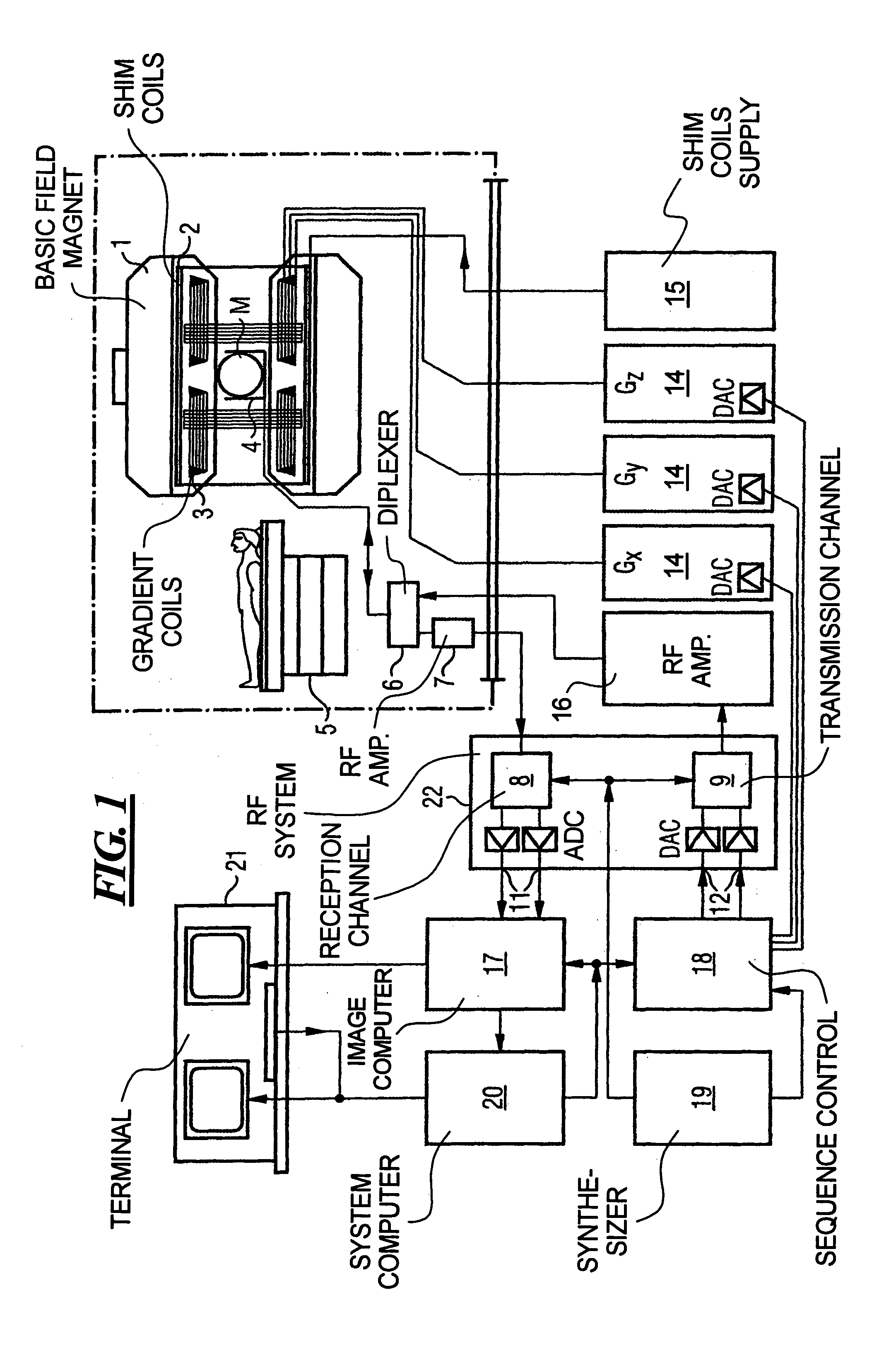
[0055]FIG. 1 schematically illustrates a magnetic resonance tomography apparatus in which gradient pulses according to the present invention are generated. The design of the magnetic resonance tomography apparatus corresponds a conventional tomography apparatus, with the exceptions discussed below. A basic field magnet 1 generates a temporally constant strong magnetic field for polarization or alignment of the nuclear spins in the examination region of the subject such as, for example, a part of a human body to be examined. The high homogeneity of the basic magnetic field necessary for the magnetic resonance data acquisition is defined in a spherical measurement volume M in which the parts of the human body to be examined are introduced. For support of the homogeneity requirements, and in particular for elimination of temporally invariable influences, shim plates made from ferromagnetic material are mounted at a suitable location. Temporally variable influences are eliminated by shi...


PUM


Abstract

In a multi-shot method for diffusion-weighted imaging in magnetic resonance tomography, the sampling of the k-matrix in the readout direction ensues in segments, and immediately after acquisition of an image data set by readout of one segment by means of echo signals, a navigation data set is acquired by readout of the middle region of the k-matrix at virtually the same time. A movement-corrected diffusion-weighted MRT image is generated by combining of the image data sets with the corresponding navigation data sets and subsequent Fourier transformation.

Description

BACKGROUND OF THE INVENTION[0001]1. Field of the Invention[0002]The present invention concerns nuclear magnetic resonance tomography as applied in medicine for examination of patients. The present invention in particular concerns a movement-corrected multi-shot method for diffusion-weighted imaging in magnetic resonance tomography.[0003]2. Description of the Prior Art[0004]MRT is based on the physical phenomenon of nuclear magnetic resonance and has been successfully used as an imaging method for over 15 years in medicine and biophysics. In this examination method, the subject is exposed to a strong, constant magnetic field (called the B0 field). The nuclear spins of the atoms in the subject, which were previously randomly oriented, thereby align. Radio-frequency energy can now excite these “ordered” nuclear spins to a specific oscillation. In MRT, this oscillation generates the actual measurement signal that is acquired by appropriate reception coils. By the use of non-homogeneous ...

Claims


Application Information

Patent Timeline
Patent Type & Authority Patents(United States)
IPC IPC(8): G01V3/00A61B5/055G01R33/561G01R33/563G01R33/565G01R33/567
CPCG01R33/56341G01R33/56509
Inventor PORTER, DAVID ANDREW
Owner SIEMENS HEALTHCARE GMBH
Learn More