
Urinary tract tissue graft compositions and methods for producing same

a technology of urinary tract and composition, which is applied in the field of tissue reconstruction and repair, can solve the problems of monetary cost to our health care system of treating children with dysfunctional bladders in the millions of dollars each year, affecting the use of bowel, and causing bowel obstruction, etc., and achieves the effects of reducing the number of bowel obstructions

Inactive Publication Date: 2006-07-18
THE BOARD OF RGT UNIV OF OKLAHOMA
View PDF7 Cites 3 Cited by
  • Summary
  • Abstract
  • Description
  • Claims
  • Application Information

AI Technical Summary

Benefits of technology

This approach allows for consistent and reliable production of functional urinary tract tissue with reduced risk of complications, improving bladder regeneration by providing a larger, functional tissue graft that resembles normal tissue architecture.

Problems solved by technology

These clinical presentations are highlighted by urinary incontinence, urinary tract infections and decreased bladder compliance that leads to increased pressure transmission to the upper urinary tract which leads to subsequent renal deterioration.
The monetary cost to our health care system of treating children with dysfunctional bladders runs into millions of dollars each year.
Deleterious side effects associated with the use of bowel include infection, intestinal obstruction, mucus production, electrolyte abnormalities, perforation and neoplasia.
The major obstacle to advancing the field of urinary tract reconstruction and rehabilitation has been the availability of a biomaterial, either permanent or biodegradable, that will function as a suitable scaffold to allow the natural process of regeneration to occur.
There are two major obstacles for in vivo or unseeded tissue engineering technology for bladder regeneration.
However, despite initial encouraging results, none of these materials have been found to be suitable for clinical use.
It has been reported that bladders augmented with dura, peritoneum, placenta and fascia contract over time, and that such tissue grafts fail to promote complete bladder wall regeneration (i.e., tissue having a urine impermeable layer and a functional muscle cell layer) (Kelami, et al, J.
The second potential limitation of the unseeded tissue engineering technique for bladder regeneration is that the size of the graft may be limited to the amount of area which can be quickly invested with bladder cells from the remaining native bladder, and therefore may not be sufficient for bladder replacement.
If the ratio of the size of the unseeded graft to the amount of native bladder tissue becomes too large, the ability of the animal to invest the graft with SMC and urothelial cells appears to be compromised.
In the absence of quickly covering the graft with bladder cells, contraction and excess scar formation becomes a concern and poor clinical outcomes may result.
In addition, while the obstacle of identifying a biomaterial that will act as a suitable scaffold for the natural process of bladder regeneration to occur is overcome by the use of SIS in unseeded tissue engineering technology, the obstacle of the limited size of a graft formed therefrom still exists.
However, over the last few years multiple problems have been encountered with different small intestinal segments, including calcifications and graft shrinkage, and therefore unreliable and inconsistent results have been obtained in the experimental use of this material for bladder augmentation.
They further demonstrated that human transitional epithelium and smooth muscle cells grown on the biodegradable polymers could then be implanted into athymic mice and grown in vivo, and that the tissue architecture became progressively more complex with time in the animal.
Unfortunately, although the in vitro technique of tissue engineering has been shown to be feasible for both synthetic and xenogenic matrices, thus far no studies have been undertaken to determine the effectiveness of the materials to facilitate the regeneration of functional bladder tissue in a large animal.
In addition, while all segments of small intestinal submucosa have been used to promote urinary bladder regeneration, multiple problems have been encountered with different small intestinal segments, including calcifications and graft shrinkage, and therefore unreliable and inconsistent results have been obtained in the experimental use of this material for bladder augmentation.
However, thus far no studies have been undertaken to determine if the effectiveness of one segment of small intestine over another and to determine if the use of one segment of small intestine over another has any effect on the consistency and reliability of the grafts formed therefrom.

Method used


Image

  • Urinary tract tissue graft compositions and methods for producing same
  • Urinary tract tissue graft compositions and methods for producing same
  • Urinary tract tissue graft compositions and methods for producing same

Examples


example 1

Materials and Methods

[0066]Isolation of Distal Ileal Segment of SIS.

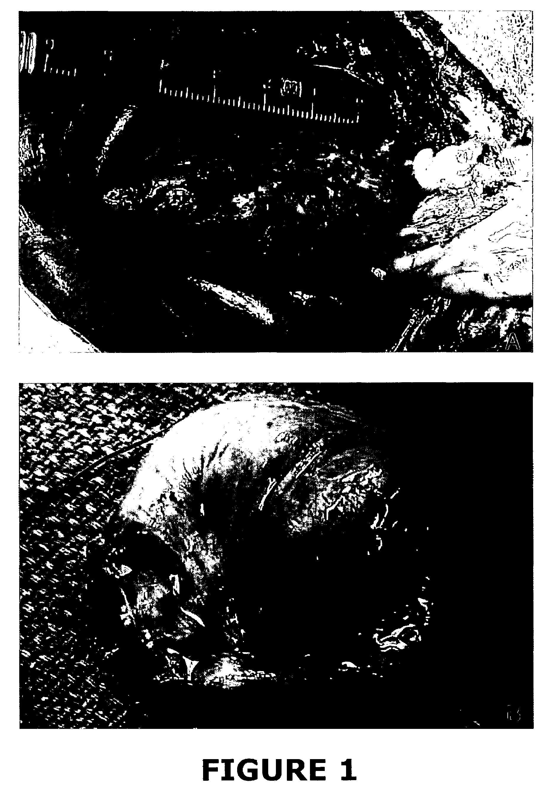
[0067]The entire small intestine of a pig greater than three years of age (i.e., a sow) was obtained, and the terminal ileum was identified. The bowel was then throroughly cleansed with water. The cleansed bowel was opened on the antimesenteric borders, and the terminal portion of the bowel was inspected. There were clear areas of Peyer's patches, and the areas just adjacent to Peyer's patches were used. These areas were thoroughly stripped of muocsa using a gauze sponge and mechanical force. The serosa muscle layers were also stripped using a gauze sponge and mechanical abrasion. The remaining submucosa layer of the small intestine was placed in 0.1% paracetic acid and 20% ethanol for twelve to twenty-four hours. The material was then thoroughly rinsed and placed in sterile water. Only bowel that was within 300 cm of the terminal ileum and only bowel that was closely associated with Peyer's patches was utilized. Pe...

example 2

Method and Materials

[0078]Tissue Samples.

[0079]Human bladder specimens were obtained from 11 patients 2 to 11 years old with primary vesicouretal reflux undergoing open operations for ureteral reimplantation. None of the patients had clinical evidence of bladder dysfunction or neuropathic bladder. A small portion of the tissue was sent for routine histology and the remainder was used to establish primary cultures. Bladder tissue was obtained and processed in conjunction with approval from the Institutional Review Board.

[0080]Tissue samples from dog bladders were obtained from five adult male beagles (weighing between 11 and 13 kg) undergoing partial cystectomy for bladder augmentation and were used for establishing primary cell cultures. Bladder tissue was obtained and processed in conjunction with approval from the Institutional Animal Care and Use Committee.

[0081]Establishment of Primary SMC and UC Cultures.

[0082]Under the dissecting microscope the bladder mucosa was dissected off...


PUM

PropertyMeasurementUnit
wall thicknessaaaaaaaaaa
sizeaaaaaaaaaa
widthaaaaaaaaaa

Abstract

A method for repairing damaged or diseased urinary tract tissue includes providing a urinary tract tissue graft composition that includes a distal ileal segment of small intestinal submucosa. The distal ileal segment of small intestinal submucosa may be utilized as an unseeded tissue graft composition, or the distal ileal segment of small intestinal submucosa may be positioned in a tissue culture frame, and smooth muscle and urothelial cells isolated from a tissue specimen of a subject and cultured are then seeded upon the distal ileal segment of small intestinal submucosa, thereby forming a urinary tract tissue graft.

Description

CROSS-REFERENCE TO RELATED APPLICATIONS[0001]This application claims the benefit under 35 U.S.C. 119(e) of U.S. Provisional Application Serial No. 60 / 338,608, filed Dec. 7, 2001, entitled “THE USE OF DISTAL ILEUM SUBMUCOSA FOR URINARY BLADDER REGENERATION”, the contents of which are hereby expressly incorporated by reference herein in their entirety.[0002]This application is also a continuation-in-part of U.S. Ser. No. 10 / 013,270, filed Dec. 10, 2001, entitled “IN VITRO ENGINEERED, REGENERATED URINARY TRACT TISSUE COMPOSITIONS AND METHODS FOR PRODUCING SAME”, the contents of which are hereby expressly incorporated by reference herein in their entirety.FEDERALLY SPONSORED RESEARCH OR DEVELOPMENT[0003]This application was supported in part by National Research Grants 1RO1DK56968-01A1 (Principal Investigator Bradley P. Kropp); 5RO1-DK54925-06 (Principal Investigator H. K. Lin) and 1RO1-CA90744-01 (Principal Investigator H. K. Lin) from the National Institutes of Health. The United Stat...

Claims


Application Information

Patent Timeline
Patent Type & Authority Patents(United States)
IPC IPC(8): C12N5/00A61F2/02A61K35/12A61K35/38
CPCA61F2/02A61K35/38A61L27/3629A61L27/3679A61L27/3813A61L27/3826A61L27/383A61L27/3882A61L27/3886C12N5/0684C12N5/0685C12N2533/92A61L2430/22C12N2502/23A61K35/12
Inventor KROPP, BRADLEY P.ZHANG, YUAN YUANCHENG, EARL Y.LIN, HSUEH-KUNGCOWAN, RICK
Owner THE BOARD OF RGT UNIV OF OKLAHOMA
Learn More