
Antigen-binding fragments specific for dendritic cells, compositions and methods of use thereof antigens recognized thereby and cells obtained thereby

Inactive Publication Date: 2006-04-18
MILTENYI BIOTEC B V & CO KG
View PDF64 Cites 66 Cited by
  • Summary
  • Abstract
  • Description
  • Claims
  • Application Information

AI Technical Summary

Benefits of technology

[0059]The invention further encompasses a method of inhibiting an interaction of a DC with a T cell by contacting a composition containing DC and T cells with an effective amount of an agent that inhibits the interaction of BDCA-2, BDCA-3, or BDCA-4 with the T cell.
[0060]The invention further encompasses a method of treating inflammation by ad

Problems solved by technology

APCs such as B cells and macrophages are typically incapable of inducing primary responses.
Animal models have demonstrated that DC tumor vaccines reverse T cell anergy and result in subsequent tumor rejection.
Studies on DC's in blood have been hampered by scarcity of the cells and the relative lack of DC-specific cell surface markers.
In addition, several attempts to generate mAb specific for human DC have failed, yielding only mAb that bind antigens expressed by both DC and other leukocytes.
These cytokines appear to play a significant role in recruiting eosinophils into the site of allergen exposure, where tissue damage and dysfunction result.
Many patients who begin the therapy do not complete the regimen, and if injections are missed for over a week, the patient must begin the entire treatment regimen again.
Antigen immunotherapy treatments present the risk of inducing potentially lethal IgE-mediated anaphylaxis and do not address the cytokine-mediated events of the allergic late phase response.
Another significant problem with antigen immunotherapy is that the risk of adverse reactions, especially anaphylaxis, significantly reduces the dosage of antigen both with respect to the amount given per administration and the amount given over a period of time.
Thus, traditional allergy immunotherapy is protracted and thus time-consuming, inconvenient, and expensive.
However, a drawback of this approach is that it merely masks the symptoms, while not providing any kind of permanent cure or protection.

Method used


Image

  • Antigen-binding fragments specific for dendritic cells, compositions and methods of use thereof antigens recognized thereby and cells obtained thereby
  • Antigen-binding fragments specific for dendritic cells, compositions and methods of use thereof antigens recognized thereby and cells obtained thereby
  • Antigen-binding fragments specific for dendritic cells, compositions and methods of use thereof antigens recognized thereby and cells obtained thereby

Examples


example 1

Generation of DC-Specific mAb

[0305]Five 6–8 week old female Balb / c mice (Simonsen Laboratories, Gilroy, Calif.) were inoculated with approximately 5×10⁵ to 1×10⁶ purified HLA-DR+lin−blood DC under anesthesia on d 0, 4, 7, 11, and 14 in the right hand footpad, and approximately 1×10⁶ HLA-A2+ Bristol-8 B lymphoblastoma cells in the left hand footpad on d-3, 0, 4, 7, 11, and 14. Both cell types were incubated with 1:100 PHA (Gibco / BRL, Gaithersburg, Md.) for 10 min at room temperature and washed with PBS before injection. This treatment provides non-specific adjuvant effects and obviates the need for adjuvants such as Freund's adjuvant.

[0306]On d 15, one day after the fifth injection of HLA-DR+lin−DC, the mouse right hand popliteal lymph nodes were removed. A lymphocyte suspension was prepared and the cells were fused to SP2 / 0 Ag14 myeloma cells using a modification of the method described by Kohler and Milstein (1975) Nature 256:495. Fused cells were plated on 96-well plates in DMEM s...

example 2

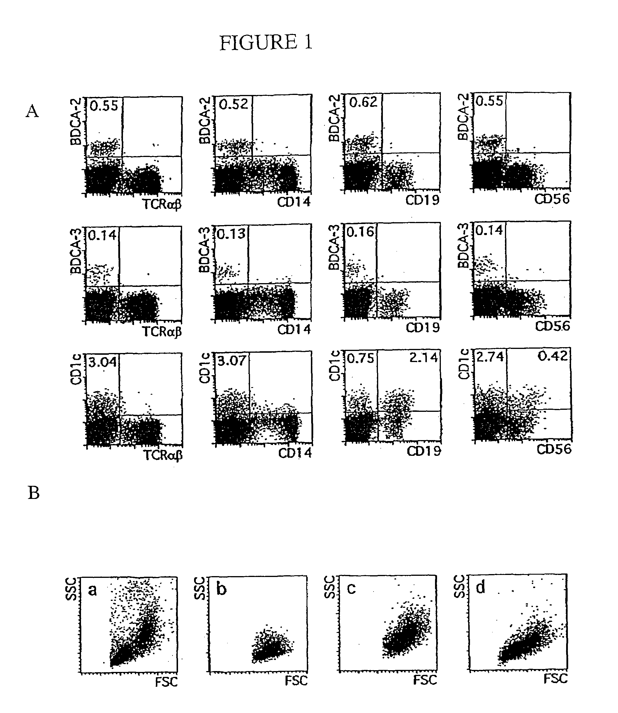
Flow Cytometric Analysis of Blood DCs

[0317]A FACScalibur (BD Biosciences) was used for one-, two-, three- or four-color flow cytometry. Data of 5×10³ to 2×10⁵ cells per sample were acquired in list mode and analyzed using CellQuest software (BD Biosciences).

[0318]The following mAb (clone names) were used in this study for flow cytometry: CD1a (HI149), CD10 (H110a), CD11a (G43-25B), CD11c (B-ly6), CD25 (M-A261), CD27 (M-T271), CD32 (FL18.26), CD38 (HIT2), CD40 (5C3), CD43 (IG10), CD54 (HA58), CD62L (Dreg 56), CD64 (10.1), CD69 (FN50), CD98 (UM7F8), anti-HLA-DQ (TU169), and anti-TCRαβ T10B9.1A-31 from Pharmingen, San Diego, Calif.; CD2 (S5.2), CD8 (SK1), CD13 (L138), CD14 (MFP9), CD19 (SJ25-C1), CD33 (P67.6), CD34 (8G12), CD45RO (UCHL-1), CD56 (NCAM16.2), CD71 (LO1.1), CD123 (9F5), ariti-IgD (TA4.1), anti-mouse IgG1 (X56), anti-mouse IgG2 (X57), and anti-mouse IgM (X54) from BD Biosciences; CD5 (CLB-T11 / 11, 6G4), CD7 (CLB-T-3A1 / 1, 7F3), CD16 (CLB-FcR gran / 1, 5D2), CD45RA (F8-11-13), C...

example 3

Cross-Inhibition, Co-Capping and Co-Internalization Analysis

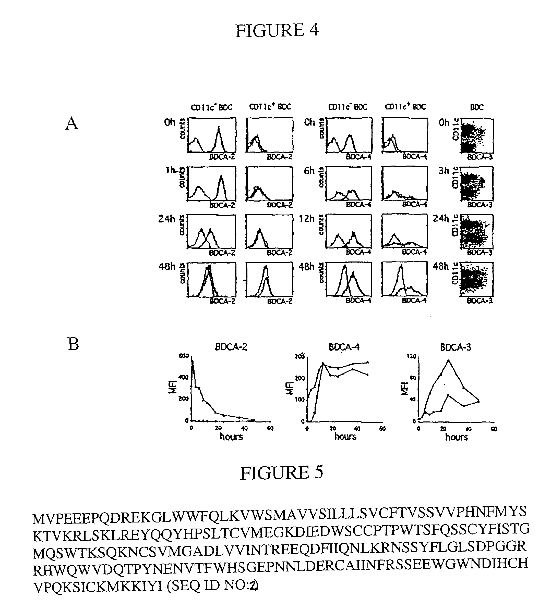
[0322]To analyze whether two different mAb clones recognize the same (or a closely related) antigen epitope, cross-inhibition binding assays were performed. Between 1×10⁶ and 2×10⁶ cells were pre-incubated with one of the two mAb clones at a concentration of about 100 μg / ml for 10 min at 4° C., and then stained with a PE-conjugate of the other mAb clone at its optimal titer for another 5 min at 4° C. PBMC were used to analyze cross-inhibition of BDCA-2-, BDCA-3-, and BDCA-4-specific mAb clones and MOLT-4 cells were used to analyze cross-inhibition of CD1c-specific mAb clones. Cell staining was analyzed by flow cytometry.

[0323]To ascertain whether AD5-5E8 and AD5-14H12 recognize the same antigen (or the same antigen-complex) a co-capping assay was performed. Briefly, BDCA-3-expressing cells were isolated from PBMC by indirect magnetic labeling with PE-conjugated AD5-14HI2 mAb and anti-PE mAb-conjugated microbeads, and isolat...


PUM

PropertyMeasurementUnit
Fractionaaaaaaaaaa
Fluorescenceaaaaaaaaaa
Chemiluminescenceaaaaaaaaaa

Abstract

The invention provides antigen-binding fragments specific for dendritic cells and effective in treatment and / or diagnosing a variety of disorders. Methods of use are also provided as are methods for screening for additional such antigen-binding fragments and the products obtained thereby.

Description

CROSS-REFERENCE TO RELATED APPLICATIONS[0001]This application claims the benefit of and priority to U.S. patent applications Ser. No. 60 / 197,205 filed Apr. 13, 2000; Ser. No. 60 / 196,824 filed Apr. 11, 2000; Ser. No. 60 / 180,775 filed Feb. 7, 2000; Ser. No. 60 / 179,003 filed Jan. 28, 2000; Ser. No. 60 / 167,076 filed Nov. 23, 1999; and Ser. No. 60 / 165,555 filed Nov. 15, 1999.STATEMENT OF RIGHTS TO INVENTIONS MADE UNDER FEDERALLY SPONSORED RESEARCH[0002]Not applicable.TECHNICAL FIELD[0003]The present invention relates to antibodies and derivatives thereof specific for subpopulations of dendritic cells (DCs). Compositions and methods of use thereof are also provided including isolation and purification of DCs and subpopulations thereof and antibody- or ligand-mediated immunotherapy. The invention also provides substantially isolated DC subpopulations. Methods of use thereof are also provided including DC-based immunotherapy, characterization of various diseases and in vivo numeric DC expan...

Claims


Application Information

Patent Timeline
IPC IPC(8): C07K16/00C07K17/00C12P21/08
CPCC07K14/7056C07K16/2851Y10S435/81
Inventor SCHMITZ, JUERGENDZIONEK, ANDRZEJBUCK, DAVID WILLIAM
Owner MILTENYI BIOTEC B V & CO KG
Learn More