Method of producing cardiac tomogram and tomograph using x-ray ct apparatus

a tomograph and x-ray technology, applied in tomography, applications, instruments, etc., can solve the problems of difficult to highly raise the scan speed to a speed of all heartbeat cycles or less, short projection data of the desired slicing position, and undesirable images. , to achieve the effect of fewer motion artifacts

Inactive Publication Date: 2006-02-28
FUJIFILM HEALTHCARE CORP
View PDF8 Cites 17 Cited by
  • Summary
  • Abstract
  • Description
  • Claims
  • Application Information

AI Technical Summary

Benefits of technology

[0011]Since the discontinuity in the projection data generated in the helical scans can be solved by the above method of producing a cardiac tomogram, a cardiac tomogram with few motion artifacts caused by heartbeat and good image quality can be obtained.
[0014]Further, the projection data at an arbitrary heartbeat time phase may be formed by transferring the starting projection angle of the divided projection data, a plurality of cardiac tomograms at heartbeat time phases at with arbitrary time interval may be formed by forming a plurality of projection data including the divided projection data having different distances of starting projection angle by an arbitrary projection angle, and image-reconstructing the respective data. Further, the step of forming a plurality of cardiac tomograms at heartbeat time phases with an arbitrary time interval includes a step of performing weighing processing in the slicing direction of the object on a plurality of projection data obtained from the divided projection data necessary for image reconstruction, a step of image-reconstructing each of the plurality of projection data which has been subjected to the weighing processing and obtaining a plurality of cardiac tomograms at different slicing positions, and a step of performing interpolation or extrapolation processing to obtain a cardiac tomogram at an arbitrary slicing position from the plurality of cardiac tomograms. Further, the step of forming a plurality of cardiac tomograms at an arbitrary heartbeat time phase with an arbitrary time interval includes a step of performing interpolation or extrapolation in the slicing direction on the plurality of projection data necessary for image reconstruction obtained from the divided projection data and acquiring arbitrary projection data, and a step image-reconstructing the arbitrary projection data and obtaining a cardiac tomogram at an arbitrary slicing position. By reconstructing the cardiac tomogram using the projection data formed in the above described manner, the number of the divided projection data and the data width can be properly adjusted even when difference between phases of the heartbeat cycle of the object and the scan cycle of the helical scans is generated, whereby a cardiac tomogram having few motion artifacts can be obtained.
[0017]With the above construction, the discontinuity of the projection data in helical scans can be solved, whereby a cardiac tomogram with few motion artifacts caused by heartbeat and good image quality can be obtained.
[0019]By reconstructing the cardiac tomograms by using thus formed projection data, the divided projection data number and the data width can be properly adjusted even when difference is generated between the phase of the heartbeat cycle of the object and that of the scan cycle of helical scans, whereby a cardiac tomogram with few motion artifacts can be obtained.

Problems solved by technology

Generally, when a heart of human body is imaged by an X-ray CT apparatus, the image is diagnostically undesirable when motion artifacts appear on the tomogram due to its heartbeat.
With a scanner mechanism of the current third-generation CT system, it is hard to highly raise its scan speed to a speed of all heartbeat cycles or less.
When the projection data for image reconstruction includes a discontinuous portion, the projection data of a desired slicing position come short.
Accordingly, if an image is reconstructed with those data, artifacts are generated on the image or the image quality is significantly deteriorated.
The other one is that when phases of a heartbeat cycle and of a scan cycle are different, the number of data necessary for reconstruction in a projection angle direction it too small to make a reconstruction image and so the image cannot be reconstructed in some cases.
However, in consideration of mechanical accuracy of the scanner which rotates the X-ray source and the X-ray detector around the object, there is a limit on the range of fine adjustment of scanning time.
Further, according to the above conventional techniques, one heartbeat is divided and a moving image is produced from images obtained at each heartbeat time phase, thereby it is hard to make a smooth moving image.

Method used


Image

  • Method of producing cardiac tomogram and tomograph using x-ray ct apparatus
  • Method of producing cardiac tomogram and tomograph using x-ray ct apparatus
  • Method of producing cardiac tomogram and tomograph using x-ray ct apparatus

Examples


first embodiment

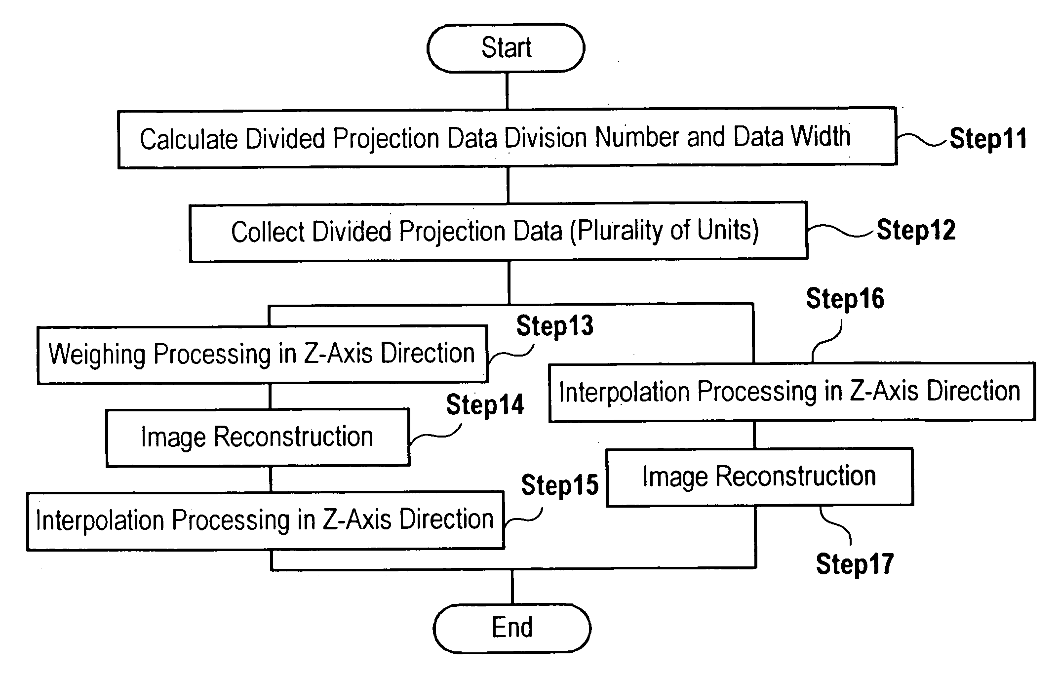
[0052]A method of producing a cardiac tomogram and tomograph using an X-ray CT apparatus according to the present invention will be described, in which a cardiac tomogram having few artifacts caused by discontinuity of projection data can be obtained when the retrospective ECG gating imaging method is applied to helical scan in which the table moves.

(1) In the Case Where the Discontinuous Portion of Projection Data is Narrow

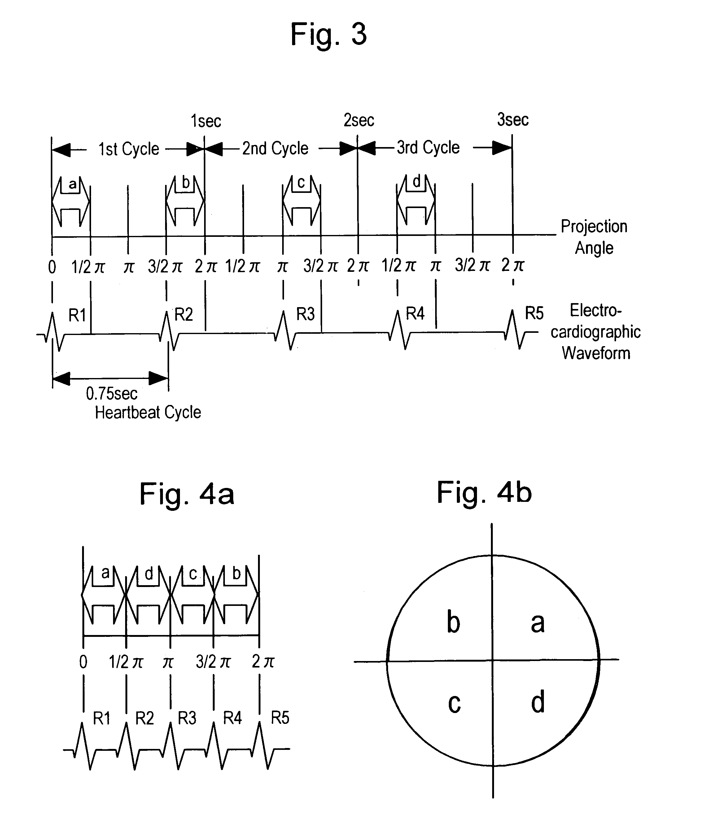
[0053]FIG. 6 shows a case where the multi-slice detector having four lines is used, a scan cycle is 1 second, a heartbeat cycle is 0.75 seconds, and the discontinuous portion of projection data is narrow.

[0054]In the figure, the ordinate axis designates a linear movement direction of a table on which the object to be examined is laid (longitudinal direction of the object, also referred to as Z-axis direction), and the abscissa axis designates a projection angle. Trajectory of the detector traces the center of each detector. The helical scan pitch is “pitch 1” in ...

second embodiment

[0070]Next, a method of producing a cardiac tomogram and tomograph according to the present invention will be described, in which a cardiac tomogram (two-dimensional image), a three-dimensional image, and a four-dimensional image (moving image) can be obtained by optimizing projection data in correspondence with phase difference between a heartbeat cycle and a scan cycle when projection data of the continuous portion obtained by the present invention described above, i.e., projection data at a matched heartbeat time phase are collected in each scan cycle.

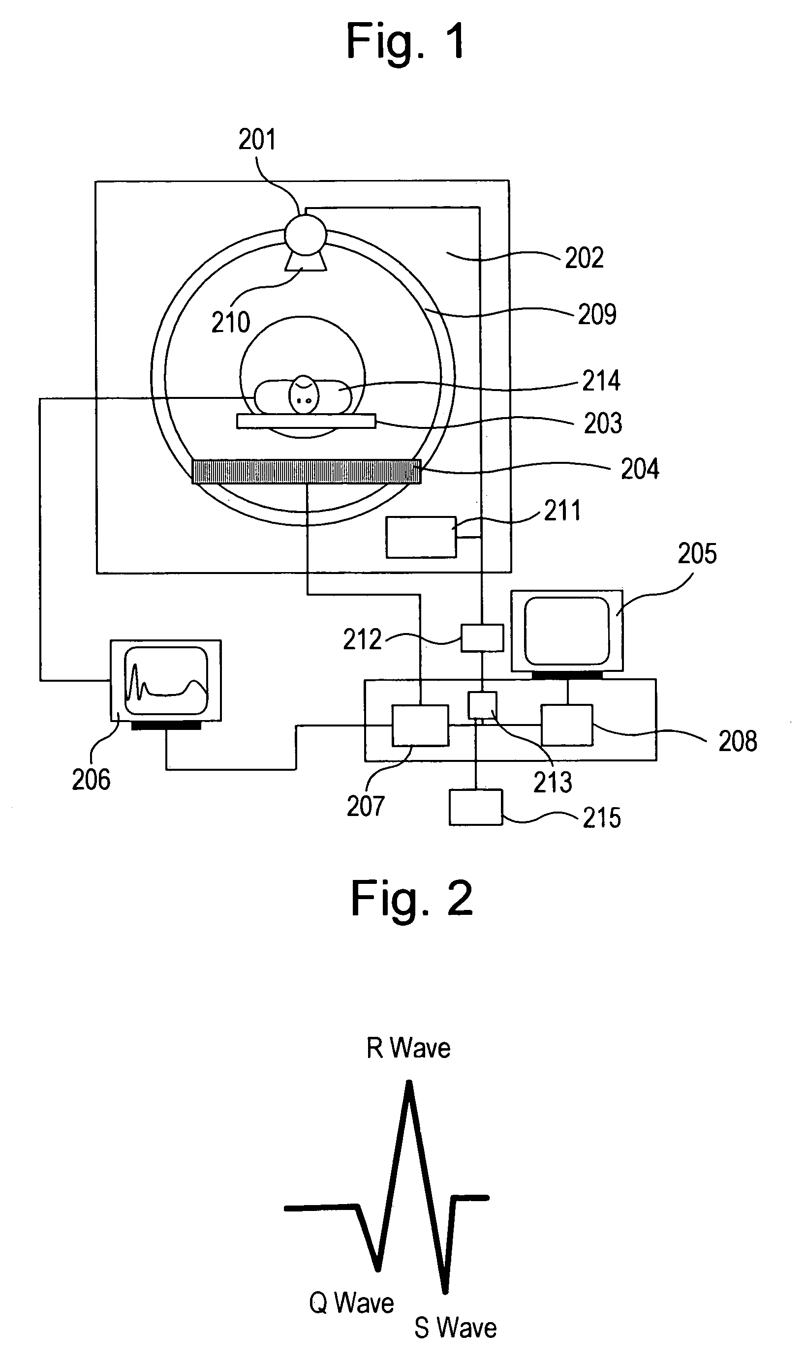
[0071]Meanwhile, the X-ray detector 204 used for creating a cardiac tomogram according to the invention is a multi-slice detector in which a plurality of detector elements are aligned in the longitudinal direction of an object to be examined so as to acquire projection data of a plurality of positions at once, and the scan system here is the helical scan.

[0072]As described in FIG. 1, X-rays are radiated from the X-ray tube 201 while...


PUM


Abstract

In an X-ray CT apparatus having a multi-slice detector, the retrospective ECG (Electrocardiography) gating imaging is applied to helical scans. Discontinuity of projection data generated in the scans is interpolated using data at a heartbeat time phase in 180-degree opposite relation to reduce motion artifacts. Further, projection data at an arbitrary slicing position and in at arbitrary heartbeat time phase are formed utilizing continuous divided projection data obtained above, and by properly combining or gathering them, a tomogram of whole heart, a three-dimensional image thereof, and further, a three-dimensional cardiac moving image in heartbeat time phases with an arbitrary time interval can be continuously and smoothly created.

Description

TECHNICAL FIELD[0001]The present invention relates to a method of producing a cardiac tomogram and tomograph using an X-ray CT apparatus, more particularly to a method and apparatus for creating a cardiac tomogram by applying a retrospective ECG (electrocardiography) gating imaging method to an X-ray CT apparatus which performs helical scan and detects projection data with a multi-slice detector.BACKGROUND OF THE INVENTION[0002]Generally, when a heart of human body is imaged by an X-ray CT apparatus, the image is diagnostically undesirable when motion artifacts appear on the tomogram due to its heartbeat. To prevent motion artifacts from being generated, it is necessary to highly speed up scanning over one heartbeat cycle to a speed of one heartbeat cycle or less. However, heart rates depend on individual bodies, and physical and mental conditions at the point of imaging in the same body. With a scanner mechanism of the current third-generation CT system, it is hard to highly raise ...

Claims


Application Information

Patent Timeline
Patent Type & Authority Patents(United States)
IPC IPC(8): A61B6/03A61B6/00
CPCA61B6/032A61B6/4085A61B6/541A61B6/027A61B6/503Y10S378/901
Inventor KOKUBUN, HIROTONAKAZAWA, TETSUOMIYAZAKI, OSAMU
Owner FUJIFILM HEALTHCARE CORP
Why Eureka
Try Eureka