
Methods, instruments and materials for chondrocyte cell transplantation

a chondrocyte and chondrocyte technology, applied in the direction of prosthesis, surgical staples, osteosynthesis devices, etc., can solve the problems of affecting the function of the chondrocyte cell transplantation process, and reducing the biomechanical properties of the functional tissue. , to achieve the effect of effective treatment of the articulating joint surface cartilag

Inactive Publication Date: 2005-03-15
VERIGEN TRANSPLANTATION SERVICE INT
View PDF106 Cites 553 Cited by
  • Summary
  • Abstract
  • Description
  • Claims
  • Application Information

AI Technical Summary

Benefits of technology

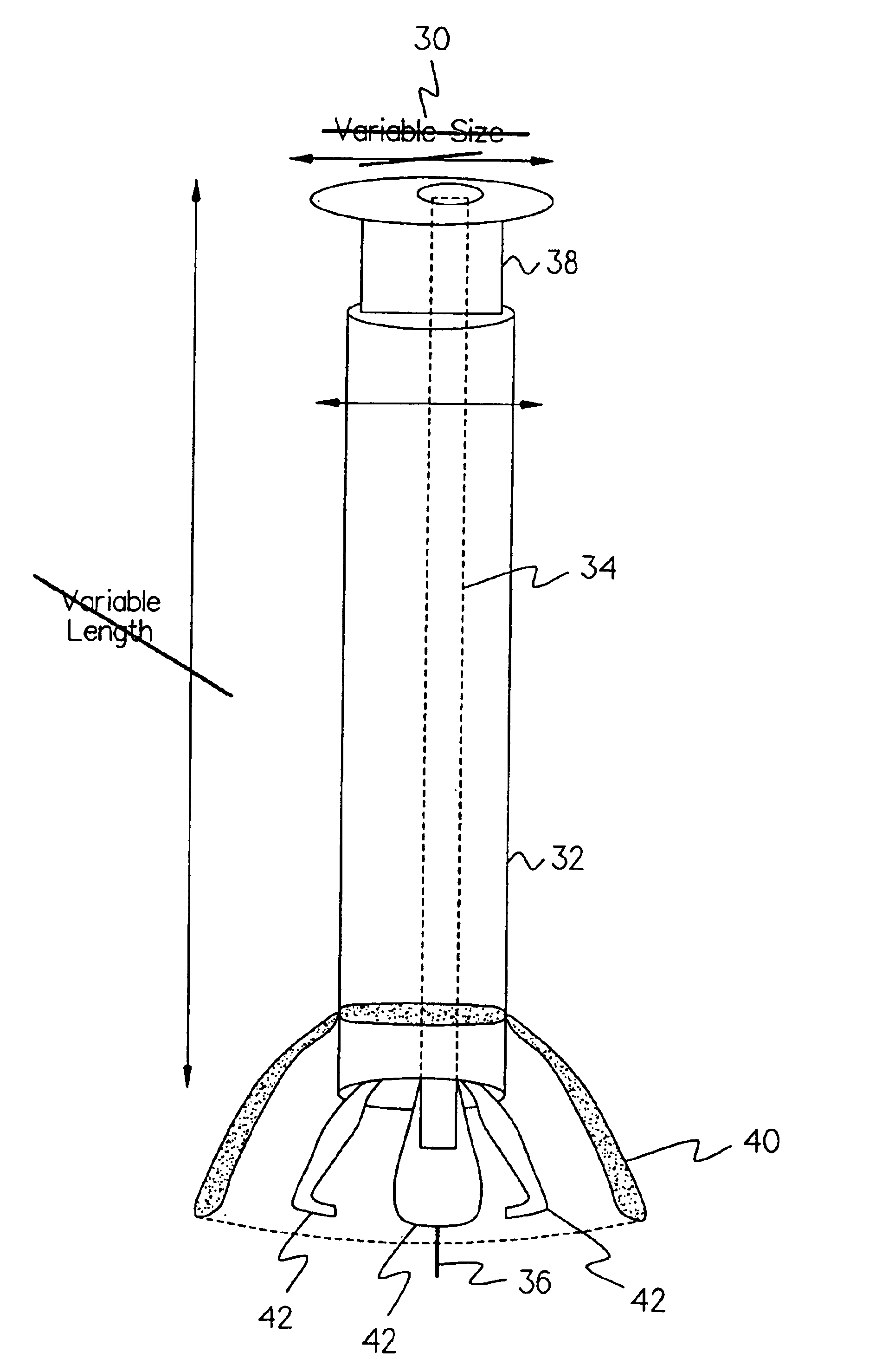
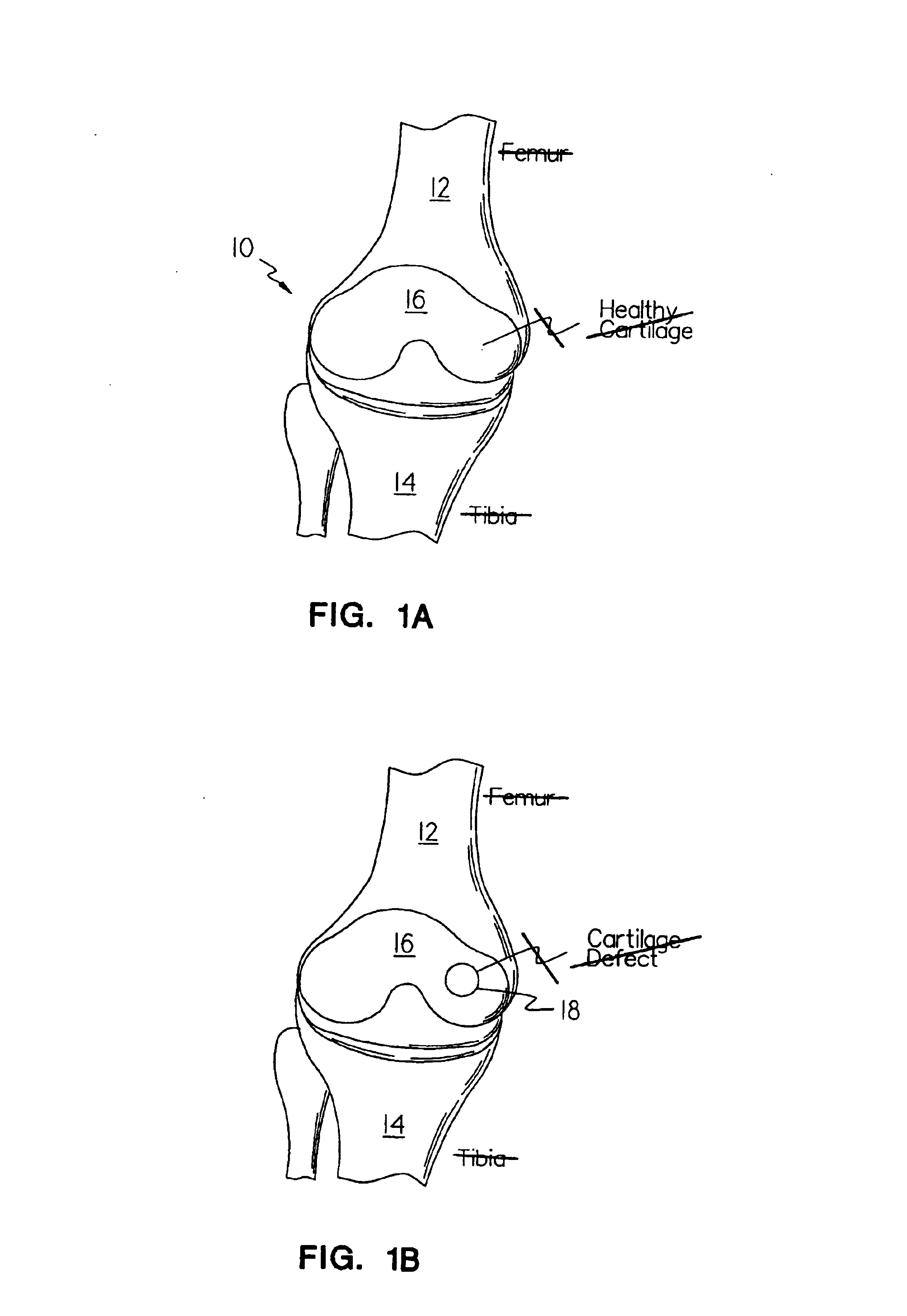
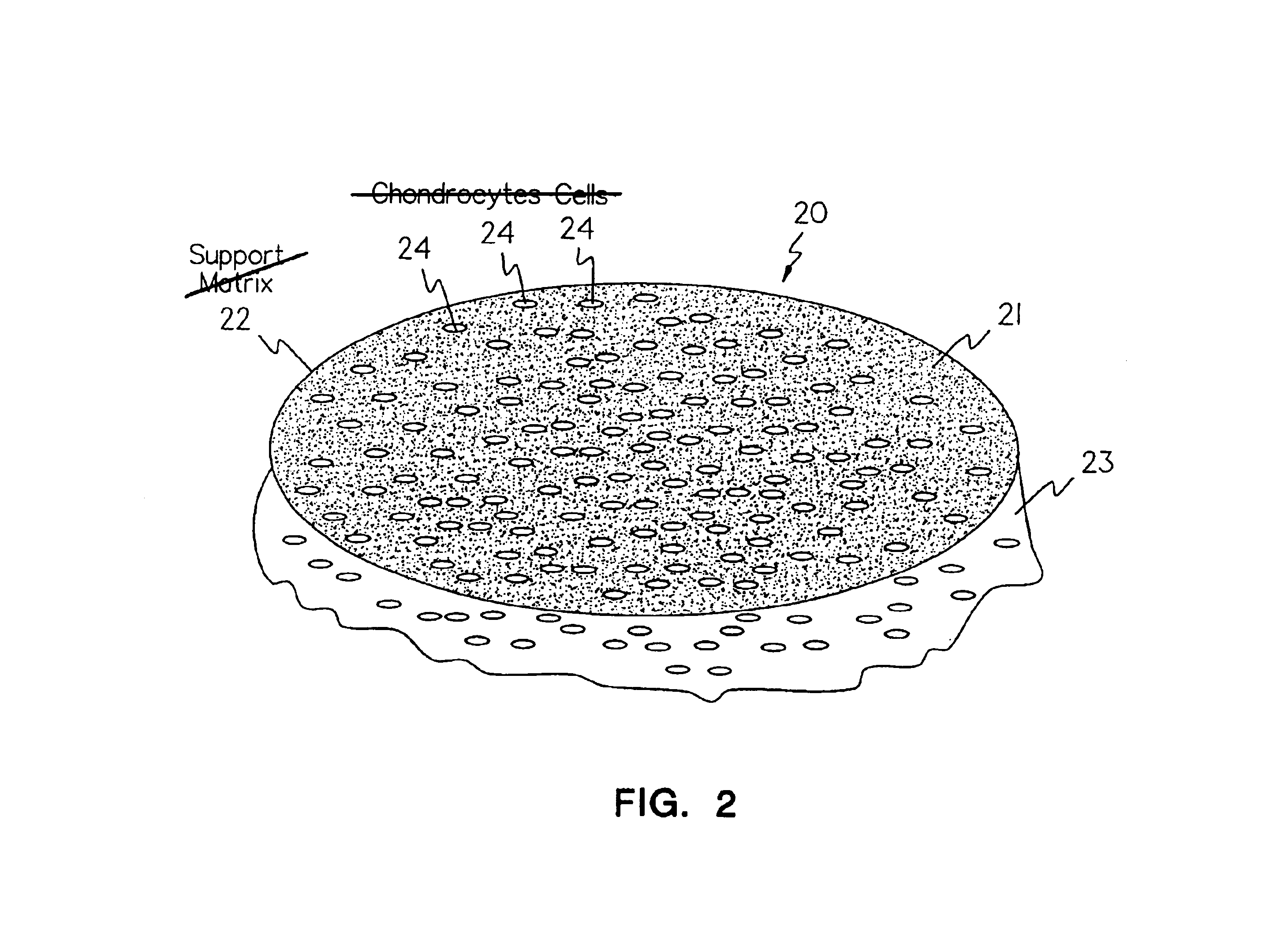
The present invention provides an implantable article including a support matrix which can support the growth and attachment of cells thereto, and a method of implanting such an article to regenerate cells at an implantation location. In one embodiment, the present invention provides a method for the effective treatment of articulating joint surface cartilage in an animal by the transplantation of an implantable article including chondrocyte cells retained on an absorbable support matrix. In

Problems solved by technology

In all instances, the major problem was the lack of biomechanical properties required for functional tissue repair.
The collagen network is accordingly weakened and subsequently develops fibrillation whereby matrix substances such as proteoglycans are lost and eventually displaced entirely.

Method used


Image

  • Methods, instruments and materials for chondrocyte cell transplantation
  • Methods, instruments and materials for chondrocyte cell transplantation
  • Methods, instruments and materials for chondrocyte cell transplantation

Examples


example 1

Chondrocyte cells were grown for three weeks in the growth media described above in a CO2 incubator at 37° C. and handled in a Class 100 laboratory at Verigen Transplantation Service ApS, Copenhagen, DK or at University of Lübeck, Lübeck, Germany. [Note that other compositions of growth media may also be used for culturing the chondrocyte cells.] The cells were trypsinised using trypsin EDTA for 5 to 10 minutes and counted using Trypan Blue viability staining in a Bürker-Türk chamber. The cell count was adjusted to 7.5×10⁵ chondrocyte cells per milliliter. One NUNCLON™ plate was uncovered in the Class 100 laboratory.

A support matrix material, specifically a Chondro-Gide® collagen membrane, was cut to a suitable size to fit into the bottom of a well in a NUNCLON™ cell culture tray. In this case a circle of a size of approximately 4 cm was placed under aseptic conditions on the bottom of the well.

After three weeks, chondrocyte cells were transferred from the growth media to the transp...

example 2

Chondrocytes were grown for three weeks in the growth media described above in a CO2 incubator at 37° C. and handled in a Class 100 laboratory at Verigen Transplantation Service ApS, Copenhagen, DK or at University of Lübeck, Germany. The cells were trypsinised using trypsin EDTA for 5 to 10 minutes and counted using Trypan Blue viability staining in a Bürker-Türk chamber. The chondrocyte cell count was adjusted to 5×10⁵ chondrocyte cells per milliliter. One NUNCLON™ plate was uncovered in the Class 100 laboratory.

The Chondro-Gide® support matrix, as in Example 1, was cut to a suitable size fitting into the bottom of a well in the NUNCLON™ cell culture tray. In this case a circle of approximately 4 cm in diameter was placed under aseptic conditions on the bottom of the well.

After three weeks, the chondrocyte cells were transferred from the growth media to the transplant media described above, and approximately 5×10⁵ cells in 5 ml transplant media were placed directly on top of the s...

example 3

Chondrocytes were grown for three weeks in the growth media described above in a CO2 incubator at 37° C. and handled in a Class 100 laboratory at Verigen Transplantation Service ApS, Copenhagen, DK or at University of Lübeck, Germany. The chondrocyte cells were trypsinised using trypsin EDTA for 5 to 10 minutes and counted using Trypan Blue viability staining in a Bürker-Türk chamber. The chondrocyte cell count was adjusted to 5×10⁵ chondrocyte cells per milliliter. One NUNCLON™ plate was uncovered in the Class 100 laboratory.

The Chondro-Gide® support matrix, as in Example 1, was cut to a suitable size fitting into the bottom of a well in the NUNCLON™ cell culture tray. In this case a circle of approximately 4 cm in diameter was placed under aseptic conditions on the bottom of the well.

After three weeks, the chondrocyte cells were transferred from the growth media to the transplant media described above, and approximately 5×10⁶ cells in 5 ml transplant media were placed directly on ...


PUM

PropertyMeasurementUnit
Flexibilityaaaaaaaaaa
Bioabsorbableaaaaaaaaaa

Abstract

A method for the effective treatment of articulating joint surface cartilage in an animal by the transplantation of an implantable article including chondrocyte cells retained to an absorbable support matrix. An instrument for placing and manipulating the implantable article at the site of implantation, and a retention device for securing the implantable article to the site of implantation. An implantable article for cartilage repair in an animal, the implantable article including chondrocyte cells retained on an absorbable support matrix, and a method of making same. An article comprising an absorbable flexible support matrix for living cells grown and adhered thereto.

Description

FIELD OF THE INVENTIONBACKGROUND OF THE INVENTIONMore than 500,000 arthroplastic procedures and total joint replacements are performed each year in the United States. Approximately the same number of similar procedures are performed in Europe. Included in this number in Europe are about 90,000 total knee replacements and around 50,000 procedures to repair defects in the knee. These numbers are essentially the same in the U.S. (Praemer A., Furner S., Rice, D. P., Musculoskeletal conditions in the United States, American Academy of Orthopaedic Surgeons, Park Ridge, Ill., 1992, 125).A method for regeneration-treatment of cartilage would be most useful and could be performed at an earlier stage of joint damage, thus reducing the number of patients needing artificial joint replacement surgery. With such preventive methods of treatment, the number of patients developing osteoarthritis would also decrease.Techniques used for resurfacing the cartilage structure in joints have mainly attempt...

Claims


Application Information

Patent Timeline
IPC IPC(8): A61F2/30A61F2/46A61L27/38A61L27/00A61L31/04A61B17/22A61B17/86A61B17/68A61F2/00A61F2/02A61F2/38A61B17/00A61B17/064A61B17/06A61B17/56A61F2/28A61K9/00A61K35/32A61K47/42A61L24/00
CPCA61F2/30756A61F2/4618A61L27/3817A61L27/3852A61L31/04C08L67/04A61B17/00491A61F2002/4627A61B17/0642A61B17/221A61B17/68A61B17/86A61B2017/00004A61B2017/0647A61B2017/2215A61F2/3094A61F2/38A61F2002/009A61F2002/30006A61F2002/30013A61F2002/30028A61F2002/30062A61F2002/30293A61F2002/30448A61F2002/30761A61F2002/30762A61F2002/30764A61F2002/30932A61F2002/4624A61F2002/4635A61F2210/0004A61F2220/005A61F2230/0091A61F2250/0015A61F2250/0024A61F2250/0051A61F2310/00365A61L2430/06A61B17/06166A61F2002/30011A61F2/4603A61L27/58A61L27/22A61L27/24
Inventor GIANNETTI, BRUNOBEHRENS, PETERASCULAI, SAMUEL
Owner VERIGEN TRANSPLANTATION SERVICE INT
Learn More