Eureka AIR delivers breakthrough ideas for toughest innovation challenges, trusted by R&D personnel around the world.

Use of adipose tissue-derived stromal cells for chondrocyte differentiation and cartilage repair

a technology of chondrocyte differentiation and cartilage repair, which is applied in the direction of skeletal/connective tissue cells, prosthesis, drug composition, etc., can solve the problems of low yield of stem cells from this source, difficulty and risk of bone marrow biopsy procedures, and the use of these cells

Inactive Publication Date: 2002-08-06
COGNATE BIOSERVICES
View PDF17 Cites 346 Cited by
  • Summary
  • Abstract
  • Description
  • Claims
  • Application Information

AI Technical Summary

Benefits of technology

FIG. 4 shows Collagen type VI expression when cells were cultured in an alginate matrix at 2 weeks without TGF-beta (control) and with TGF-beta.

Problems solved by technology

The major limitations to the use of these cells are the difficulty and risk attendant upon bone marrow biopsy procedures and the low yield of stem cells from this source.
Since the subchondral bone tissue is both innervated and vascularized, damage to this tissue is often painful.
The repair reaction induced by damage to the subchondral bone usually results in the formation of fibrocartilage at the site of the full-thickness defect.
Fibrocartilage, however, lacks the biomechanical properties of articular cartilage and fails to persist in the joint on a long term basis.
Partial-thickness defects are caused by mechanical arrangements of the joint which in turn induce wearing of the cartilage tissue within the joint.
In the absence of innervation and vasculature, partial-thickness defects do not elicit repair responses and therefore tend not to heal.
Although painless, partial-thickness defects often degenerate into full-thickness defects.

Method used


Image

  • Use of adipose tissue-derived stromal cells for chondrocyte differentiation and cartilage repair
  • Use of adipose tissue-derived stromal cells for chondrocyte differentiation and cartilage repair
  • Use of adipose tissue-derived stromal cells for chondrocyte differentiation and cartilage repair

Examples


example 1

In vitro Chondrogenesis using Dexamethasone

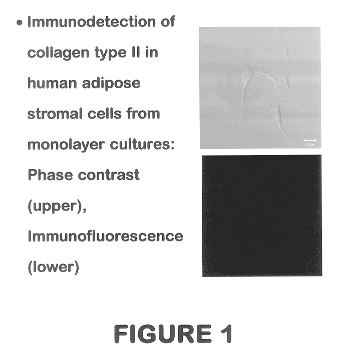
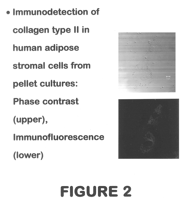
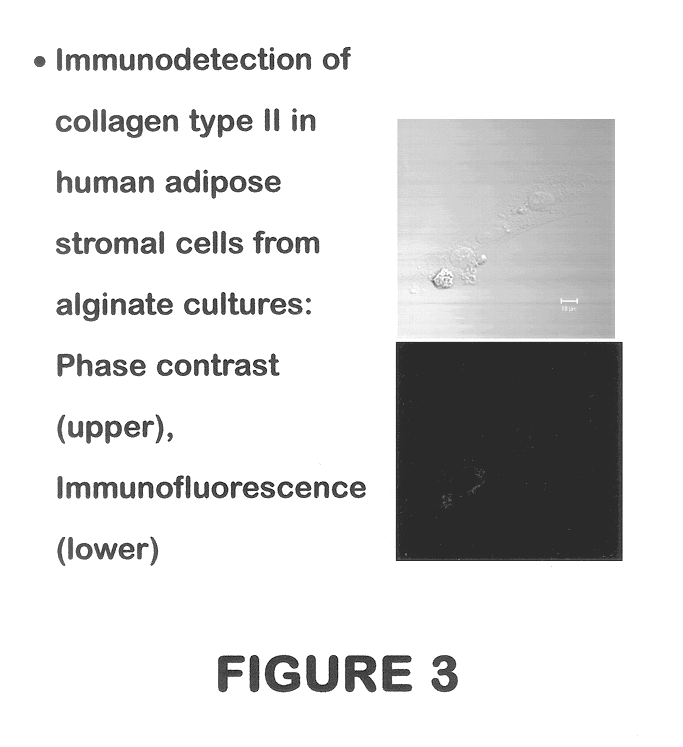
Stromal cells are isolated from human subcutaneous adipose tissue according to methods described in "Methods and Composition of the Differentiation of Human Preadipocytes into Adipocytes" Ser. No. 09 / 240,029, filed Jan. 29, 1999, now U.S. Pat. No. 6,153,432. These cells are plated at a density of 500 to 20,000 cells per cm.sup.2. The present invention contemplates that the creation of a precartilage condensation in vitro promotes chondrogenesis in mesenchymal progenitor cells derived from human adipose tissue. This is accomplished by methods including, but not limited to:

(1) The pellet culture system, which was developed for use with isolated growth plate cells (Kato et al. (1988) PNAS 85:9552-9556; Ballock & Reddi, J. Cell Biol. (1994) 126(5):1311-1318) and has been used to maintain expression of the cartilage phenotype of chondrocytes placed in culture (Solursh (1991) J. Cell Biochem. 45:258-260).

(2) The alginate suspension method, where ...

example 2

Preparation of Synthetic Cartilage Patch

Following proliferation, the chondrogenic cells still having chondrogenic potential may be cultured in an anchorage-independent manner, i.e., in a well having a cell contacting, cell adhesive surface, in order to stimulate the secretion of cartilage-specific extracellular matrix components.

Heretofore, it has been observed that chondrogenic cells proliferatively expanded in an anchorage-dependent manner usually differentiate and lose their ability to secrete cartilage-specific type II collagen and sulfated proteoglycan. (Mayne et al. (1984) Exp. Cell. Res. 151(1): 171-82; Mayne et al. (1976) PNAS 73(5): 1674-8; Okayama et al. (1976) PNAS 73(9):3224-8; Pacifici et al. (1981) J. Biol Chem. 256(2): 1029-37; Pacifici et al. (1980) Cancer Res. 40(7): 2461-4; Pacifici et al. (1977) Cell 4:891-9; von der Mark et al. (1977) Nature 267(5611):531-2; West et al. (1979) Cell 17(3):491-501; Oegama et al. (1981)J. Biol. Chem. 256(2):1015-22; Benya et al. (19...

example 3

Surgical Repair of Articular cartilage Defect

Cartilage defects in mammals are readily identifiable visually during arthroscopic examination orduring open surgery of the joint. Cartilage defects may also be identified inferentially by using computer aided tomography (CAT scanning); X-ray examination, magnetic resonance imaging (MRI), analysis of synovial fluid or serum markers or by any other procedures known in the art. Treatment of the defects can be effected during an arthroscopic or open surgical procedure using the methods and compositions disclosed herein.

Accordingly, once the defect has been identified, the defect may be treated by the following steps of (1) surgically implanting at the pre-determined site, a piece of synthetic articular cartilage prepared by the methodologies described herein, and (2) permitting the synthetic articular cartilage to integrate into pre-determined site.

The synthetic cartilage patch optimally has a size and shape such that when the patch is impla...


PUM

PropertyMeasurementUnit
concentrationaaaaaaaaaa

Abstract

Methods and compositions for directing adipose-derived stromal cells cultivated in vitro to differentiate into cells of the chondrocyte lineage are disclosed. The invention further provides a variety of chondroinductive agents which can be used singly or in combination with other nutrient components to induce chondrogenesis in adipose-derived stromal cells either in cultivating monolayers or in a biocompatible lattice or matrix in a three-dimensional configuration. Use of the differentiated chondrocytes for the therapeutic treatment of a number of human conditions and diseases including repair of cartilage in vivo is disclosed.

Description

The present invention relates to methods and compositions for directing adipose derived stromal cells cultivated in vitro to differentiate into cells of the chondrocyte lineage and particularly to such directed lineage induction prior to, or at the time of, their implantation into a recipient or host for the therapeutic treatment of pathologic conditions in humans and other species.Mesenchymal stem cells (MSCs) are the formative pluripotent blast or embryonic-like cells found in bone marrow, blood, dermis, and periosteum that are capable of differentiating into specific types of mesenchymal or connective tissues including adipose, osseous, cartilaginous, elastic, muscular, and fibrous connective tissues. The specific differentiation pathway which these cells enter depends upon various influences from mechanical influences and / or endogenous bioactive factors, such as growth factors, cytokines, and / or local microenvironmental conditions established by host tissues.In prenatal organism...

Claims


Application Information

Patent Timeline
Patent Type & Authority Patents(United States)
IPC IPC(8): C12N5/06A61K35/12C12N5/02C12N5/077
CPCA61L27/3804A61L27/3852C12N5/0653C12N5/0655A61K35/12A61L2430/06C12N2533/74C12N2500/38C12N2501/39C12N2502/1305C12N2506/1384A61P19/04A61P19/08C12N2500/30C12N2500/98C12N2501/999
Inventor HALVORSEN, YUAN-DI C.WILKISON, WILLIAM O.GIMBLE, JEFFREY MARTIN
Owner COGNATE BIOSERVICES
Why Eureka
Eureka Blog
Learn More