
Systems and methods for the multispectral imaging and characterization of skin tissue

Inactive Publication Date: 2000-06-27
THE TRUSTEES OF COLUMBIA UNIV IN THE CITY OF NEW YORK +1
View PDF33 Cites 556 Cited by
  • Summary
  • Abstract
  • Description
  • Claims
  • Application Information

AI Technical Summary

Benefits of technology

All pixels in the neighborhood of each pixel of the image contribute to the CWTs centered within the border and interior regions of each digital image. Each pixel in each region serves as a center of a "mother" wavelet. Each mother wavelet is scaled to span six different "scale wavelets," which play the role of local band-pass spatial filters that can selectively zoom in on different scales in the lesion structures of interest. See, for example, Daubechies, I., "The Wavelet transform, time-frequency localization and signal analysis.", IEEE Trans Inform Theory 36:961-1005 (1990); Aldroubi, A. et al., Wavelet in Medium and Biology, C&C Press, NY, pp 11-15 (1996). The fine-course filtering that the scale wavelets provide also reduces background noise.

Problems solved by technology

Early detection of malignant melanoma is difficult because early melanomas, those having a Breslow thickness less than 1 mm, share many diagnostic features with benign lesions, such as dysplastic nevi or atypical melanocytic nevi.
The effectiveness of clinical imaging can be compromised, however, by specular reflection by the skin.
Because the clinical and dermoscopic applications of these rules are subjective, they are not very reliable.
Since the unaided eye cannot perceive light outside of the visible region or low-contrast structure in visible-light images, information which may be useful in diagnosing a lesion may not be directly observable.
Practical limitations on the number of picture elements or pixels per unit area of image determine the achievable spatial resolution of the digital image.
Because some of the steps in the computer-aided analysis of which we are aware depend on subjective judgments of an individual, such analysis may provide highly variable results.
However, such devices have had poor resolution and / or poor signal-to-noise characteristics which prevent the acquisition of digital images of melanocytic skin lesions of sufficient quality for effective application of machine vision techniques for lesion diagnosis.
Existing imaging systems and processes also tend to suffer from an inability to provide the required repeatability of the value of extracted lesion parameters, due in part to a lack of standardization with respect to spatially varying artifacts, so that the parameters, therefore, lack invariance to lighting and image exposure conditions, for example.
Obtaining high signal-to-noise ratios in images recorded in narrow filter bandpasses, when exposure times are sufficiently short that the skin is effectively "frozen" during the exposure sequence, has also been difficult.
In addition, since the optimum wavelengths for automatic characterization may not be the optimum wavelengths for visual observation, it may be difficult to reconstruct high-fidelity color images from the digital images for visual interpretation by a clinician.
The assessment of wounds and burns through the appearance of color images present similar challenges.
Existing technology for the imaging of skin in vivo for these purposes is also inadequate.
However, since the size of some lesions exceeds 100,000 pixels, this would be computationally very intensive.
However, since this requires more computation and causes some border blurring, the need for preprocessing due to the presence of dark hair is preferably indicated interactively by an operator, and images preprocessed only when necessary.
There may be so little contrast between the lesion and the normal skin that direct segmentation is not possible.
The description of lesion texture is particularly vulnerable to subjective judgement.
Clinical evaluation of melanocytic lesions is, however, a problem since reliable differentiation between early malignant melanoma with Breslow thickness less than 1 mm and atypical melanocytic nevus is difficult even for experienced dermatologists.

Method used


Image

  • Systems and methods for the multispectral imaging and characterization of skin tissue
  • Systems and methods for the multispectral imaging and characterization of skin tissue
  • Systems and methods for the multispectral imaging and characterization of skin tissue

Examples


Embodiment Construction

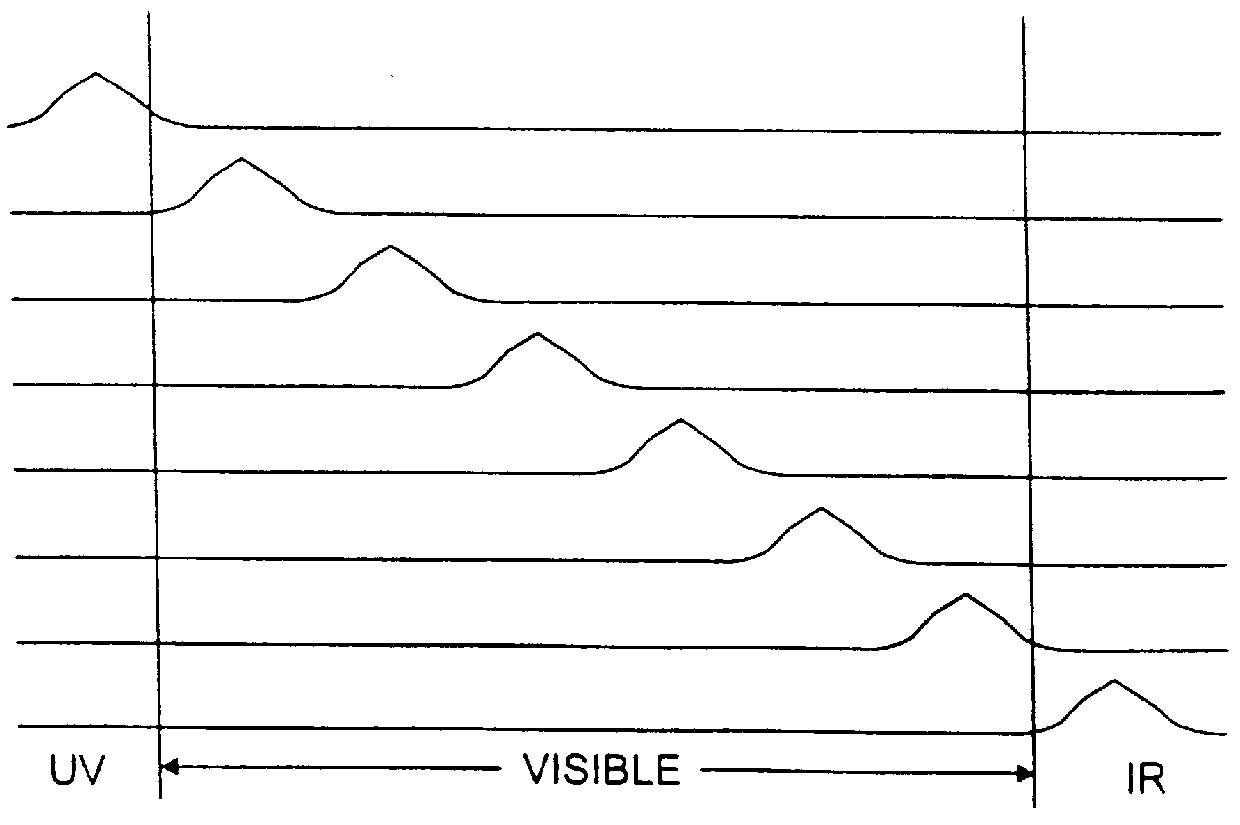
FIG. 1(a) is a schematic illustration of a method and system 1 in accordance with the present invention, by which images of the skin 2 are acquired by a camera nearly simultaneously at a plurality of spectral bands, .lambda..sub.i, i=1,2, . . . M, that are preferably effectively non-overlapping, as shown schematically in FIG. 1(b). The skin is illuminated by a source of white light 3, which is filtered by narrow passband filters 4. The filtered light is preferably conveyed to the skin 2 through a fiberoptic illuminator 5. The light re-emitted by the illuminated skin through reflection, scattering or fluorescence is imaged by a low-noise, high-resolution monochrome camera 6, which is preferably an electronic charge-coupled ("CCD") camera. Digital images output by the camera 6 are provided to a computer 12 for processing.

The computer 12 includes a digital interface 12a, a memory 12b and a digital processor 12c. A display 19 is preferably provided as well. The computer 12 includes an i...


PUM


Abstract

Systems and methods for the multispectral imaging of skin tissue enables automatic characterization of the condition of a region of interest of the skin, based on direct digital imaging of the region of interest or the digitization of color photographic slides of the region of interest, illuminated by appropriately filtered light. Preferably, a digital image at a low spectral band is automatically segmented and that segmented mask is used to segment the other images by a digital processor. Parameters are estimated through wavelet maxima representations. Parameters related to the texture, asymmetry, blotchiness and border irregularities are also automatically estimated. The region of interest is automatically characterized by the digital processor, based on those parameters. The region of interest may include a skin lesion, in which case the present invention enables the characterization of the lesion as malignant or benign. The region of interest may also include wounds or burns.

Description

This invention relates to methods and systems for the computer controlled analysis of digital images of skin tissue at a plurality of wavelengths, which may include those outside of the red-green-blue bands. The methods and systems further include the automatic characterization of the condition of the skin tissue, based on automatically computed values of parameters which are functions of characteristics of the skin tissue, derived the digital images. Skin lesions can be analyzed for determining whether the lesion is a melanoma, for example. Systems for digitally imaging and analyzing skin tissue are disclosed, as well.Melanoma is a usually fatal skin cancer, unless it is detected and surgically removed in its earliest stages. Early detection of malignant melanoma is difficult because early melanomas, those having a Breslow thickness less than 1 mm, share many diagnostic features with benign lesions, such as dysplastic nevi or atypical melanocytic nevi.To aid in the analysis of lesi...

Claims


Application Information

Patent Timeline
IPC IPC(8): G06T7/00A61B5/103G06T5/00G06T7/40
CPCA61B5/0071A61B5/442A61B5/444A61B5/445A61B5/726G06T7/0012G06T7/408G06T7/402G06T2207/30096G06T2207/10064G06T2207/20064G06T2207/20148G06T2207/30088G06T7/136G06T7/42G06T7/90
Inventor GUTKOWICZ-KRUSIN, DINAELBAUM, MAREKGREENEBAUM, MICHAELJACOBS, ADAMBOGDAN, ALEXANDRU
Owner THE TRUSTEES OF COLUMBIA UNIV IN THE CITY OF NEW YORK
Learn More