Method and system for generating a synthetic elastrography image

a synthetic elastography and image technology, applied in image enhancement, instruments, applications, etc., can solve the problems of low frame rate, computational complexity, and inability to readily find swe in many ultrasound equipmen

Pending Publication Date: 2022-11-17
KONINKLJIJKE PHILIPS NV
View PDF0 Cites 3 Cited by
  • Summary
  • Abstract
  • Description
  • Claims
  • Application Information

AI Technical Summary

Problems solved by technology

Thus, SWE may not be readily available in many ultrasound equipment, and where it is, it is computationally demanding and has a low frame rate.

Method used


Image

  • Method and system for generating a synthetic elastrography image
  • Method and system for generating a synthetic elastrography image
  • Method and system for generating a synthetic elastrography image

Examples


Embodiment Construction

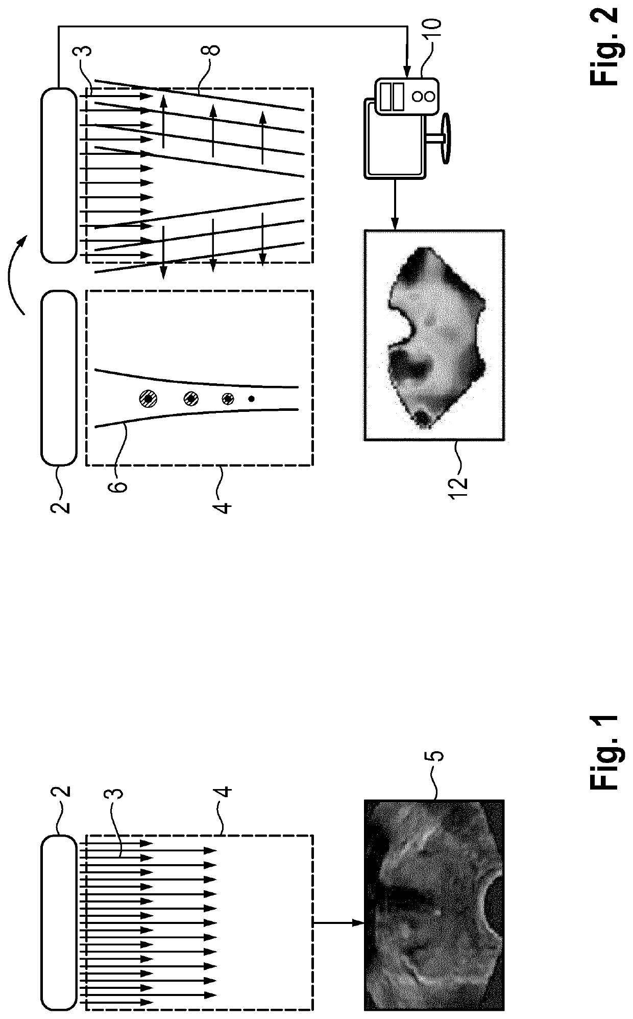
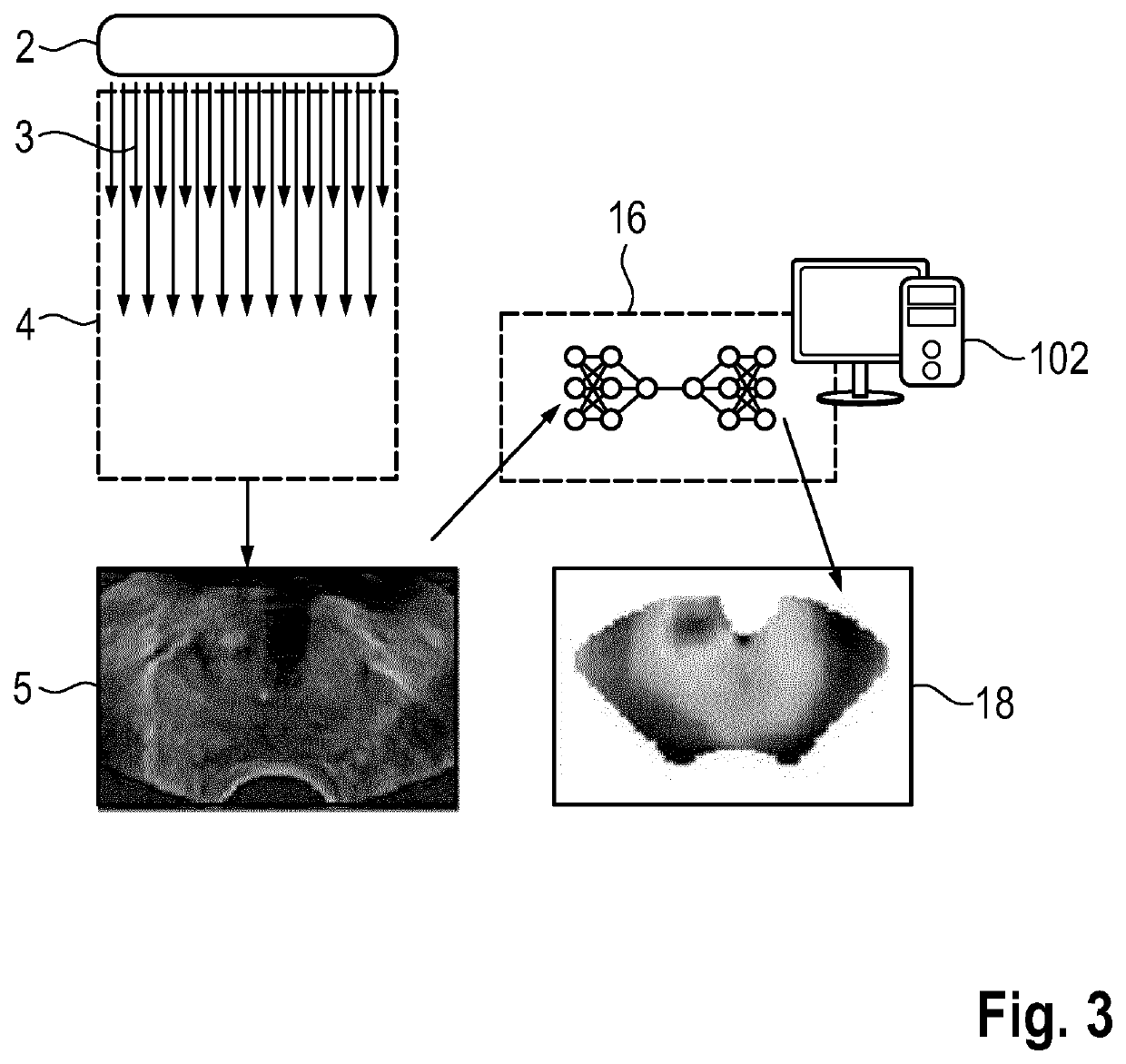
[0061]FIG. 1 schematically depicts the process of B-mode ultrasound: An ultrasound probe 2, usually comprising an array of ultrasound transducers, transmits a series of ultrasound pulses 3, for example as compressive wave fronts, into a region of interest 4, usually within a human or animal body. By recording the echoes and performing suitable signal processing such as beam-forming, a B-mode ultrasound image 5 of the region of interest is acquired. This can be done with a high frame rate, especially for 2D images.

[0062]FIG. 2 illustrates conventional SWE imaging. An ultrasound probe 2 generates a sequence of acoustic radiation force “push” pulses 6 into the region of interest 4. The “push” pulse results in laterally travelling shear-waves 8, which are recorded by ultrafast imaging by the ultrasound probe 2 and using further ultrasound transmission pulses 3. The recorded echoes are transferred to a computational unit 10, which processes the ultrafast imaging recordings and generates ...


PUM


Abstract

The invention relates to a method for generating a synthetic elastography image (18), the method comprising the steps of (a) receiving a B-mode ultrasound image (5) of a region of interest; (b) generating a synthetic elastography image (18) of the region of interest by applying a trained artificial neural network (16) to the B-mode ultrasound image (5). The invention also relates to a method for training an artificial neural network (16)5 useful in generating synthetic elastography images, and a related computer program and system.

Description

FIELD OF THE INVENTION[0001]The invention relates to a computer-implemented method for generating a synthetic elastography image, a method for training an artificial neural network useful in generating synthetic elastography images from B-mode ultrasound images, to a method for training the artificial neural network or a second artificial neural network to provide a confidence map, and a related computer program and system.BACKGROUND OF THE INVENTION[0002]Tissue elasticity or stiffness is an important marker in biomedicine, for example in cancer imaging or the assessment of musculoskeletal pathologies. Whether the tissue is hard or soft may provide important diagnostic information. For example, cancerous tumours will often be harder than the surrounding tissue, and liver stiffness is usually indicative of various disease conditions, including cirrhosis and hepatitis. To image the elastic properties of tissue, one needs to record how it behaves when deformed. In ultrasound elastograp...

Claims


Application Information

Patent Timeline
Patent Type & Authority Applications(United States)
IPC IPC(8): A61B8/00A61B8/08
CPCA61B8/4483A61B8/485A61B8/52G06T5/50G06N3/061G06N3/084G06T2207/20084G06T2207/20081G06T2207/10132G06N3/045G06T11/006G06T2211/441
Inventor WILDEBOER, ROGIER RUDOLFVAN SLOUN, RUUD JOHANNES GERARDUS
Owner KONINKLJIJKE PHILIPS NV
Why Eureka
Try Eureka