
Methods for Identifying Haemoglobin S or C in a Biological Sample and Kits Thereof

a technology of haemoglobin and kit, which is applied in the field of identifying the presence or absence of haemoglobin s (hbs) and/or or haemoglobin c (hbc) in a blood sample, can solve the problems of destroying organs, reducing the oxygen-rich blood supply of tissues, and polymerizing defective haemoglobin proteins within the rb

Pending Publication Date: 2022-09-08
INDIAN INSTITUTE OF SCIENCE
View PDF0 Cites 0 Cited by
  • Summary
  • Abstract
  • Description
  • Claims
  • Application Information

AI Technical Summary

Benefits of technology

The present patent provides an in vitro method for identifying the presence or absence of hemoglobin S (HbS) or hemoglobin C (HbC) in a blood sample. The method involves measuring the absorbance of the blood sample at different wavelengths and calculating the ratio of the absorbance to a reference absorbance. The method can be performed using a physiologically acceptable buffer and a detergent. The technical effect of the invention is to provide a reliable and accurate method for identifying the presence or absence of HbS or HbC in a blood sample, which can aid in the diagnosis of sickle cell trait or sickle cell disease.

Problems solved by technology

This leads to polymerization of defective haemoglobin proteins within the RBC to form elongated rope-like fibers that distort the normal biconcave disc shape RBCs to sickle or crescent shape under hypoxic conditions.
These abnormal sickle-shaped RBCs are rigid and sticky, so they may get trapped in small blood vessels (capillaries) that trigger acute painful events, depriving tissues of oxygen-rich blood and damaging organs, most notably, the spleen.
The trapped RBCs eventually rupture, resulting in the reduced lifespan of the RBCs in SCD patients (10-20 days), as opposed to healthy individuals (120 days).
However, they cannot be used to differentiate between SCT and SCD conditions.
Though these tests are highly specific and provide both qualitative and quantitative results, they are expensive, time-consuming, and require specialized laboratories with trained personnel to operate the device.
These confirmatory tests cannot be tested at point-of-care.
Since these affected areas are remote, current testing methods (confirmatory test to identify SCT / SCD) involve transporting the blood samples to a centralized laboratory, which is burdensome and time-consuming Hence there is a strong need for point-of-care, portable, low cost, rapid testing methods, kits, and devices capable of performing identification of HbS and HbC and identification of SCT and SCD or other sickle cell conditions.

Method used


Image

  • Methods for Identifying Haemoglobin S or C in a Biological Sample and Kits Thereof
  • Methods for Identifying Haemoglobin S or C in a Biological Sample and Kits Thereof
  • Methods for Identifying Haemoglobin S or C in a Biological Sample and Kits Thereof

Examples


example 1

tion of Threshold Values Using the Ratio Method (Known Samples)

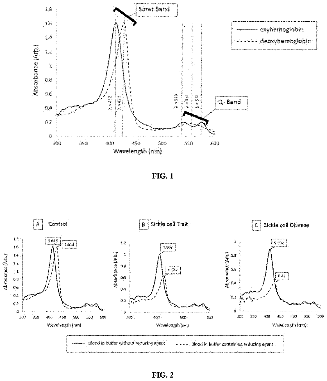
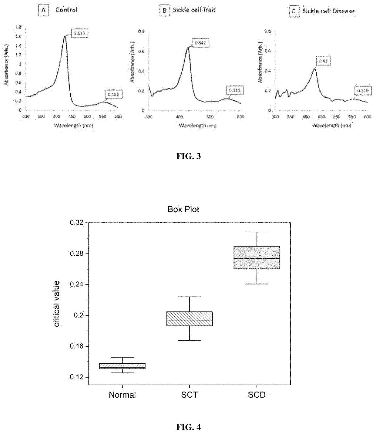
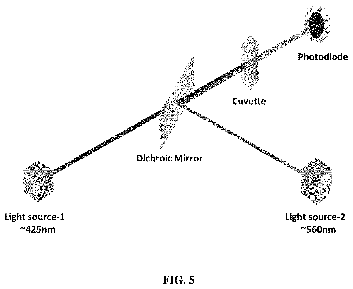
[0136]In this study, known samples were used to analyze the absorption at 427 nm and 555 nm under deoxygenated conditions using 2.365M phosphate buffer.

[0137]Sample Collection:

[0138]Venous blood samples were collected in K2EDTA coated collection tubes and stored at 2° C.-8° C. until the tests were carried out.

[0139]Buffer Preparation:

[0140]Phosphate buffer of 2.365M concentration was prepared by adding monobasic salt 1.178 M (160.3 g / L) and dibasic salt 1.187 M (206.8 g / L). Saponin and sodium metabisulfite at 2% final concentration were added to the buffer. The freshly prepared buffer was used for testing within 4 hours of preparation.

[0141]Testing:

[0142]5 μL of blood sample was added to the tube containing 1 mL of buffer, mixed well and incubated at room temperature for 15 minutes. The sample-buffer mixture loaded into a glass cuvette of 2 mm pathlength and measured the optical absorption spectrum using a spectrophotome...

example 2

tion of Threshold Values Using the Ratio Method (Known Samples)

[0147]In this study, known samples were used to analyze the absorption at 427 nm and 555 nm under deoxygenated conditions using 2.083M phosphate buffer.

[0148]Sample Collection:

[0149]Venous blood samples were collected in K2EDTA coated collection tubes and stored at 2° C.-8° C. until the tests were carried out.

[0150]Buffer Preparation:

[0151]Phosphate buffer of 2.083 M concentration was prepared by adding Monobasic salt 1.178 M (160.3 g / L) and dibasic salt 0.905 M (156.2 g / L). Saponin and sodium metabisulfite at 2% final concentration were added to the buffer. The freshly prepared buffer was used for testing within 4 hours of preparation.

[0152]Testing:

[0153]5 μL of blood sample was added to the tube containing 1 mL of buffer, mixed well and incubated at room temperature for 15 minutes. The sample-buffer mixture loaded into a glass cuvette of 2 mm pathlength and measured the optical absorption spectrum using a spectrophotom...

example 3

of Blood Samples Using the Ratio Method (Blind Study)

[0157]In this study, unknown samples were analyzed using 2.365M phosphate buffer to identify the presence or absence of HbS and the presence or absence of SCD or SCT.

[0158]Sample Collection:

[0159]Venous blood samples were collected in K2EDTA coated collection tubes and stored at 2° C.-8° C. until the tests were carried out.

[0160]Buffer Preparation:

[0161]Phosphate buffer of 2.365M concentration was prepared by adding monobasic salt 1.178 M (160.3 g / L) and dibasic salt 1.187 M (206.8 g / L). Saponin and sodium metabisulfite at 2% final concentration were added to the buffer. The freshly prepared buffer was used for testing within 4 hours of preparation.

[0162]Testing:

[0163]5 μL of blood sample was added to the tube containing 1 mL of buffer, mixed well and incubated at room temperature for 15 minutes. The sample-buffer mixture loaded into a glass cuvette of 2 mm pathlength and measured the optical absorption spectrum using a spectropho...


PUM

PropertyMeasurementUnit
wavelengthaaaaaaaaaa

Abstract

The present disclosure provides in vitro methods for identifying the presence or absence of haemoglobin S (HbS) or haemoglobin C (HbC) in a blood sample, kits and devices thereof. The inventors have found that HbS shows a substantial decrease in absorption under deoxygenated conditions compared to oxygenated conditions. The inventors expect HbC to show a similar decrease in absorption under deoxygenated conditions compared to oxygenated conditions. The methods, kits and devices of the disclosure employ this decrease in absorption under deoxygenated conditions to identify the presence or absence of HbS or HbC in a blood sample. The methods, kits and devices of the present disclosure are simple, low cost, and provide a rapid way to identify the presence or absence of HbS or HbC in a blood sample in a point-of-care setting.

Description

TECHNICAL FIELD[0001]The present disclosure relates to the field of identifying the presence or absence of haemoglobin S (HbS) and / or or haemoglobin C (HbC) in a blood sample. More particularly, the disclosure relates to a method comprising measuring absorbance of the blood sample under different conditions, calculating a ratio of absorbance or a percent reduction in absorbance under said conditions, and identifying the presence or absence HbS and / or HbC based on said ratio or percent reduction.BACKGROUND OF THE DISCLOSURE[0002]Sickle cell disease (SCD) is a genetic disorder associated with the haemoglobin protein of Red Blood Cells (RBCs). SCD is inherited in an autosomal recessive pattern and is considered among the most commonly inherited diseases worldwide. The cause of SCD is a point mutation in the beta-globulin gene (of haemoglobin), which results in the substitution of glutamic acid with valine (sixth position of beta-globin chain). Haemoglobin with this defect is referred t...

Claims


Application Information

Patent Timeline
Patent Type & Authority Applications(United States)
IPC IPC(8): G01N33/72G01N21/31G01N33/49
CPCG01N33/721G01N21/31G01N33/49G01N2333/805G01N2800/22G01N21/314G01N33/6893
Inventor SRINIVASAN, RAJESHV R, EUGENE CHRISTOKATARE, PRATEEKVENUKUMAR, ARAVINDGORTHI, SAI SIVANAMBISON, NISANTH KM
Owner INDIAN INSTITUTE OF SCIENCE
Learn More