
Hyperspectral imaging in automated digital dermoscopy screening for melanoma

a technology of automated digital dermoscopy and melanoma, applied in the direction of image enhancement, diagnostics using spectroscopy, instruments, etc., can solve the problems of substantial number of unnecessary biopsies, inability to detect melanoma clinically, and inability to perform clinically significant melanoma detection

Pending Publication Date: 2022-03-31
THE ROCKEFELLER UNIV
View PDF0 Cites 3 Cited by
  • Summary
  • Abstract
  • Description
  • Claims
  • Application Information

AI Technical Summary

Benefits of technology

The patent describes a computer-aided dermoscopic screening system for melanoma that uses artificial intelligence to analyze images of melanoma and nevi. The system generates screening algorithms based on these images and combines them with other clinical information to create a risk score that can be used to classify melanoma or nevi. The system is trained using hyperspectral imaging, which uses multiple colors to capture more information about melanoma. The system is also composed of non-deep machine learning algorithms, which are more accountable and accurate compared to simply using deep learning methods. Overall, this system has been shown to have high sensitivity and specificity in detecting melanoma.

Problems solved by technology

With current methods, a considerable number of unnecessary biopsies are performed because only a fraction of all biopsied, suspicious lesions are actually melanomas.
The detection of melanoma clinically can be visually challenging and often relies on the identification of hallmark features including asymmetry, irregular borders, and color variegation to identify potentially cancerous lesions.
In clinical practice, only 11% of all biopsied, suspicious lesions are melanomas and thus a substantial number of unnecessary biopsies are performed.
Thus, despite evidence that early detection decreases mortality, considerable uncertainty surrounds the effectiveness of state of-the-art technology in routine melanoma screening.
In this context, clinical melanoma screening is a signal-detection problem, which guides the binary decision for or against biopsy.
However, deep-learning dermatology algorithms cannot show a physician how a decision was arrived at, diminishing enthusiasm in the medical community.
Dermoscopy, in which a liquid interface or cross-polarizing light filters allow visualization of subsurface features, including deeper pigment and vascular structures, has been shown to be superior to examination with the naked eye; however, it remains limited by significant inter-physician variability and diagnostic accuracy is highly dependent on user experience.
Even more concerning, the use of dermoscopy by inexperienced dermatologists may result in poorer performance compared to examination with the naked eye.
The primary challenge with clinical application of these technologies is obtaining a near perfect sensitivity, as a false negative, or Type II melanoma screening error, can have a potentially fatal outcome.
However, there is still significant debate as to whether MelaFind is a useful tool to guide dermatologists, the concern being that the device almost always recommends biopsy.
However, when implemented in a melanoma screening clinic, the SIAscope did not improve the diagnostic abilities of dermatologists.
Further studies demonstrated poor correlation between SIAscopy analysis and histopathology in both melanoma and nonmelanoma lesions and worse accuracy than dermoscopy.
Of note, direct comparison of devices to other systems on the market is limited, as diagnostic performance of a device varies with the difficulty of lesions included in analysis, as well as the proportion of atypical nevi in the benign set.
There are a variety of proprietary DDA instruments on the market, although none have yet demonstrated a reproducibly high sensitivity and specificity for melanoma detection.
Deep learning, automated processing diagnostic devices are rapidly transforming healthcare due to their remarkable predictive power, yet careful considerations of biases in training data present ethical concerns and limit adoption of artificial intelligence.
Black box deep learning approaches such as convolutional neural networks (CNN) may be inappropriate for stand-alone diagnostic medical decision-making because these algorithms cannot be held liable for screening errors and neither can physicians who use them without understanding the underlying computational diagnostic mechanics.

Method used


Image

  • Hyperspectral imaging in automated digital dermoscopy screening for melanoma
  • Hyperspectral imaging in automated digital dermoscopy screening for melanoma
  • Hyperspectral imaging in automated digital dermoscopy screening for melanoma

Examples


Embodiment Construction

[0041]In the following detailed description, numerous specific details are set forth in order to provide a thorough understanding of the invention. However, it will be understood by those skilled in the art that the present invention may be practiced without these specific details. In other instances, well-known methods, procedures, and components have not been described in detail so as not to obscure the present invention.

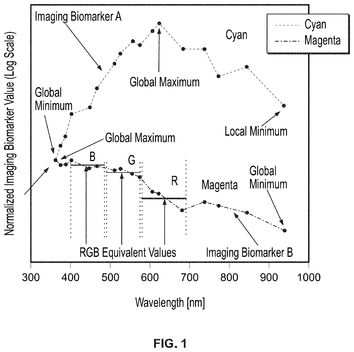
[0042]FIG. 1 shows the spectral dependence of two imaging biomarkers on one sample lesion over the entire spectrum, as a function of wavelength, providing evidence that a machine learning algorithm utilizing a range of wavelengths may achieve higher sensitivity and specificity compared to RGB equivalent values. The two imaging biomarkers selected for analysis were the most melanoma-predictive RGB biomarkers identified in the aforesaid U.S. Patent Application Publication No. 2018 / 0235534 (i.e., “optimum imaging biomarkers”).

[0043]The optimum imaging biomarker value...


PUM


Abstract

Hyperspectral dermoscopy images obtained in N wavelengths in the 350 nm to 950 nm range with a hyperspectral imaging camera are processed to obtain imaging biomarkers having a spectral dependence. Machine learning is applied to the imaging biomarkers to generate a diagnostic classification.

Description

CROSS REFERENCE TO RELATED APPLICATIONS[0001]This application is a continuation of PCT International Application No. PCT / US2020 / 012724, International Filing Date Jan. 8, 2020, claiming the benefit of U.S. Provisional Application Ser. No. 62 / 789,652, filed on Jan. 8, 2019 and entitled HYPERSPECTRAL IMAGING IN AUTOMATED DIGITAL DERMOSCOPY SCREENING FOR MELANOMA, which is incorporated in its entirety herein by reference.BACKGROUND OF THE INVENTION[0002]Early melanoma detection decreases morbidity and mortality. Early detection classically involves dermoscopy to identify suspicious lesions for which biopsy is indicated. Biopsy and histological examination then diagnose benign nevi, atypical nevi, or cancerous growths. With current methods, a considerable number of unnecessary biopsies are performed because only a fraction of all biopsied, suspicious lesions are actually melanomas. Thus, there is a need for more advanced noninvasive diagnostics to guide the decision to biopsy.[0003]The d...

Claims


Application Information

Patent Timeline
Patent Type & Authority Applications(United States)
IPC IPC(8): A61B5/00G06T7/00
CPCA61B5/444A61B5/0075A61B5/7267A61B5/7253G06T2207/20084G06T2207/30088G06T2207/30096G06T2207/10036G06T2207/20081G06T7/0012G01N21/31G01N2021/4764G01N2201/0627G06V10/82
Inventor GAREAU, DANIEL
Owner THE ROCKEFELLER UNIV
Learn More