Method for producing a corneal epithelial cell population
- Summary
- Abstract
- Description
- Claims
- Application Information
AI Technical Summary
Benefits of technology
Problems solved by technology
Method used
Image

Examples
reference example 1
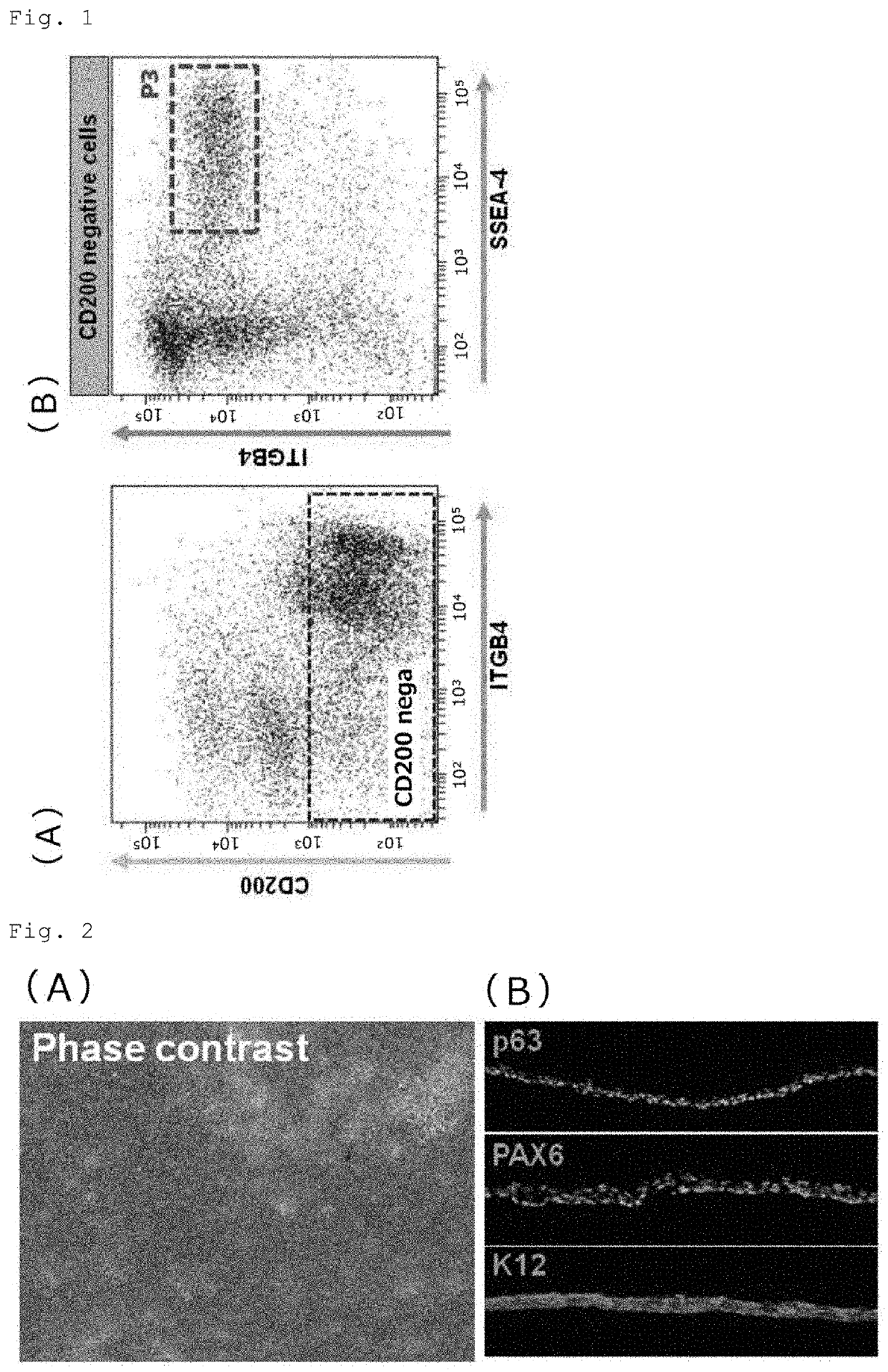
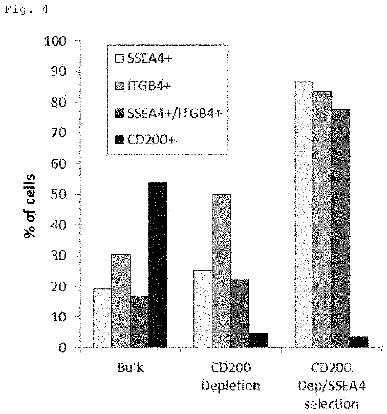
Separation of CD200-Negative / SSEA-4-Positive / ITGB4-Positive Cells by FACS Sorting and Analysis of Corneal Epithelial Cell Markers
[0061](1) Induced Differentiation of iPS Cells into Periocular Cells
[0062]For induced differentiation of iPS cells into periocular cells, the protocol for self-formed ectodermal autonomous multi-zone (SEAM) formation (Hayashi et al. Nature. 2016 Mar. 17; 531(7594):376-80.) was used. Human iPS cells (201B7) were seeded at a density of 300 to 440 cells / cm2 on 6- or 12-well plates coated with laminin 511E8 (i-Matrix; Nippi) at a concentration of 0.5 μg / cm2, maintained for 8 to 10 days in StemFit medium (Ajinomoto), and cultured for 4 weeks in a differentiation medium (DM; GMEM (Life Technologies) supplemented with 10% knockout serum replacement (KSR, Life technologies), 1 mM sodium pyruvate (Life Technologies), 0.1 mM non-essential amino acids (Life Technologies), 2 mM L-glutamine (Life Technologies), 1% penicillin-streptomycin solution (Life Technologies) an...
reference example 2
Purification of Corneal Epithelial Cells by MACS
[0067]The periocular cell suspension prepared in Reference Example 1 (1) and (2) was stained with a PE-Cy7-labeled anti-CD200 antibody (BD Pharmingen), an FITC-labeled anti-SSEA-4 antibody (BioLegend) and a PE-labeled anti-CD104 antibody (anti-ITGB4 antibody) (BioLegend or BD Pharmingen). The stained cells were resuspended in MACS buffer (PBS with 0.5% BSA and 2 mM EDTA) and processed on a magnetic-activated cell sorter (Miltenyi Biotec). For depletion of CD200-positive cells, the stained cells were labeled with anti-Cy7 MicroBeads (Miltenyi Biotec) at 4° C. for 15 minutes, washed with MACS buffer, and applied to an MS or LS column (Miltenyi Biotec) in a magnetic field for removal of labeled cells and collection of unlabeled cells. For isolation of SSEA-4-positive cells, the collected unlabeled cells were labeled with anti-FITC MicroBeads (Miltenyi Biotec) at 4° C. for 15 minutes, washed with MACS buffer, and appli...
reference example 3
[0070]Purification of Corneal Epithelial Cells with Use of Laminin E8s
(1) Laminin E8s
[0071]Laminin 111E8 (hereinafter referred to as “LN111E8”, and other laminin E8s are similarly abbreviated), LN211E8, LN332E8, LN411E8 and LN511E8, that is, 5 kinds of laminin E8s were used. The LN511E8 was a commercial product (i-Matrix; Nippi), and other laminin E8s were received from Professor Kiyotoshi Sekiguchi from Osaka University.
(2) Plate Coating
[0072]To 12-well plates (BD Falcon), 1 mL / well of PBS was added, and the E8 fragment of each laminin isoform was added such that the coating concentration would be 0.5 μg / cm2. The plates were allowed to stand at 37° C. for 1 to 3 hours.
(3) FACS Analysis of Non-Adherent Cells
[0073]As described in Reference Example 2 (1), the periocular cell suspension prepared in Reference Example 1 (1) and (2) was stained with a PE-Cy7-labeled anti-CD200 antibody (BD Pharmingen), an FITC-labeled anti-SSEA-4 antibody (BioLegend) and a PE-labeled anti-CD104 antibody (...
PUM
 Abstract
Description
Claims
Application Information
 - R&D
- Intellectual Property
- Life Sciences
- Materials
- Tech Scout
- Unparalleled Data Quality
- Higher Quality Content
- 60% Fewer Hallucinations
Browse by: Latest US Patents, China's latest patents, Technical Efficacy Thesaurus, Application Domain, Technology Topic, Popular Technical Reports.
© 2025 PatSnap. All rights reserved.Legal|Privacy policy|Modern Slavery Act Transparency Statement|Sitemap|About US| Contact US: help@patsnap.com