
Compositions and methods for the treatment of heart disease

a technology for heart disease and compositions, applied in the field of molecular biology and medicine, can solve the problems of reducing the clinical value of heart disease, so as to improve the cardiac ejection fraction and increase the cardiac output.

Pending Publication Date: 2021-10-14
MT SINAI SCHOOL OF MEDICINE
View PDF0 Cites 0 Cited by
  • Summary
  • Abstract
  • Description
  • Claims
  • Application Information

AI Technical Summary

Benefits of technology

This patent describes a treatment for adult humans that involves introducing a vector into the patient to treat cardiac conditions. The treatment results in improved cardiac function and output, including increased cardiomyocyte hyperplasia (the growth of heart muscle cells) and better cardiac ejection fraction (the amount of blood pumped out of the heart with each contraction). Overall, the treatment has the potential to improve heart health in adults and has been shown to be safe and effective in animal studies.

Problems solved by technology

The vast morbidity and mortality of heart disease is, in large part, attributed to the inability of adult human cardiomyocytes to divide in a clinically sufficient manner.
Studies centered on the use of stem cells have remained controversial given the marginal results in regards to improvement in cardiac contractile function noted in clinical trials of cardiovascular cell therapy.
Further, there has not been conclusive evidence in these trials that cell types utilized actually differentiate to functional cardiomyocytes.
One of the main challenges in using cardiac regeneration as a treatment of injury such as myocardial infarction arises from the observation that cardiomyocytes naturally exhibit very low levels of turnover in the healthy human heart.
However, such feat has—to the inventors' knowledge—never been achieved with adult human cardiomyocytes.
As such, data in large animals from pigs, sheep, dogs, and even primates cannot perfectly predict efficacy of a given therapy in humans.

Method used


Image

  • Compositions and methods for the treatment of heart disease
  • Compositions and methods for the treatment of heart disease
  • Compositions and methods for the treatment of heart disease

Examples


example 1

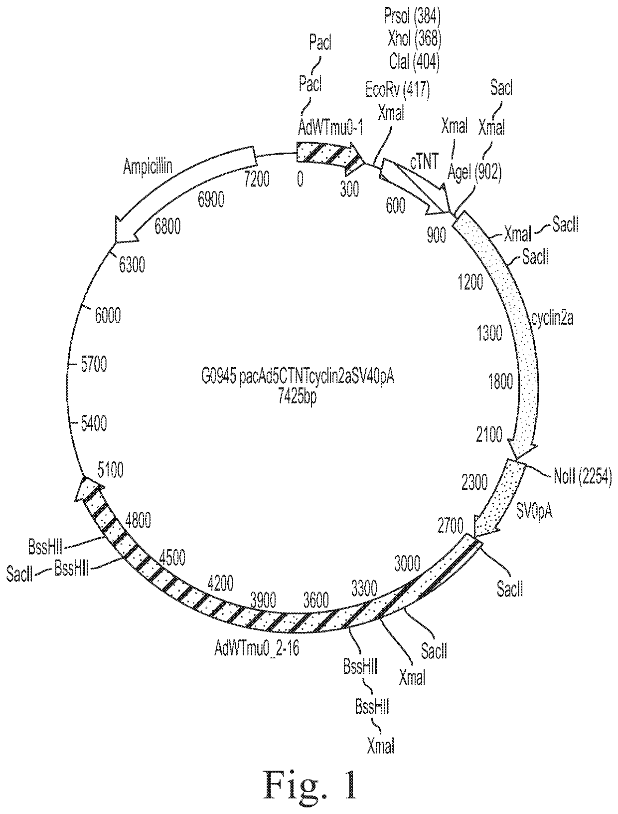
a Therapeutic Use Grade, Human CCNA2 Adenovirus Vector

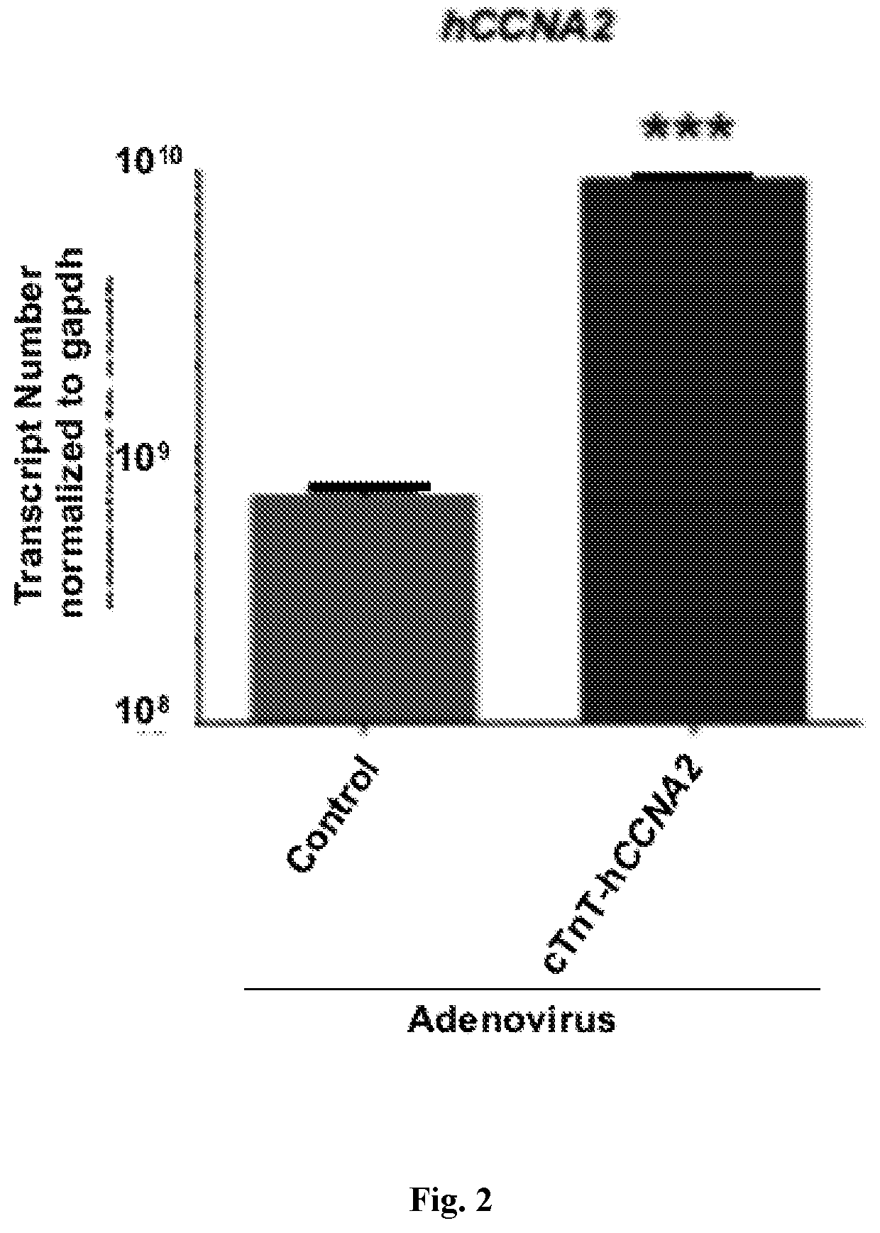
[0076]This Example demonstrates that a cTnT-CCNA2 adenovirus vector can be used to induce expression of CCNA2 in cultured adult human cardiomyocytes.

A. Methods

I. Culture of Adult Human Cardiomyocytes

[0077]Cardiomyocytes from adult human heart tissue (55-year-old male who died of a non-cardiac cause) were isolated after enzymatic digestion at Anabios, San Diego, Calif. and were processed within 24 h. Adult human cardiomyocytes were subjected to the following culture conditions: Cells were washed with serum free DMEM media twice and 105 cells were seeded in 100-mm untreated polystyrene plates (Fisher Scientific). Non-adherent cells were collected every 24 h and centrifuged at 20 g for 2 min at room temperature. The cell pellet was washed with serum-free DMEM and seeded on new polystyrene plates in modified Cardiomyocyte Culture Media22 (mod CMC) formulated by adding 13% FBS, 2.5% horse serum, lx nonessential amino acid, 1 mM sodium...

example 2

Therapeutic Use Grade, Human CCNA2 Adenovirus Vector for Promoting Cytokinesis in Adult Human Cardiomyocytes

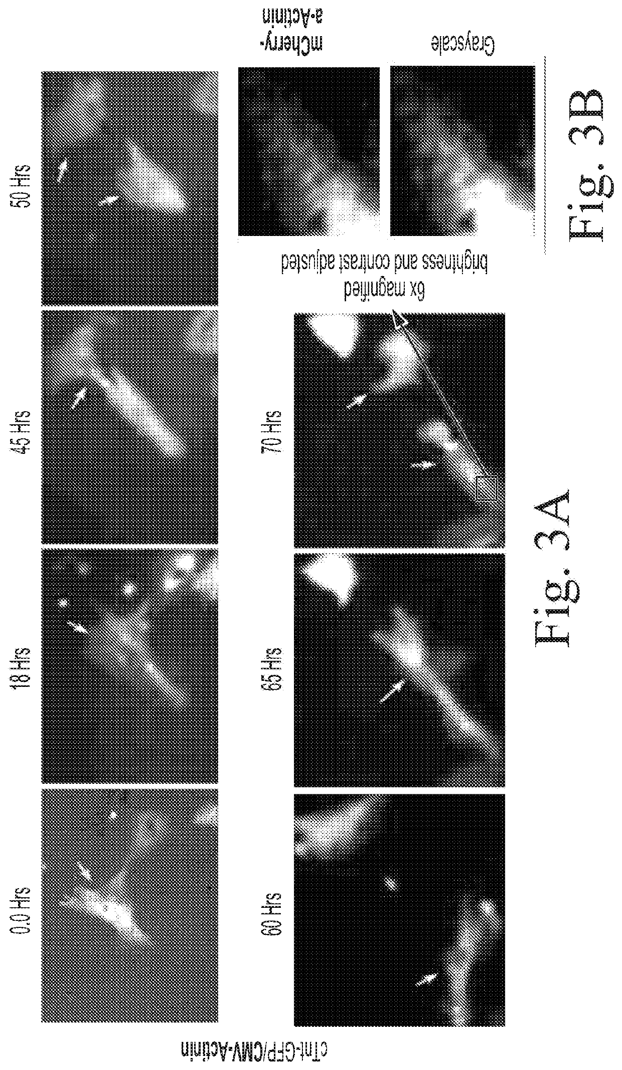
[0080]This Example illustrates that adenoviral vector mediated expression of CCNA2 induces cytokinesis in cultured adult human cardiomyocytes.

A. Methods

I. Time-Lapse Microscopy

[0081]To capture cell division events in cardiomyocytes in vitro, live cell epifluorescence time-lapse microscopy were carried out using a Zeiss AxioVision Observer Z1 (Carl Zeiss, Thornwood, N.Y., USA) inverted epifluorescence microscope in a humidified chamber in the presence of 5% CO2 at 37° C. Multiple random points with cells expressing eGFP (green) and mCherry (red) were selected in the test and control groups. The positions were marked with the “position-list” tool in the AxioVision microscopy software (AxioVision Release 4.7, Carl Zeiss). After the first cycle of imaging, only the channel for Texas red was used (for detection of mCherry) to acquire images for 72 h. The fluorescein isothiocyanate ...


PUM

PropertyMeasurementUnit
time-aaaaaaaaaa
timeaaaaaaaaaa
temperatureaaaaaaaaaa

Abstract

The disclosure relates to compositions and methods for promoting cardiomyocyte cytokinesis and cardiomyocyte proliferation and for use in cardiac regenerative therapy. Embodiments of the disclosure are particularly useful for promoting cytokinesis in adult cardiomyocytes. In embodiments, the disclosure relates to the expression of human cyclin A2 (CCNA2) under the control of a cardiac Troponin T (cTNT) promoter to promote cytokinesis of adult human cardiomyocytes.

Description

STATEMENT REGARDING FEDERALLY SPONSORED RESEARCH[0001]This invention was made with government support under grant numbers HL067048, HL088255, and HL088867 awarded by the National Institutes of Health. The government has certain rights in the invention.BACKGROUNDField of the Invention[0002]The present description relates generally to the field of molecular biology and medicine. More particularly, the methods and compositions herein are useful for promoting cell cycle regulation of heart cells for the treatment of cardiovascular disease.Description of Related Art[0003]Heart disease remains a leading global cause of death in the industrialized world. The vast morbidity and mortality of heart disease is, in large part, attributed to the inability of adult human cardiomyocytes to divide in a clinically sufficient manner. Scar formation via fibrosis is therefore the primary response to cardiac injury. A multitude of molecular and cellular approaches have been investigated over the past 15...

Claims


Application Information

Patent Timeline
Patent Type & Authority Applications(United States)
IPC IPC(8): A61K48/00A61K38/18C12N15/86C12N7/00A61K35/34A61P9/04A61K9/00
CPCA61K48/0058A61K38/18C12N15/86C12N2750/00043A61K35/34A61P9/04A61K9/0029C12N7/00C12N2710/10343C07K14/4738C07K14/47C12N9/12A61K9/0019
Inventor CHAUDHRY, HINA W.
Owner MT SINAI SCHOOL OF MEDICINE
Learn More