Chimeric Antigen Receptor and Method for Treating Cancers
a technology of chimeric antigen and receptor, applied in the field of immunology and pharmacy, to achieve the effect of easy melting, high stringency hybridization, and easy distinguishability
- Summary
- Abstract
- Description
- Claims
- Application Information
AI Technical Summary
Benefits of technology
Problems solved by technology
Method used
Image

Examples
example 1
uction, Process, and Methods
[0087]Buffy coat was obtained from the Hong Kong Red Cross Blood Transfusion Service. Peripheral blood mononuclear cells (PBMCs) were isolated from buffy coat by using Ficoll-Paque PLUS (GE Healthcare). T cells were isolated from PBMCs by using CD3 / CD28 Dynabeads (Thermo). T cells isolated from PBMCs were cultured in initiate medium consisting of AIM-V medium (Thermo) supplemented with 5% human serum (Sigma), 2 mM L-glutamine (Thermo) and 50 U / ml IL-2 (Peprotech), or expansion medium consisting of AIM-V medium supplemented with 5% human serum, 2 mM L-glutamine and 300 U / ml IL-2.
[0088]All the cell lines mentioned below were obtained from ATCC, ECACC or Chinese Academy of Sciences Cell Bank.
[0089]293T cells (ATCC #CRL-3216) were cultured in DMEM medium (Thermo) supplemented with 10% FBS (Thermo), 100 U / ml penicillin (Thermo) and 100 ug / ml streptomycin (Thermo).
[0090]Chronic myelogenous leukemia cell line-K562(ATCC #CCL-243), was cultured in IMEM medium (The...
example 2
n of NKG2D Ligands in Various Cancer Cell Lines
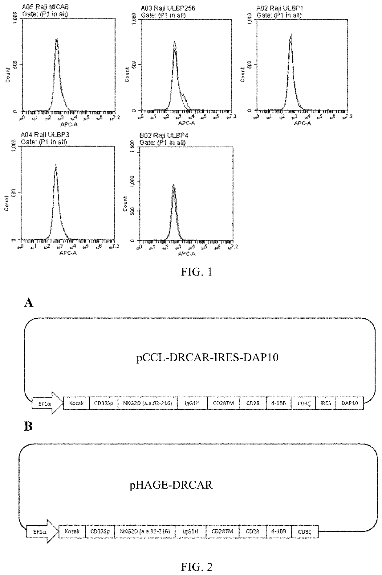
[0109]Detect the expression of NKG2D ligand (human MICA / B and human ULBP1-ULBP6) on different cancer cell lines to determine whether CAR with NKG2D as the antigen domain can be used to kill these cell lines.
[0110]To detect human MICA / B, resuspend 1×10⁶ cells to be tested in 0.5 ml PBS buffer, and used monoclonal mouse anti-human MICA / B (R&D Cat #MAB13001) followed by biotin goat anti-mouse IgG (H+L) and then use streptavidin-APC staining.
[0111]In order to detect human ULBP2 / 5 / 6, resuspend 1×10⁶ cells to be tested in 0.5 ml PBS buffer, and used monoclonal mouse anti-human ULBP2 / 5 / 6 (R&D Cat #MAB1298) followed by biotin Goat anti-mouse IgG (H+L) and then used streptavidin-APC staining.
[0112]To detect human ULBP1, 1×10⁶ cells to be tested were resuspended in 0.5 ml PBS buffer, and used monoclonal mouse anti-human ULBP1 (R&D Cat #MAB1380) followed by biotin goat anti-mouse IgG (H+L) and then used streptavidin-APC staining.
[0113]To detect hu...
example 3
ion of Lentiviral Vector Expressing DRCAR
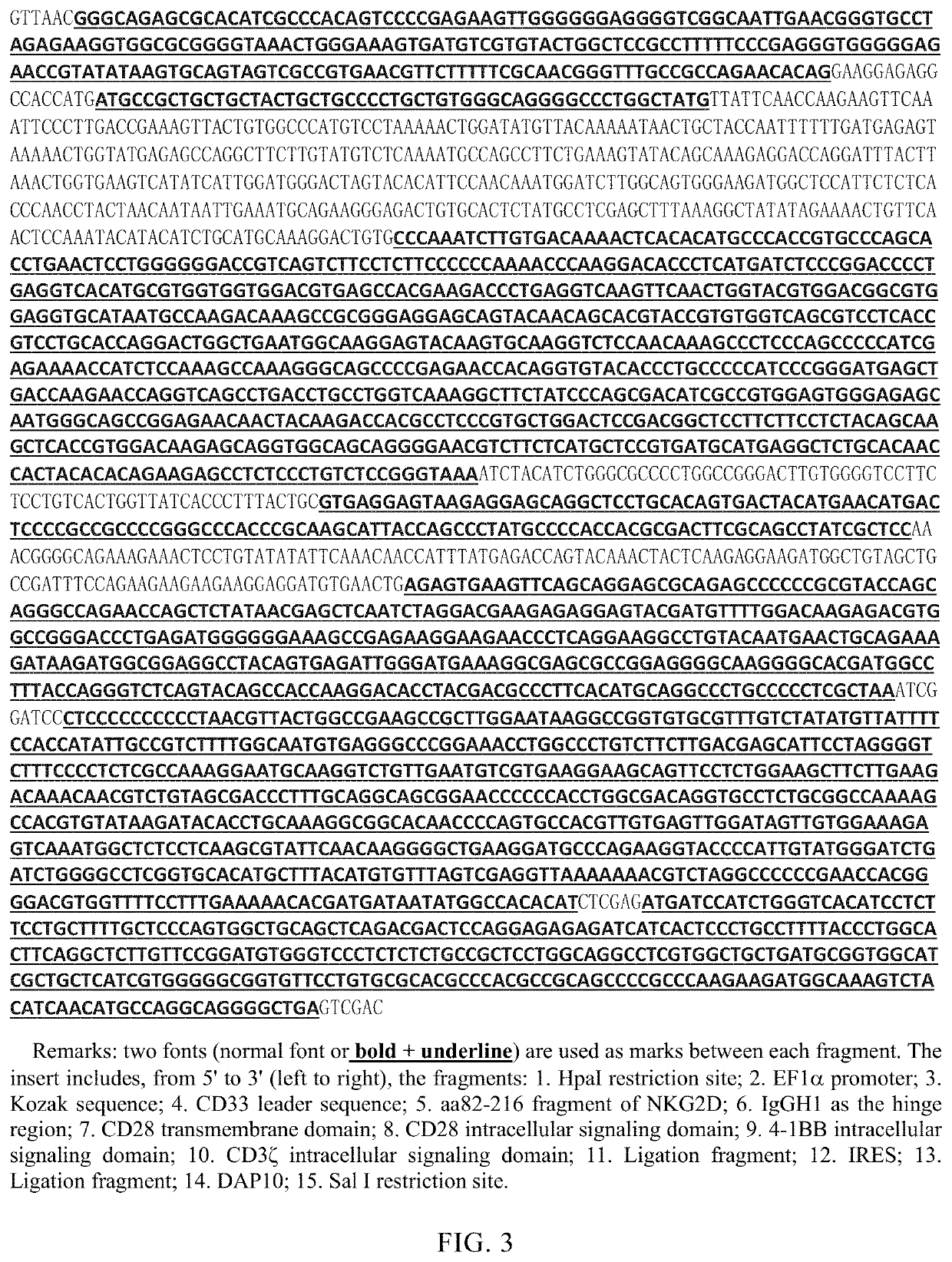
[0119]1. Construction of pCCL-DRCAR-IRES-DAP10
[0120]pCCL-DRCAR-IRES-DAP10 has the structure shown in FIG. 2A. pCCL-DRCAR-IRES-DAP10 was constructed by the following method.
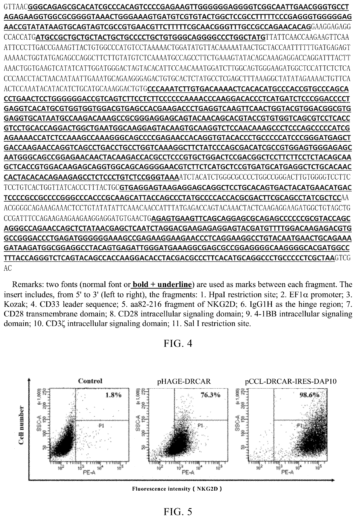
[0121]Synthesized a nucleic acid insert encoding DRCAR and DAP10, which has a nucleotide sequence as shown in FIG. 3. The insert includes, from 5′ to 3′, the fragments: 1. HpaI restriction site; 2. EF1α promoter; 3. Kozak sequence; 4. CD33 leader sequence; 5. aa82-216 fragment of NKG2D; 6. IgGH1 as the hinge region; 7. CD28 transmembrane domain; 8. CD28 intracellular signaling domain; 9. 4-1BB intracellular signaling domain; 10. CD3ζ intracellular signaling domain; 11. Ligation fragment; 12. IRES; 13. Ligation fragment; 14. DAP10; 15. Sal I restriction site.
[0122]The above insert fragment and plasmid Pax5 (Addgene, plasmid #35003) were double digested with restriction enzymes HpaI and Sal I. After the digested product was cut and recovered, it was ligated with T4 ligase ove...
PUM
 Abstract
Description
Claims
Application Information
 - R&D
- Intellectual Property
- Life Sciences
- Materials
- Tech Scout
- Unparalleled Data Quality
- Higher Quality Content
- 60% Fewer Hallucinations
Browse by: Latest US Patents, China's latest patents, Technical Efficacy Thesaurus, Application Domain, Technology Topic, Popular Technical Reports.
© 2025 PatSnap. All rights reserved.Legal|Privacy policy|Modern Slavery Act Transparency Statement|Sitemap|About US| Contact US: help@patsnap.com