Method and Device for Examining the Neurovascular Coupling at the Eye of a Patient

a neurovascular coupling and eye technology, applied in the field of vascular medicine, can solve the problems of increasing the exposure of the retina to light, limiting the range of applications and adaptivity, and inability to capture the function of vascular regulation, so as to avoid errors in the assessment of endothelial function and improve diagnostic reliability

Pending Publication Date: 2021-04-22
IMEDOS SYST GMBH
View PDF0 Cites 1 Cited by
  • Summary
  • Abstract
  • Description
  • Claims
  • Application Information

AI Technical Summary

Benefits of technology

[0024]Advantageously, the method integrates the examination of endothelial function in the large retinal vessels, using the vessel response describing the neurovascular coupling as a reference for the evaluation of the vessel response describing the endothelial function. It is irrelevant how said reference is implemented. An example is the formation of a quotient from the percentage maximum dilation of the large vessels and the percentage maximum change of the vessel response of the neurovascular coupling. Just the information about the strength of the neurovascular coupling is already sufficient to evaluate the examined vascular endothelial function. Thus, the influence of neuronal coupling on the examination of the vascular endothelial function can be eliminated, thereby avoiding errors in the assessment of endothelial function and significantly improving diagnostic reliability.

Problems solved by technology

The fields of application of the Doppler- or OCT-based systems provide statements that describe the vascular or flow state and thus are of little importance for vascular diagnostics outside ophthalmology and are unable to capture the function of vascular regulation.
This considerably increases the retina's exposure to light.
However, another substantial disadvantage of the disclosed solutions also consists in the fixed illumination-side arrangement of a light modulator in the joint illumination-side beam path, which likewise only allows flexible temporal modulation and substantially restricts the range of applications and adaptivity, ultimately having the same disadvantages as the DVA, except for the advantage of being able to also perform measurements on small retinal vessels that are, however, significantly larger than capillaries.
Although this method does not allow representation of the perfusion of the optic nerve head, if perfusion is understood to mean the capillary blood flow, but it provides a measure of the blood volume and, thus, of the capillary vessel diameter and the capillarization of the examined tissue volumes.
The disadvantage of this method is that, in contrast to the description in the aforementioned paper, it cannot provide functional statements about the regulation of capillary perfusion.
A first major drawback of the prior art is the lack of possibilities to examine neurovascular coupling in the retina.
Methods for examining neurovascular coupling in the brain are purely experimental, invasive, very costly and not suitable for clinical use.
A further disadvantage of the prior art is that the results of studies on vascular endothelial function are highly scattered and thus the correlation between endothelial dysfunction and cardiovascular risk factors, events and diseases is unclear, which makes clinical use for the individual assessment of endothelial function and the diagnosis of endothelial dysfunction faulty and unreliable.

Method used


Image

  • Method and Device for Examining the Neurovascular Coupling at the Eye of a Patient
  • Method and Device for Examining the Neurovascular Coupling at the Eye of a Patient
  • Method and Device for Examining the Neurovascular Coupling at the Eye of a Patient

Examples


Embodiment Construction

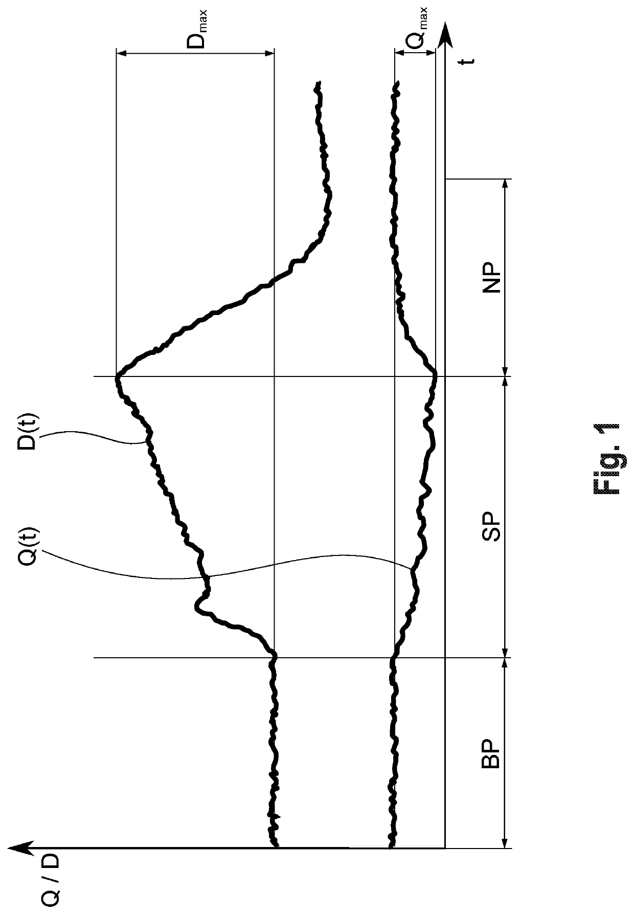
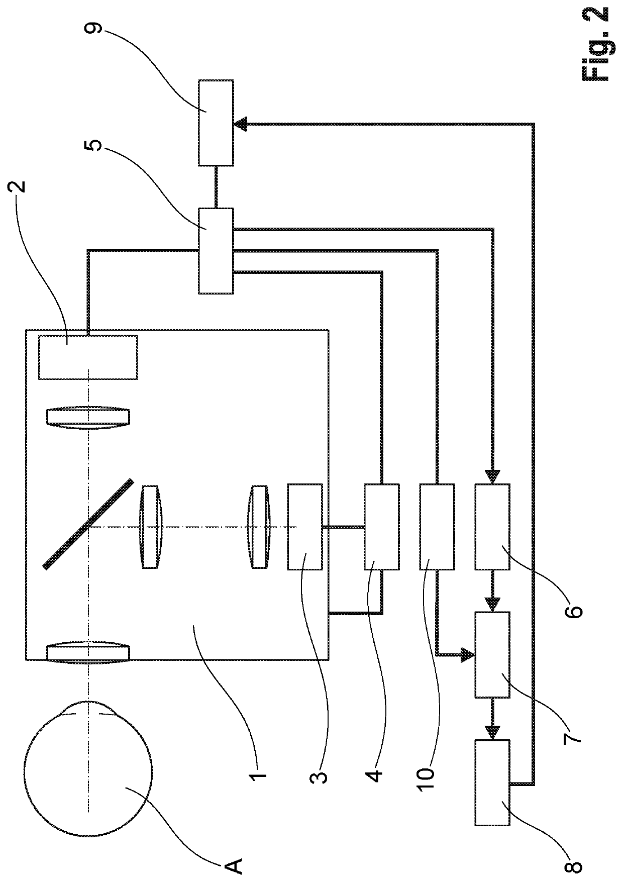
[0057]In a method for examining neurovascular coupling at the eye A, an imaging method is used to record an image sequence of images of the fundus of the eye, preferably over a baseline phase BP, a stimulation phase SP in which the fundus is stimulated with a flickering light, and a posterior phase NP, see FIG. 1.

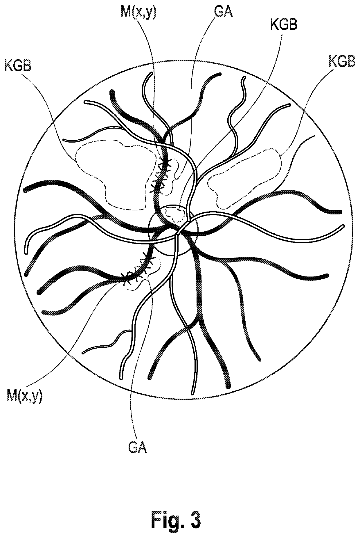
[0058]A signal is derived from the image sequence for at least one capillary vessel region KGB, which represents a capillary vessel response (signal of a measured variable) of the capillary vessels to the stimulation of the retina and whose maximum change during the stimulation phase SP represents an evaluation criterion (biomarker) for NVC. Basically, such a capillary vessel response (vascular signal) can be the changes in capillary blood flow or capillary blood velocity, in capillary vessel diameter or capillary blood volume in the retina or on the optic nerve head during the stimulation phase SP.

[0059]It is irrelevant by which imaging method the at least one image sequen...


PUM


Abstract

A method and a device for examining the neurovascular coupling at the eye (A) of a patient, wherein an imaging method is used to record an image sequence of images of the fundus of the eye (A), preferably over a baseline phase (BP), a stimulation phase (SP) in which the fundus is stimulated with a flickering light, and a posterior phase (NP). Signals, in particular an averaged quotient signal (Q(t)) representing a vessel response of the vessels of the capillary vessel region to the stimulation, are derived from the image sequence for at least one capillary vessel region of the fundus. The absolute or percentage maximum change (Qmax) of the averaged quotient signal (Q(t)) is then used as an evaluation criterion for the neurovascular coupling.

Description

RELATED APPLICATIONS[0001]This Application is a U.S. National Stage Under 35 USC § 371 of International Application PCT / DE2019 / 100295, filed on Mar. 29, 2019, which in turn claims priority to German Patent Application DE 10 2018 107 623.1, filed Mar. 29, 2018, both of which are incorporated herein by reference in their entirety.FIELD OF THE INVENTION[0002]The area of application of the invention relates to the entire field of vascular medicine, e.g. in ophthalmology, neurology, cardiology, nephrology, diabetology, and hypertensiology.BACKGROUND OF THE INVENTION[0003]It is known from studies that microvascular changes are frequently of a systemic nature, i.e. they occur similarly in the vessels, in particular the microcirculation vessels of all organs in the human and animal body, and, depending on the organ, lead to different manifestations of cardiovascular conditions, such as atherosclerosis, arteriosclerosis, cardiac insufficiency, renal insufficiency, eye conditions, e.g. retino...

Claims


Application Information

Patent Timeline
Patent Type & Authority Applications(United States)
IPC IPC(8): A61B3/12A61B3/10A61B3/00
CPCA61B3/1233A61B3/0008A61B3/1241A61B3/102
Inventor VILSER, WALTHARDSKORSETZ, MARTINRIEMER, THOMAS
Owner IMEDOS SYST GMBH
Why Eureka
Try Eureka