
Implantable drug delivery device

a drug delivery and implantable technology, applied in medical science, organic active ingredients, pharmaceutical non-active ingredients, etc., can solve the problems of high difficult control of active agent release from implantable devices, and discontinuation of treatment, so as to reduce the initial burst of drug, prevent moisture ingress, and improve the effect of controlled releas

Pending Publication Date: 2020-07-02
JUNIPER PHARMA UK LTD
View PDF5 Cites 0 Cited by
  • Summary
  • Abstract
  • Description
  • Claims
  • Application Information

AI Technical Summary

Benefits of technology

The patent is about an implantable device that can be used to continuously release a drug for several days, weeks, or even longer periods of time. The device has a protective barrier made of a water-insoluble polymer that prevents moisture from entering while still allowing the drug to be released at a controlled rate. The device is easy to manufacture and is flexible for implantation. This technology has applications in treating medical conditions and can save money and reduce waste by using a lower drug loading.

Problems solved by technology

However, transdermal patches and topical administration can lead to problems such as skin irritation, resulting in discontinuation of treatment (Cohn, Therapeutic Advances in Urology, 2016, Vol. 8(2) 83-90).
Although implantable devices hold the above benefits, controlling the release of the active agent from the implantable device can be problematic, particularly when looking to achieve steady sustained release over an extended period of time.
A particular problem with prior art implantable devices is that a high initial burst of the drug is released.
This initially excessive release can cause a number of undesirable drug-related side effects.
However, the manufacture of a fractionated core can be challenging, for example due to the complexity of joining the segments together.

Method used


Image

  • Implantable drug delivery device
  • Implantable drug delivery device
  • Implantable drug delivery device

Examples


example 2

In-Vitro Dissolution of Example 1

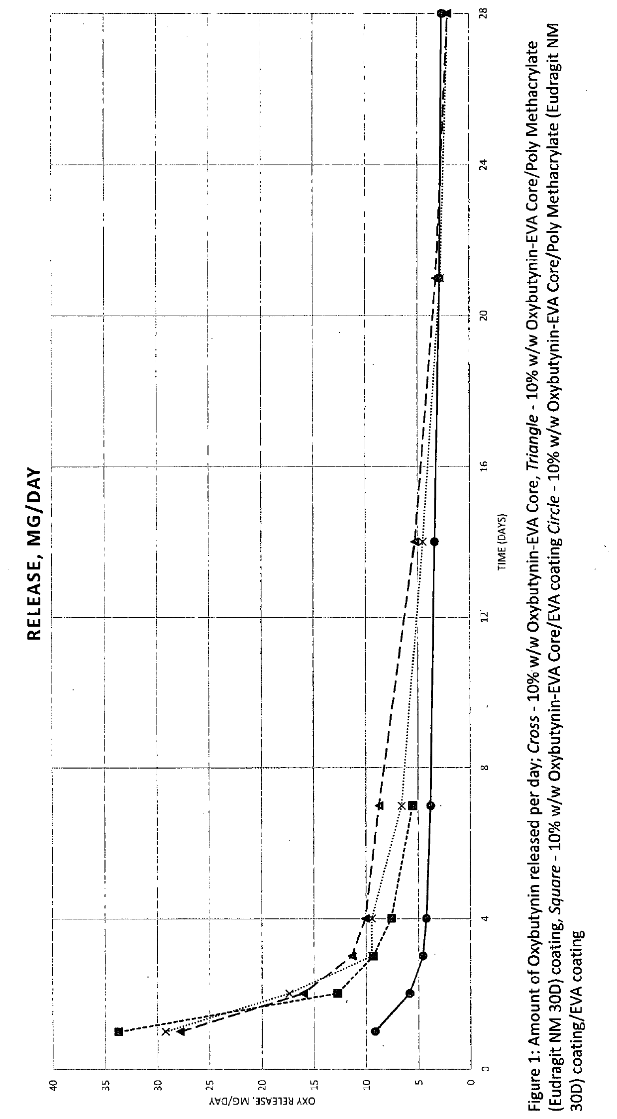
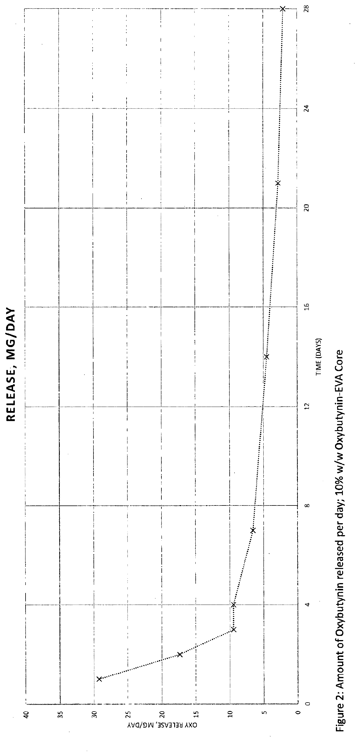
[0066]In-vitro dissolution was performed on the intravaginal rings of Example 1 in 200 mL acetate buffer pH 4.1 at 37° C., and 100 rpm over a 28 day period using shaking incubator. Dissolution samples were typically taken after 1, 2, 3, 4, 7, 14, 21, 28 days. Dissolution samples were analysed by HPLC.

Example 3: 10% w / w Oxybutynin-EVA (28% VA Content) Core / Eudragit NE 30D Coating / EVA (40% VA Content) Coating

[0067]A 10% w / w blend of Oxybutynin base and EVA (28% VA content) was prepared and hot melt extruded at 110° C., to produce a solid solution, and pelletised. The pellets were injection moulded using a barrel temperature of 95° C. and a tool temperature of 35° C. to form a ring. Eudragit NE 30D solution / dispersion was deposited on the 10% w / w Oxybutynin-EVA core to form a coating with an approximate thickness of 50-100 μm. A solution of EVA (40% VA content) was deposited on the Eudragit NE 30D coating to form a coating with an approximate thickness ...

example 4

In-Vitro Dissolution of Example 3

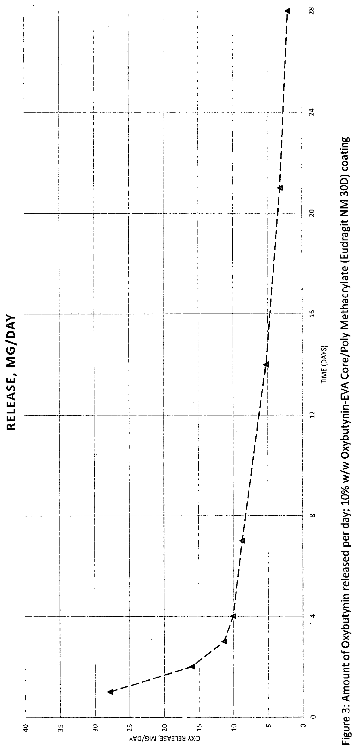
[0068]In-vitro dissolution was performed on the intravaginal ring of Example 3 in 200 mL acetate buffer pH 4.1 at 37° C., and 100 rpm over a 28 day period using shaking incubator. Dissolution samples were typically taken after 1, 2, 3, 4, 7, 14, 21, 28 days. Dissolution samples were analysed by HPLC.

Example 5: 20% w / w Oxybutynin-EVA (28% VA Content) Core / Eudragit NM 30D Coating / EVA (40% VA Content) Coating

[0069]A 20% w / w blend of Oxybutynin base and EVA (28% VA content) was prepared and hot melt extruded at 110° C., to produce a solid solution, and pelletised. The pellets were injection moulded using a barrel temperature of 90° C. and a tool temperature of 35° C. to form a ring. Eudragit NM 30D solution / dispersion was deposited on the 20% w / w Oxybutynin-EVA core to form a coating with an approximate thickness of 50-100 μm. A solution of EVA (40% VA content) was deposited on the Eudragit NM 30D coating to form a coating with an approximate thickness o...

example 6

In-Vitro Dissolution of Example 5

[0070]In-vitro dissolution was performed on the intravaginal ring of Example 5 in 200 mL acetate buffer pH 4.1 at 37° C., and 100 rpm over a 28 day period using shaking incubator. Dissolution samples were typically taken after 1, 2, 3, 4, 7, 14, 21, 28 days. Dissolution samples were analysed by HPLC.


PUM

PropertyMeasurementUnit
thicknessaaaaaaaaaa
thicknessaaaaaaaaaa
thicknessaaaaaaaaaa

Abstract

Disclosed herein are drug delivery devices and their use as a means of achieving controlled release of one or more active pharmaceutical ingredients. In particular, disclosed herein is an implantable drug delivery device comprising an inner core comprising one or more active pharmaceutical ingredients distributed throughout a first water-insoluble polymer; an intermediate coating positioned around the inner core, said intermediate coating comprising an acrylate polymer; and an outer coating positioned around the intermediate coating, said outer coating comprising a second water-insoluble polymer; wherein the acrylate polymer is formed from one or more monomers of formula (I).

Description

FIELD OF THE INVENTION[0001]The present invention relates to implantable drug delivery devices and their use as a means of achieving controlled release of one or more active pharmaceutical ingredients.BACKGROUND OF THE INVENTION[0002]Implantable drug delivery devices are known, and are understood to be devices that are implanted in a bodily orifice (e.g. intravaginal implantation, such as an intravaginal ring), or in bodily tissue (e.g. subcutaneous implantation). Administering a drug by way of an implantable device can offer benefits over oral administration, particularly when looking to achieve more localised drug delivery in order to minimise undesirable systemic effects. Alternative means of achieving more localised drug delivery using non-implantable devices include transdermal patches and topical gels. However, transdermal patches and topical administration can lead to problems such as skin irritation, resulting in discontinuation of treatment (Cohn, Therapeutic Advances in Ur...

Claims


Application Information

Patent Timeline
Patent Type & Authority Applications(United States)
IPC IPC(8): A61K9/00A61K47/32A61K31/216
CPCA61K9/0036A61K31/216A61K47/32A61K9/0024A61K47/34
Inventor RIGBY-SINGLETON, SHELLIEBARKER, IANLUK, SHENPANKAJ LAD, RAJANKUMAR, SANDEEP
Owner JUNIPER PHARMA UK LTD
Learn More