
Method for amplifying cd8+t cells and cell subpopulations thereof in-vitro

a technology of cd8+t cells and cell subpopulations, which is applied in the field of cell biology, can solve the problems of difficult amplifaction, tumor recurrence and metastasis, and the vaccin that can stimulate the effective response of cd8+t cells in the normal body is not ideal in the tumor-bearing body,

Pending Publication Date: 2020-02-27
SHANGHAI INNOVATIONAL CHANGAN BIOLOGICAL TECH CO LTD
View PDF5 Cites 2 Cited by
  • Summary
  • Abstract
  • Description
  • Claims
  • Application Information

AI Technical Summary

Benefits of technology

The present invention provides a method for amplifying CD8+ T cells and functional cell subpopulations thereof, particularly PD-1+CD8+ T cells, using a combination of TLR agonists and specific cytokines. This method significantly enhances the efficiency of CD8+ T cell amplification and activation. The amplified CD8+ T cells exhibit increased functionality, including the ability to secrete CD107a and IFN-γ. This patent offers a more effective method for amplifying specific CD8+ T cell subpopulations that are difficult to amplify under conventional conditions.

Problems solved by technology

However, the vaccine that can stimulate effective response of CD8+ T cells in the normal body is not ideal in the tumor-bearing body, which is associated with an immunosuppressive microenvironment formed by anergy or depletion of CD8+ T cells in local parts of tumors due to the fact that a plurality of immunoregulatory cells or factors are highly enriched or highly expressed in local parts of tumors.
Blocking these pathways can partially restore the function of depleted T cells, but fail to reverse the phenotype of depleted T cells into non-depleted T cells, leading to tumor recurrence and metastasis, which cannot achieve the purpose of radical cure.
However, specific functional cell subpopulations such as PD-1+CD8+ T cells and TEM CD8+ T cells necessary for anti-tumor immunity are difficult to amplify under conventional culture conditions, and there has been to date no method for rapidly amplifying PD1+CD8+ T cells in vitro.
It has been reported in the literature that the PD1+CD8+ T cells sorted from cancer patients have limited in-vitro amplification efficiency, and the amplified cells generally have a capacity of secreting IFN-γ of less than 20%.
Thus, it is difficult to meet the needs of anti-tumor immunity and anti-infection immunity.

Method used


Image

  • Method for amplifying cd8+t cells and cell subpopulations thereof in-vitro
  • Method for amplifying cd8+t cells and cell subpopulations thereof in-vitro
  • Method for amplifying cd8+t cells and cell subpopulations thereof in-vitro

Examples


example 1

of Peripheral Blood Mononuclear Cells

[0033]1. 20 ml of human peripheral blood anticoagulated by heparin was placed in a centrifuge tube. The peripheral blood was diluted with normal saline in a ratio of 1:1 and evenly mixed.

[0034]2. 15 ml of a lymphocyte isolation solution (Ficoll) was added to a new 50 mL centrifuge tube, then the evenly mixed blood dilution solution was slowly added to the upper layer of the lymphocyte isolation solution along the tube wall in a volume ratio of the Ficoll to the blood dilution solution of 1:2 to form a clear separation therebetween, and the resulting solution was centrifuged at 3000 rpm for 30 min.

[0035]3. After centrifugation was completed, a mononuclear cell layer was transferred into a new 50 ml centrifuge tube and washed once with 30 ml of an X-VIVO-15 medium, centrifuged at 800 g for 5 min, and the supernatant was discarded.

[0036]4. 20 ml of the X-VIVO-15 medium was added, the resulting mixture was evenly mixed by blowing and suction, centrif...

example 2

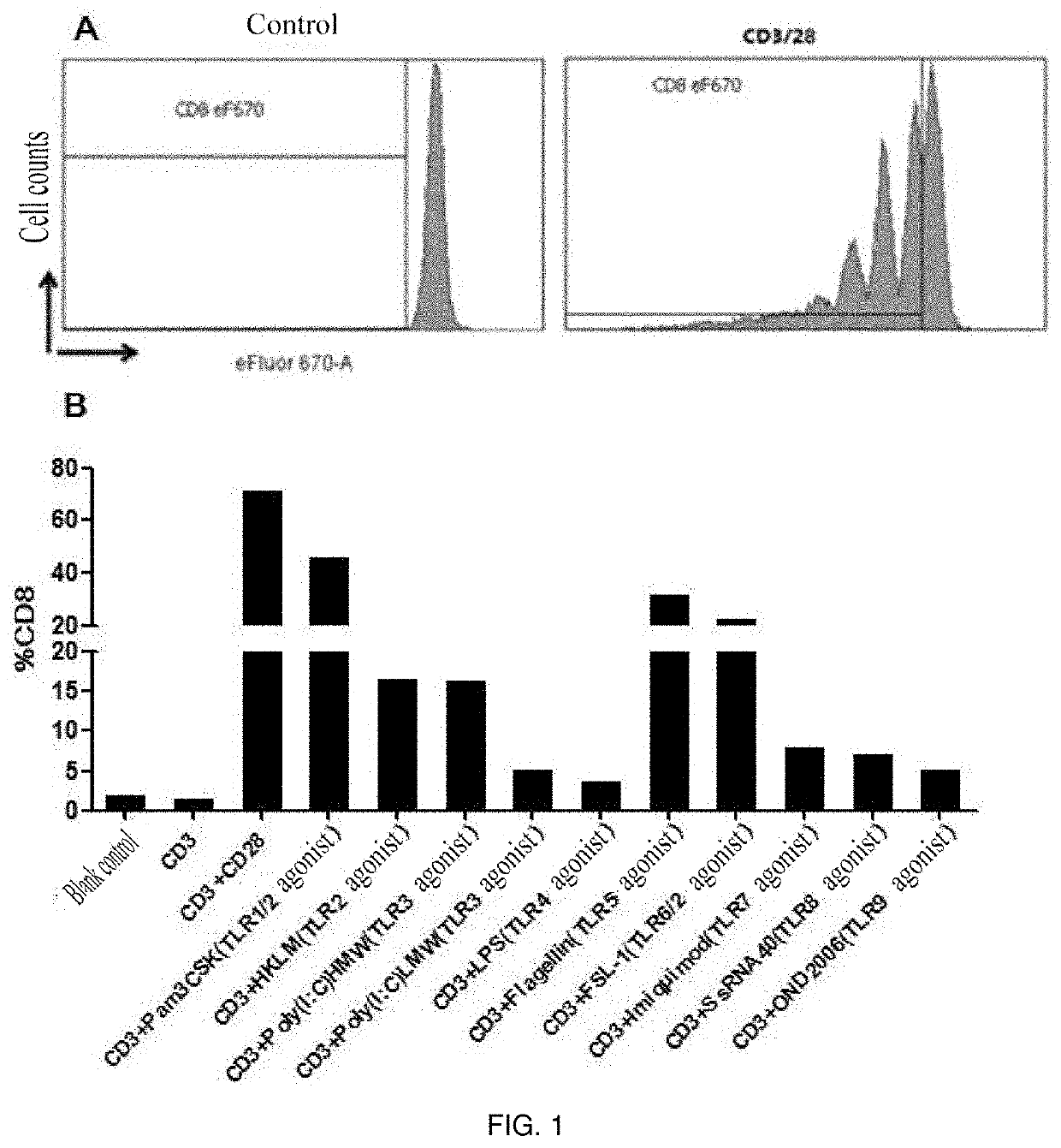
tion of CD8+ T Cells was Effectively Stimulated by TLR Agonists

[0037]1. Peripheral blood mononuclear cells (PBMC) of healthy subjects were isolated according to Example 1.

[0038]2. CD8+ T cells were sorted using an EasySep™ Negative Selection Human CD8+ T cell Enrichment kit (Stemcell: 19053).

[0039]3. The sorted CD8 cells were stained using Cell Proliferation Dye eFluor 670 (ebioscience: 65-0840-90).

[0040]4. The cells were placed in a 96-well U-shaped bottom plate with 1×10⁵ cells per well.

[0041]5. The following stimulants and TLR agonists (invivoGen: tlr-kit1hw, human TLR1-9 Agonist KIT) were added in groups:

[0042](1) blank control;

[0043](2) anti-human CD3 antibody;

[0044](3) anti-human CD3 antibody+anti-human CD28 antibody;

[0045](4) anti-human CD3 antibody+TLR1 / 2 agonist-Pam3CSK4 (0.1-1 μg / ml);

[0046](5) anti-human CD3 antibody+TLR2 agonist-HKLM (108 cells / ml);

[0047](6) anti-human CD3 antibody+TLR3 agonist-Poly(I:C) (10 ng-10 ug / ml);

[0048](7) anti-human CD3 antibody+TLR3 agonist-Poly...

example 3

tion of CD8+ T Cell Subpopulations was Effectively Stimulated by TLR Agonists

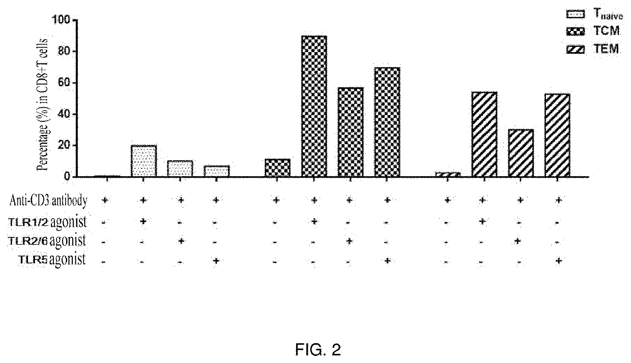
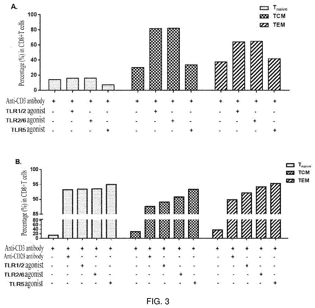
[0058]TLR agonists were capable of effectively stimulating the proliferation of CD8+ T cell subpopulations, i.e. central memory T cells (TCM) and effector memory T cells (TEM).

[0059]1. Peripheral blood mononuclear cells (PBMC) of healthy subjects were isolated.

[0060]2. CD8+ T cells were sorted using an EasySep™ Negative Selection Human CD8+ T cell Enrichment kit (stem cell: 19053).

[0061]3. The sorted CD8 cells were stained using Cell Proliferation Dye eFluor 670 (ebioscience: 65-0840-90).

[0062]4. The cells were placed in a 96-well U-shaped bottom plate with 1×10⁵ cells per well.

[0063]5. The following stimulants and TLR agonists (invivoGen: tlr-kit1hw, human TLR1-9 Agonist KIT) were added in groups:

[0064](1) anti-human CD3 antibody;

[0065](2) anti-human CD3 antibody+TLR1 / 2 agonist-Pam3CSK4 (0.1-1 μg / ml);

[0066](3) anti-human CD3 antibody+TLR6 / 2 agonist-FSL-1 (1 ng-1 μg / ml); and

[0067](4) anti-human CD3 antibody...


PUM

PropertyMeasurementUnit
fluorescentaaaaaaaaaa
temperatureaaaaaaaaaa
concentrationsaaaaaaaaaa

Abstract

Disclosed is a method for rapidly amplifying CD8+ T cells and functional cell subpopulations thereof in vitro. A TLR1 / 2 agonist, a TLR2 / 6 agonist and a TLR5 agonist or a combination of above agonists is added to a conventional culture system for in-vitro amplification of CD8+ T cells. Recombinant cytokines IL-2, IL-7 and IL-15 as well as magnetic beads coated with an anti-human CD3 antibody and an anti-human CD28 antibody can be further added to the culture system for continuous co-stimulation.

Description

TECHNICAL FIELD[0001]The present invention belongs to the field of cell biology, and particularly relates to a method for inducing in vitro amplification of CD8+ T cells and functional cell subpopulations thereof.BACKGROUND[0002]CD8+ T cells are important effector cells involved in anti-tumor and anti-infection immunity. Activated CD8+ T cells can rapidly release high concentrations of cytokines such as IFN-γ and IL-2, and have extensive immunomodulatory effects. In acute infection, CD8+ T cells are activated and substantially proliferate by antigenic stimulation and then differentiate into effector CD8+ T cells (CTL), which kill infected cells by secreting porforin and granzyme (Granzyme B) or induce apoptosis of infected cells by the Fas-FasL pathway. When pathogens are eliminated, a small part of effector CD8+ T cells differentiate into memory CD8+ T cells. When the same antigens appear again, the memory CD8+ T cells in the body will rapidly proliferate and differentiate into eff...

Claims


Application Information

Patent Timeline
Patent Type & Authority Applications(United States)
IPC IPC(8): C12N5/0783
CPCC12N2501/2302C12N2501/999C12N2501/2307C12N2501/998C12N2501/2315C12N5/0638C12N2509/10C12N5/0636C12N2501/51C12N2501/515C12N2501/056C12N2501/052
Inventor XU, JIANQINGZHANG, XIAOYANQIU, CHENLI
Owner SHANGHAI INNOVATIONAL CHANGAN BIOLOGICAL TECH CO LTD
Learn More