
Tissue repair by stem cell recruitment and differentiation

Inactive Publication Date: 2019-04-25
THE TRUSTEES OF COLUMBIA UNIV IN THE CITY OF NEW YORK
View PDF3 Cites 0 Cited by
  • Summary
  • Abstract
  • Description
  • Claims
  • Application Information

AI Technical Summary

Benefits of technology

The present patent provides a method for treating tissue defects by delivering a scaffold with a matrix material, a chemotactic factor, and a chondrogenic factor to the defect. The scaffold can be formed ex vivo or in vivo and can be delivered to the defect using a progenitor cell. The method can also involve delivering the scaffold to the defect and allowing the factors to release over time. The scaffold can be made from fibrin, collagen, or other matrix materials. The method can provide improved tissue regeneration and can be used to treat various tissue defects such as bone, cartilage, and tendon.

Problems solved by technology

Despite recent attempts using matrix metalloproteinases (MMPs), biomaterials, and / or growth factors, no therapy exists to induce seamless healing of inner meniscus tears.
As such, tears in the inner avascular region cannot be repaired by conventional methods and no regenerative therapy currently exists for avascular tears.
In contrast, tears in the inner avascular region, similar to articular cartilage, cannot be repaired due to poor intrinsic healing capacity and the tears frequently extended into the middle-third region.
Tears can often be accompanied by meniscus deterioration.
Tears can frequently progress to meniscus deterioration.
Poor healing of a tear occurring in the avascular region can deteriorate the meniscus.
However, meniscectomy significantly increases the incidence of osteoarthritis (OA) later in life by increasing joint contact stress.
But the use of allografts can have disadvantages, such as donor shortage, pathogen transmission, immunorejection, and tissue mis-match.
Despite the findings, previous studies lacked biochemical and mechanical characterization of the healed tissues and produced controversial experimental outcomes.
Seamless integration of cartilaginous matrix has not been achieved for inner meniscus tears.
Similarly, infusion of autologous MSCs into sheep meniscal defects engrafted and improved healing as evaluated by some, but far from comprehensive, parameters.
However, several experimental studies have not shown an advantage to using stem cell transplantation in meniscus healing.

Method used


Image

  • Tissue repair by stem cell recruitment and differentiation
  • Tissue repair by stem cell recruitment and differentiation
  • Tissue repair by stem cell recruitment and differentiation

Examples


example 1

Fibrochondrogenic Differentiation of Mesenchymal Stem / Progenitor Cells

[0229]The following example shows that treatment with a profibrogenic factor for a limited duration, followed by chondrogenic stimulation can induce step-wise differentiation of bone marrow or synovium MSCs into fibrochondrocyte-like cells.

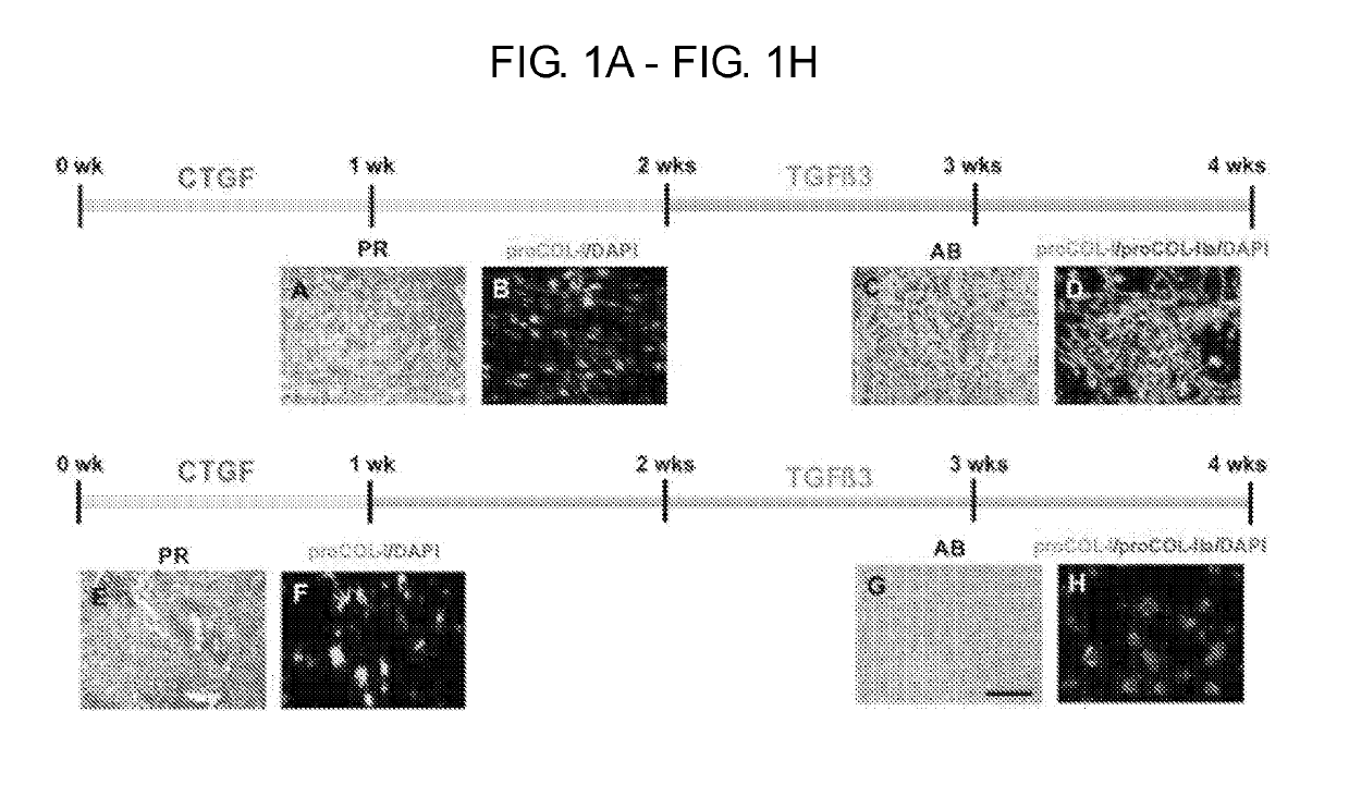
[0230]More specifically, the following example shows a novel strategy to induce seamless healing of inner meniscus repairs (see e.g., FIG. 1). Chemotaxis or other profibrogenic factors (e.g., CTGF) are applied to the torn meniscus via fibrin glue to recruit synovium MSCs into the torn site, followed by formation of fibrous integration. Then the intermediate fibrous matrix is remodeled to cartilaginous tissue by inducing chondrogenic differentiation with growth factor treatment (e.g., TGF-β3). Consistent in meniscus development, the following example demonstrates premature COL-I rich fibrous tissue (E16 & P8w) differentiates into fibrocartilage (Pazin+JOR 2014) (see e.g., FIG. 1)...

example 2

Parameter Determination for Multiple Cytokines / Growth Factors

[0233]The following example determines optimal doses, sequence, and duration of multiple cytokines / growth factors to induce temporally controlled stem cell recruitment and step-wise differentiation for inner meniscus healing in vitro.

[0234]Various delivery strategies were applied, including local delivery, controlled release, and exogenous treatment, to find the optimal doses, sequence, and duration of each growth factor to lead to the seamless healing of inner meniscus tears.

[0235]It is presently thought that the stem / progenitor cells giving rise to meniscus fibrochondrocytes follow a step-wise differentiation: intermediate fibrogenic differentiation followed by chondrogenic differentiation (see e.g., FIG. 1). Given that collagenous fibrous matrix is the default filler for tissue repair, integration and remodeling, the induction of intermediate fibrous matrix in the torn meniscus is presently thought to provide initial in...

example 3

ize Tissue and Cellular Phenotypes in the Healed Meniscus

[0241]The following examples characterize tissue and cellular phenotypes in the healed meniscus using histology, qRT-PCR, nano-indentation, computerized histomorphometry, and immunohistochemistry.

[0242]Previous studies have rarely characterized healed meniscus tissues thoroughly and completely. It is presently thought that chondrocyte-like cell population and cartilaginous extracellular matrix are significant for long-term stability and functions in inner meniscus healing. This study shows avascular zone-specific distribution of extracellular matrix in the native meniscus can be recapitulated in a healed meniscus. This Example describes in-depth characterization of cell and tissue phenotypes in the healed meniscus tissue using multi-level analysis methods. Given the importance of mechanically stable integration for functional restoration of meniscus, pull-out tests are performed to analyze the integration strength, and nanoind...


PUM

PropertyMeasurementUnit
Timeaaaaaaaaaa
Timeaaaaaaaaaa
Timeaaaaaaaaaa

Abstract

Provided herein are compositions and methods for healing cartilage tissue defects or injury by forming fibrochondrocyte cells or fibrochondrocyte-like cells from recruited progenitor cells, such as mesenchymal stem cells.

Description

CROSS REFERENCE TO RELATED APPLICATIONS[0001]The present application claims the benefit of priority to PCT International Application No. PCT / US16 / 22234 filed 13 Mar. 2016, which claims the benefit of U.S. Provisional Application Ser. No. 62 / 303,915 filed 4 Mar. 2016 and U.S. Provisional Application Ser. No. 62 / 133,171 filed 13 Mar. 2015; each of which are incorporated herein by reference in their entireties.STATEMENT REGARDING FEDERALLY SPONSORED RESEARCH OR DEVELOPMENT[0002]Not Applicable.MATERIAL INCORPORATED-BY-REFERENCE[0003]Not Applicable.FIELD OF THE INVENTION[0004]The present disclosure generally relates to repair of cartilaginous tissue by temporal control of stem cell recruitment and differentiation.BACKGROUND OF THE INVENTION[0005]The knee meniscus is an important fibrocartilaginous tissue that stabilizes and absorbs impact on the knee. The knee meniscus is semi-lunar and wedge-shaped fibrocartilaginous tissue between the distal femoral condyle and the proximal tibial plat...

Claims


Application Information

Patent Timeline
IPC IPC(8): A61K35/28C12N5/0775A61L24/00A61L27/22A61K38/18A61L27/38
CPCA61K35/28C12N5/0663A61L24/0031A61L27/225A61K38/1841A61K38/1825A61L27/3869A61L2400/06A61K38/18A61K38/1875A61K2300/00
Inventor MAO, JEREMY J.LEE, CHANG HUN
Owner THE TRUSTEES OF COLUMBIA UNIV IN THE CITY OF NEW YORK
Learn More