Devices and methods for image-guided percutaneous cardiac valve implantation and repair

Inactive Publication Date: 2019-01-03
INNOSCION
View PDF2 Cites 0 Cited by
  • Summary
  • Abstract
  • Description
  • Claims
  • Application Information

AI Technical Summary

Benefits of technology

The present invention provides a trans-apical approach for cardiac valve replacement that avoids the issues of prior art techniques and apparatus. It requires minimal invasive surgery and built-in imaging, but it also requires considerable operating room time. Both femoral and apical or atrial approaches may use separate intra-procedure ultrasound imaging. The invention allows for safe and reliable deployment of cardiac valves and significantly expands the use of transcatheter valves. The invention also provides an ultrasound imaging system that can be easily replaced in real time within a catheter. The system can be calibrated and used to guide the placement of the valve and the need for a periport to prevent blood loss. The invention also allows for the easy removal of the aortic filter and the collapsed filter through the prosthetic valve.

Problems solved by technology

A trans-femoral approach to cardiac valve replacement and repair has been very successful, but the primary concern has been a high incidence rate of stroke.
The femoral approach also makes mitral valve intervention challenging since the operator may perform a transeptal puncture and orient the catheter tip and device almost perpendicular to the location of the mitral valve.
Given the sharp turn needed to access the vicinity of the mitral valve, this alignment is difficult and may need several attempts.

Method used


Image

  • Devices and methods for image-guided percutaneous cardiac valve implantation and repair
  • Devices and methods for image-guided percutaneous cardiac valve implantation and repair
  • Devices and methods for image-guided percutaneous cardiac valve implantation and repair

Examples


Embodiment Construction

[0074]The aspects summarized above can be embodied in various forms. The following description shows, by way of illustration, combinations and configurations in which the aspects can be practiced by the various image guided catheter-like devices discussed and utilized in sequence in a method for replacing or repairing a defective heart valve. It is understood that the described aspects and / or embodiments are merely examples. It is also understood that other aspects and / or embodiments can be utilized, and that structural and functional modifications can be made, without departing from the scope of the present disclosure. Furthermore, modifications to the method(s) of repair and replacement of cardiac valves should not be considered limited in scope to the disclosure but may involve known processes and devices and procedures to those of ordinary skill in the art.

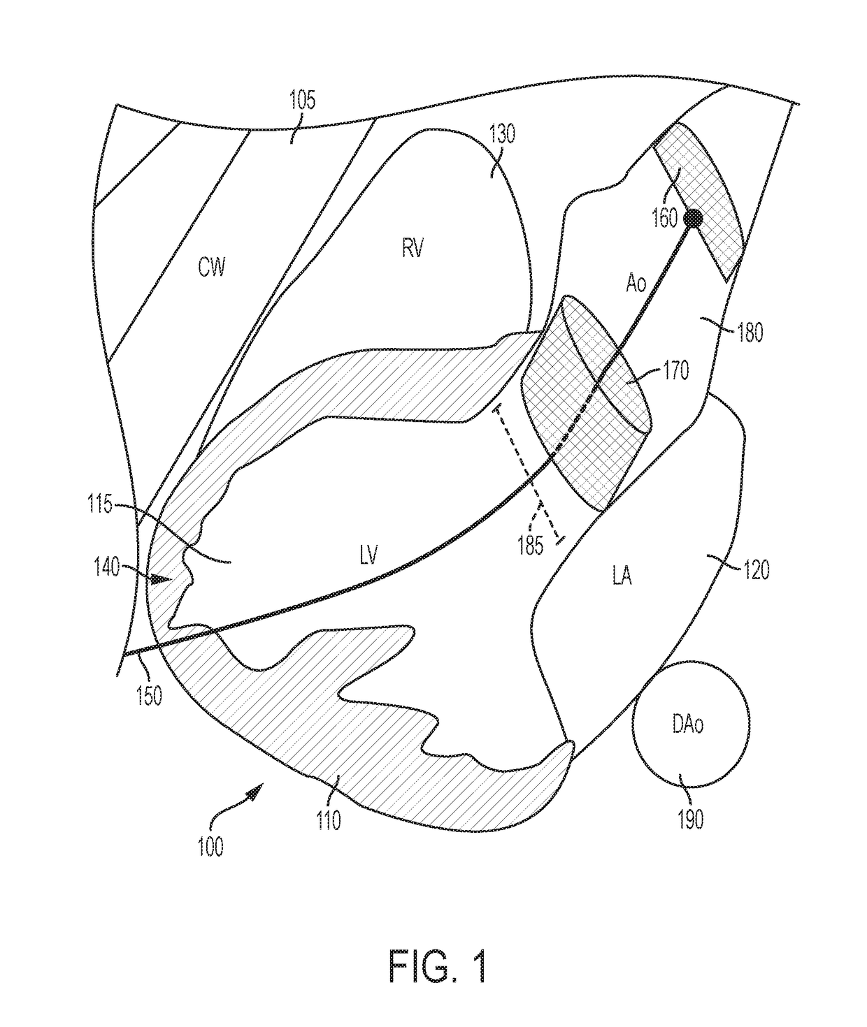
[0075]Referring to FIG. 1, there is shown a drawing in two dimensions of the heart 100 and chest wall (CW) 105 which may be ...


PUM


Abstract

Devices and methods for ultrasound image-guided percutaneous cardiac valve implantation and repair comprise, in combination, a plurality of devices including but not limited to an ultrasound-image guided catheter, a pericardial sheath, a cardiac valve delivery system and an ascending aortic filter. An image-guided catheter is utilized to introduce via an introducer needle and a guide wire a pericardium portal for permitting entry from the chest wall to inside the pericardial space between the pericardial outer lining and inner lining. The pericardium portal permits the use of ultrasound vision to locate a site proximate the left ventricular apex for introduction of a sheath via a myocardium needle into the left ventricular space at an angle and avoiding any coronaries or vessels. A first delivery system permits placement of at least one aortic filter which collects any emboli, particulate matter, plaque and prevents such matter travelling via the ascending aorta to the brain, causing a stroke. A second delivery system permits placement of an aortic and / or mitral valve replacement or repair using several types of repair tools.

Description

[0001]This application claims the right of priority to U.S. Provisional Patent Application Ser. No. 62 / 527,905 of the same title and by the same inventor filed Jun. 30, 2017 and is a continuation-in-part of U.S. patent application Ser. No. 15 / 845,756 filed Dec. 18, 2018, which is a continuation-in-part of U.S. patent application Ser. No. 14 / 865,151 filed Sep. 25, 2015 by the same inventor (now U.S. Pat. No. 9,855,021 issued Jan. 18, 2018) and U.S. patent application Ser. No. 15 / 845,756 claims the right of priority to U.S. Provisional Patent application Ser. No. 62 / 526,170 filed Jun. 28, 2017 and U.S. Provisional Patent Application Ser. No. 62 / 590,464 filed Nov. 24, 2017.TECHNICAL FIELD[0002]The invention relates to the field of cardiac valve replacement and repair and, in particular, to a plurality of devices and a sequential method for implanting via ultrasound and other image guidance a percutaneous cardiac valve, for example, the aortic valve between the left ventricle outflow tr...

Claims


Application Information

Patent Timeline
IPC IPC(8): A61B34/20A61B8/08A61B8/12A61B8/00A61B17/34A61F2/24
CPCA61B34/20A61B8/0841A61B8/12A61B8/445A61B17/3403A61F2/2427A61B17/3478A61B8/0883A61B2034/2063A61B2017/00243A61B2017/3413A61B2017/00575A61M25/09A61B17/3421A61B2017/00247A61B2017/00783A61B2090/3782A61F2/013A61F2230/0067A61B2017/00606A61B2017/00592A61B17/0057A61B10/04A61B2017/00619A61B2017/00623A61B2017/12018A61B2017/06171A61B2017/06052A61F2/0105
Inventor ABRAHAM, THEODORE P.
Owner INNOSCION
Why Eureka
Try Eureka