Method for detecting circulating tumor cells and uses thereof

a tumor cell and detection method technology, applied in the field of biochemistry, can solve the problems of ctcs detection remaining a technical challenge, affecting the diagnosis or prognosis of cancer, and affecting the use of ctcs for cancer diagnosis or prognosis,

Inactive Publication Date: 2018-08-30
AGENCY FOR SCI TECH & RES +1
View PDF0 Cites 2 Cited by
  • Summary
  • Abstract
  • Description
  • Claims
  • Application Information

AI Technical Summary

Problems solved by technology

Methods for reliable detection of cancer and the monitor of cancer progression mainly involve the use of endoscopies, radioactive scannings and tissue biopsy, which are expensive and invasive procedures that impose certain health risks to the patient.
However, the detection of CTCs remains as a technical challenge that hinders the use of CTCs for the diagnosis or prognosis of cancers.
However, the detection of CTCs based on the expression of these epithelial markers is inaccurate as some cells that over-express these epithelial markers are benign epithelial cells which lack clinical utility value.
In addition, certain CTCs, such as CTCs from lung cancers, have been demonstrated to have low or no expression of epithelial markers, thus hinder the detection of such CTCs.
Moreover, tumor cells generally undergo epithelial-to-mesenchymal transition (EMT) when shedding from the primary tumor, thus the current technology that relies on the epithelial markers will generally under-estimate the presence of CTCs with mesenchymal phenotype (i.e. false negative results).
Further, the existing technologies for the detection of CTCs generally involve DNA extraction, amplification and sequencing, which are laborious and time-consuming.
These techniques counteract with the clinical needs for tests with short turnaround time.

Method used


Image

  • Method for detecting circulating tumor cells and uses thereof
  • Method for detecting circulating tumor cells and uses thereof
  • Method for detecting circulating tumor cells and uses thereof

Examples


example 1

pecificity of Mutation-Specific Antibodies

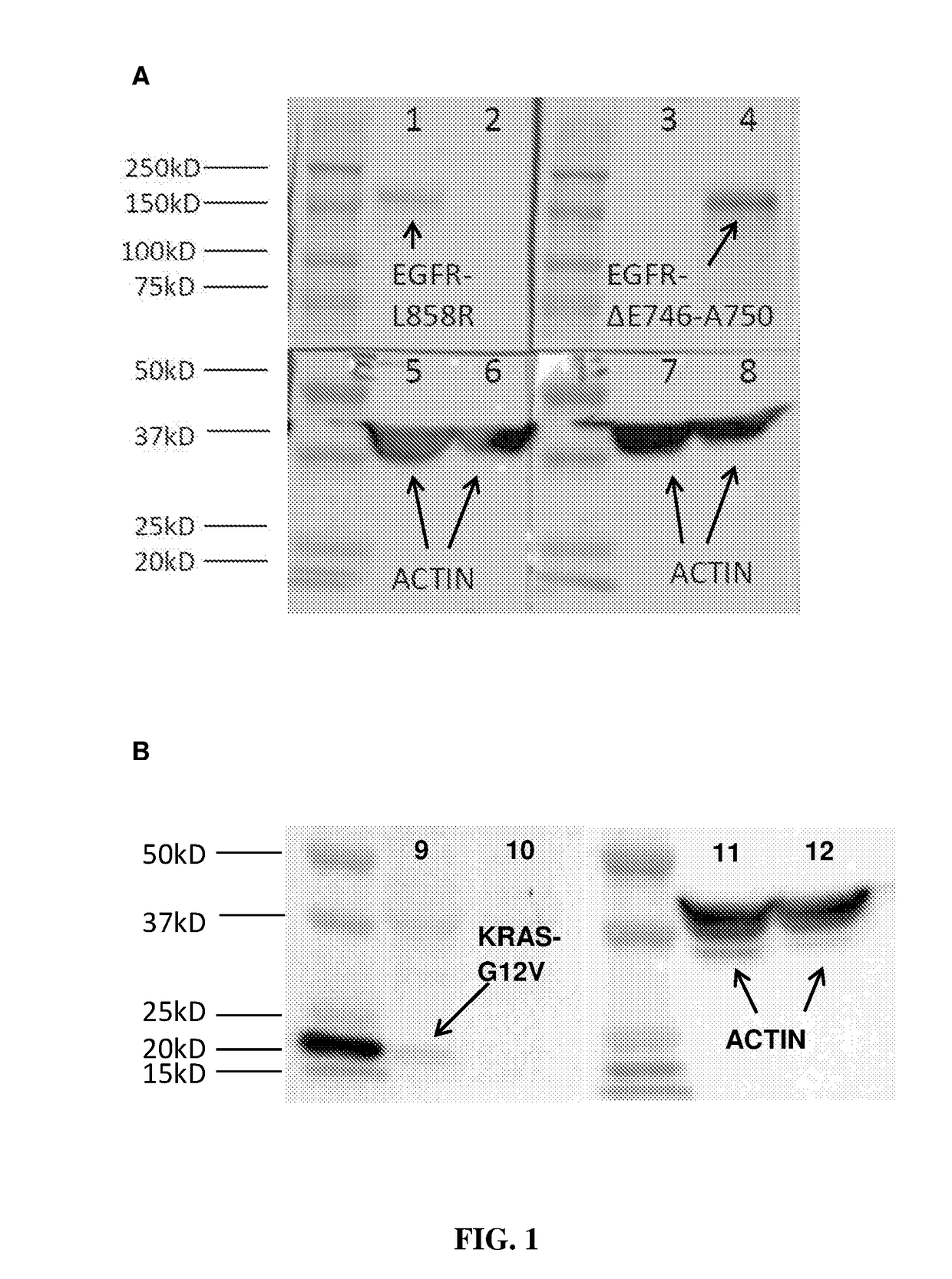
[0067]In order to test the specificity of the antibodies, Western blot analysis was carried out using EGFR L858R and EGFR exon19 (ΔE746-A750) antibodies on H1975 (with known EGFR L858R mutation) and PC9 (with known EGFR exon19 (ΔE746-A750) mutation), and using KRAS G12V antibody on SW620 (with known KRAS G12V mutation) and PC9 (known as negative for KRAS-G12V), according to the following protocol:

[0068]1. The cell lysate from PC9, H1975 and SW620 cancer lines were harvested using RIPA buffer added with protease inhibitor and incubated at 4° C. for 30 minutes.

[0069]2. The lysate was sonicated for three times for 30 seconds.

[0070]3. The protein was denatured at 99° C. for 5 minutes in laemmli buffer.

[0071]4. Equal amount of samples were loaded to the polyacrylamide gel and electrophoresis was performed at constant 20 mA for 1 hour in Tris Glycine SDS running buffer.

[0072]5. The protein was transferred to polyvinylidene difluoride (PVDF) membra...

example 2

Tumor Cells Using Mutation-Specific Antibodies

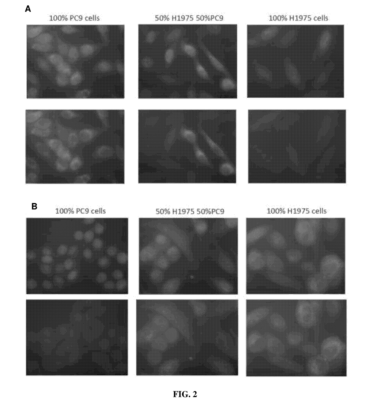
[0078]Immuno-fluorescent experiment was carried out to detect tumor cells in the samples using mutation-specific antibodies EGFR L858R and EGFR exon19 (ΔE746-A750) according to the following protocol:

[0079]1. Cytospin was performed on approximately 100,000 cells.

[0080]2. The cells were incubated with FcR blocking reagent for 15 mins at 4° C. followed by CD45 staining for 30 minutes at 4° C.

[0081]3. The cells were washed for three times followed permeabilization with 0.1% Triton-X for 10 mins at room temperature.

[0082]4. The blocking buffer supplemented with goat serum was added to the sample and incubate for 1 hour.

[0083]5. Primary antibody was added to the sample followed by overnight incubation at 4° C.

[0084]6. Secondary antibody was added to the sample followed by 1 hour incubation at room temperature.

[0085]7. DAPI stain was added to the sample and the slide was mounted with coverslip before visualization on the microscope.

[0086]A ser...

example 3

Tumor Cells in Blood Samples from Healthy Subjects Spiked with Tumor Cells

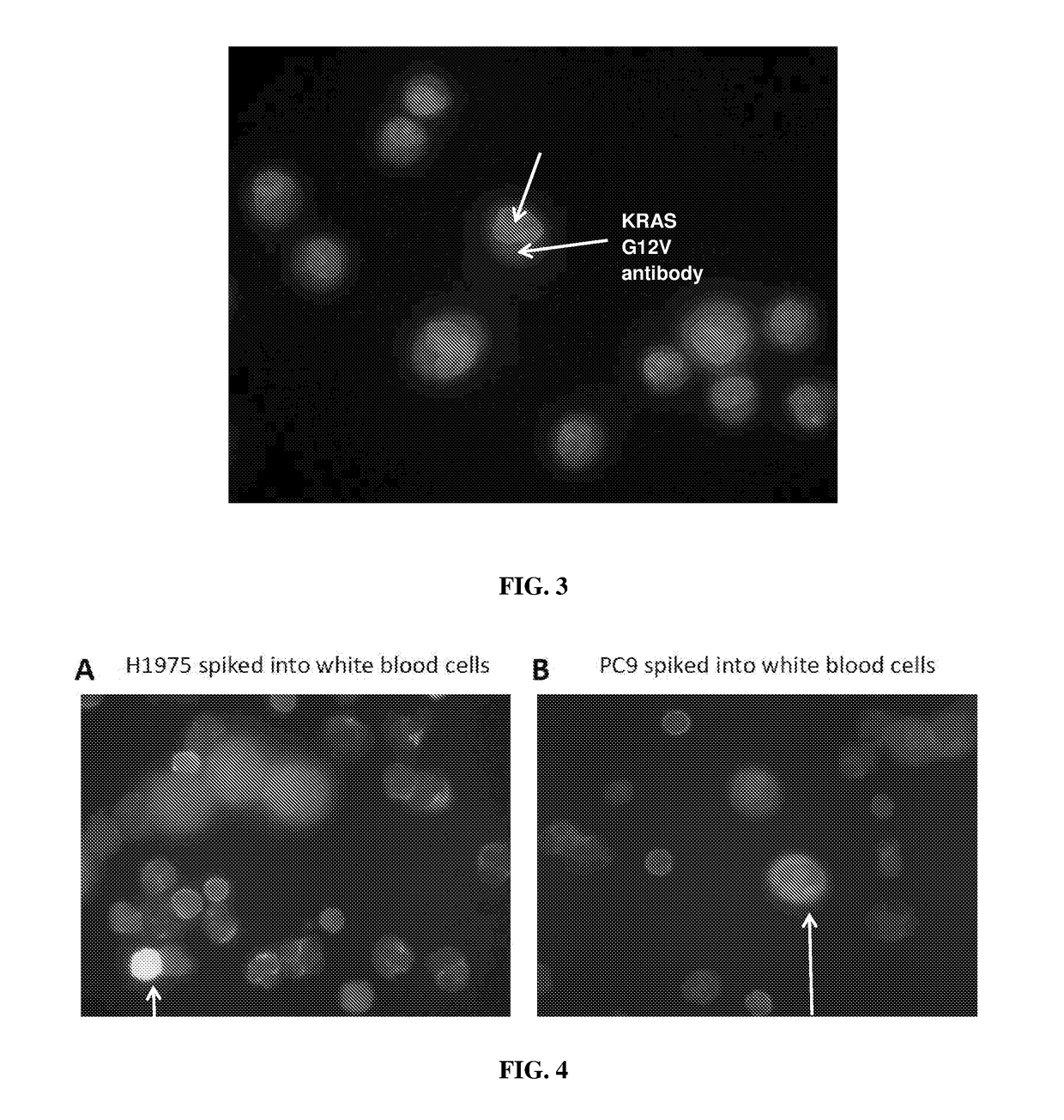
[0088]To simulation samples containing CTCs, blood samples from healthy subjects were collected and spiked with cancer cells H1975 or PC9. The ratio of the spiked cancer cells and the healthy blood cells is 1:9 (10% of cancer cells and 90% of healthy blood cells). The cells were mixed gently and immunofluorescence assay was carried out according to the following protocol:

[0089]1. Cytospin was performed on approximately 100,000 cells.

[0090]2. The cells were incubated with FcR blocking reagent for 15 mins at 4° C. followed by CD45 staining for 30 minutes at 4° C.

[0091]3. The cells were washed for three times followed permeabilization with 0.1% Triton-X for 10 mins at room temperature.

[0092]4. The blocking buffer supplemented with goat serum was added to the sample and incubate for 1 hour.

[0093]5. Primary antibody was added to the sample followed by overnight incubation at 4° C.

[0094]6. Secondary antibody was add...


PUM

PropertyMeasurementUnit
timeaaaaaaaaaa
timeaaaaaaaaaa
timeaaaaaaaaaa

Abstract

A method for the detection and / or diagnosis of cancer in a patient is disclosed. The method involves detecting circulating tumor cells (CTCs) in a sample obtained from the patient using an antibody specifically binding to an antigen in a protein expressed by the CTCs, wherein the antigen comprises a mutation and wherein the mutation renders the patient receptive or resistance to an anti-cancer drug. Claimed methods include the use of an antibody specific for the EGFR L858R or EGFR exon 19 (ΔE746-A750) mutations for detecting lung cancers and the use of an antibody specific for the K-Ras G12V, K-Ras G12C, K-Ras G12S, K-Ras G12D, or K-Ras G13D mutations for detecting colorectal cancers.

Description

CROSS-REFERENCE TO RELATED APPLICATIONS[0001]This application claims the benefit of priority of Singapore provisional application No. 10201506501X, filed on 18 Aug. 2015, the contents of which are being hereby incorporated by reference in their entirety for all purposes.FIELD OF THE INVENTION[0002]The present invention relates to the field of biochemistry. Particularly, the present invention relates to the method of detecting tumor cells, more particularly circulating tumor cells (CTCs) of interest. The present invention also relates to use of the method of detecting CTCs in the detection and / or diagnosis and treatment of cancer.BACKGROUND OF THE INVENTION[0003]Cancer is the second leading cause of death worldwide, accounting for 8.2 million deaths in 2012. Cancer mortality can be significantly reduced if detected and treated early. In addition, during cancer treatment, it is essential to monitor treatment efficacy and cancer progression in order to determine the suitable treatment ...

Claims


Application Information

Patent Timeline
Patent Type & Authority Applications(United States)
IPC IPC(8): G01N33/574
CPCG01N33/5748G01N33/574G01N33/57484
Inventor KONG, SAY LI
Owner AGENCY FOR SCI TECH & RES
Why Eureka
Try Eureka