Devices, methods, and systems relating to super resolution imaging

Inactive Publication Date: 2018-03-29
NORTHWESTERN UNIV
View PDF0 Cites 19 Cited by
  • Summary
  • Abstract
  • Description
  • Claims
  • Application Information

AI Technical Summary

Benefits of technology

The patent text describes a need for improved methods of non-invasive imaging of internal polymer structural information and embedded materials, as current methods are insufficient for non-invasive imaging of internal polymer structural information and embedded materials. The text describes various super-resolution optical imaging techniques that can offer unique advantages in the visualization and characterization of cellular genetic material, especially in biomedical applications. However, these techniques often require extrinsic labels, which have multiple weaknesses such as introducing inaccuracy in spatial localization and requiring additional labeling processes. The text proposes the development of improved super-resolution methods of imaging cellular genetic material that do not require extrinsic labels and are able to better resolve samples with extrinsic labeling for improved diagnostic and prognostic imaging.

Problems solved by technology

While electron microscopy (EM) and scanning probe microscopy (SPM), are widely successful and commonly adopted methods for high resolution imaging of various materials, these methods are insufficient for non-invasive imaging of internal polymer structural information and embedded materials.
While both these methods can provide information on the nanoscopic scale, they often require harsh sample preparation than may either damage or destroy the imaged sample.
However, the spatial resolution of conventional optical imaging methods is fundamentally limited by optical diffraction, far below that of EM and SPM techniques.
Extrinsic agents can have multiple weaknesses, including (1) they require additional labeling processes, (2) they modify physical properties of the test sample material, and (3) they introduce inaccurate spatial localization caused by the physical dimension of the tagged fluorescent and linker molecule (4), due to spectral overlap, a limited number of labels may be resolved or may confound imaging signals leading to inaccuracy.
The combination of these weaknesses reduces the appeal of extrinsic fluorescent contrast agents with traditional imaging methods.

Method used


Image

  • Devices, methods, and systems relating to super resolution imaging
  • Devices, methods, and systems relating to super resolution imaging
  • Devices, methods, and systems relating to super resolution imaging

Examples


example 1

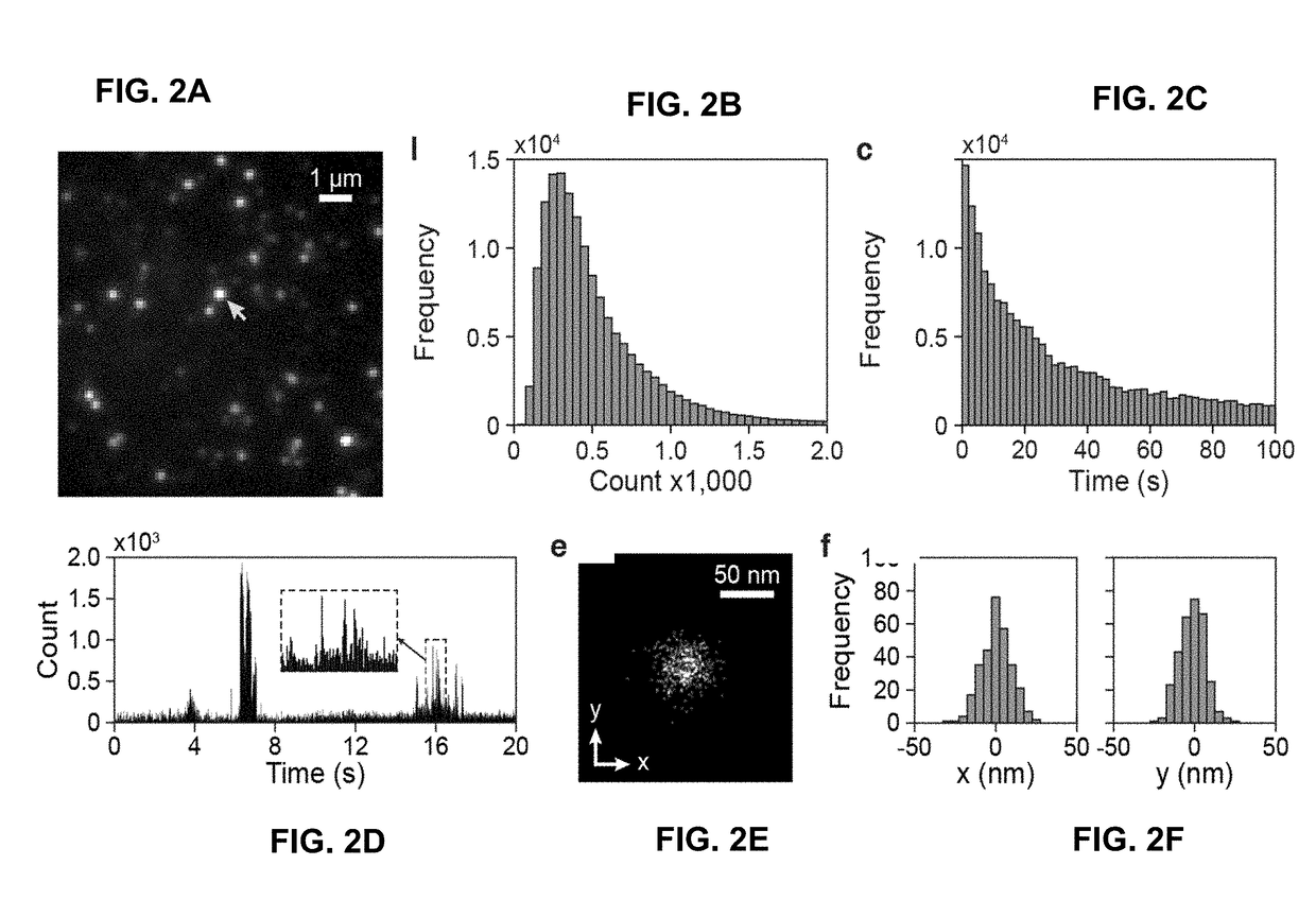
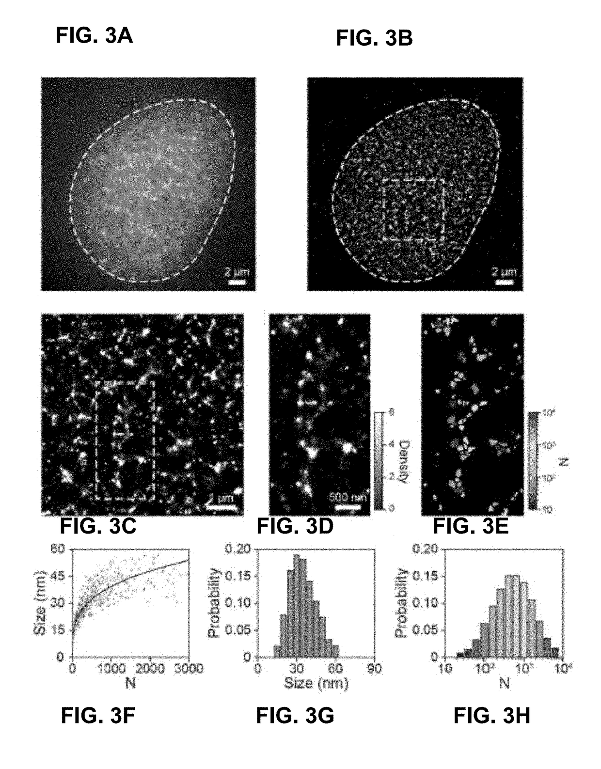
[0097]Certain examples determine stochastic fluorescence switching in nucleic acids under visible light illumination. By combining the principle of photon-localization microscopy, certain examples provide optical super-resolution imaging of native, unmodified DNA molecules; a technique referenced herein as DNA-PLM. Super-resolution imaging is then conducted from isolated, unstained chromosomes and nuclei, revealing nanoscopic features of chromatin without the need for exogenous labels. This paves the way for unperturbed, label-free nanoscale imaging of chromatin structure.

[0098]Results

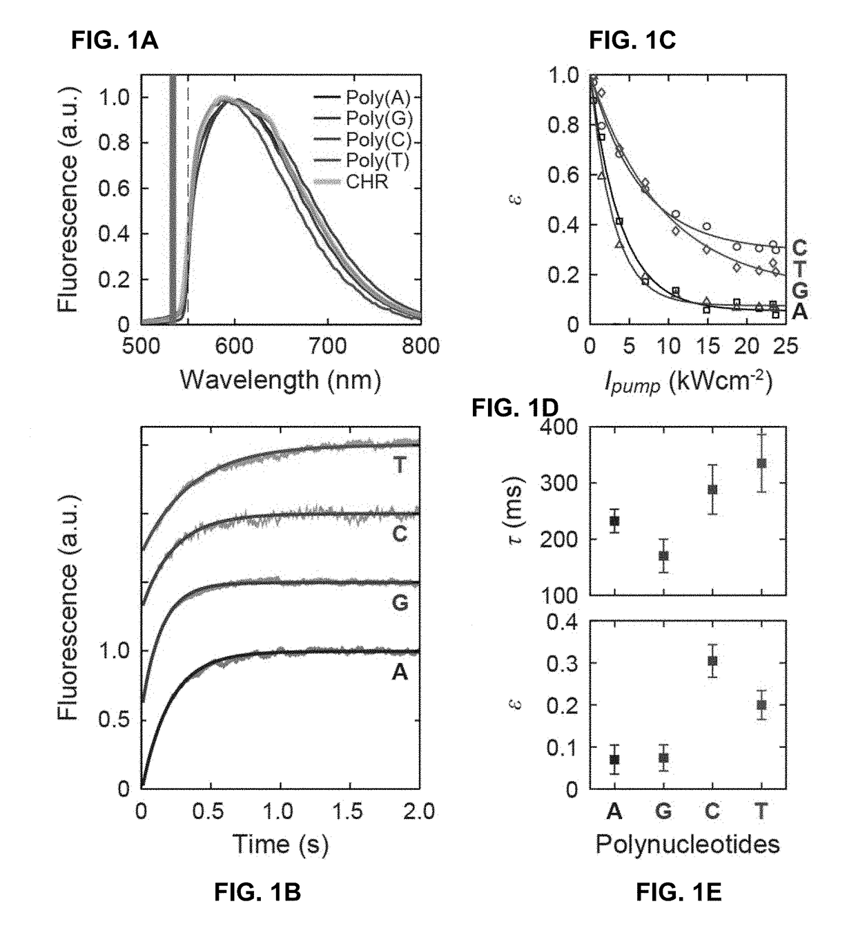
[0099]In our study, we first used short single-stranded polynucleotides (e.g., 20-bp poly-A, G, C, and T, IDT) as model systems to investigate the fluorescence excitation and photo-switching of DNA molecules. Although nucleic acids have significantly weaker absorption for visible versus UV light, they exhibit low, but detectable, absorption due to the electron delocalization effect, in part arising fro...

example 2

[0119]Nucleic acids have significantly weaker absorption for visible versus UV light. However, they exhibit low, but detectable, absorption in the visible range due to the electron delocalization arising from the aromatic rings. This is a fundamental property critical to their molecular function and stability. Visible light absorption by nucleic acids have been measured and the data are readily available. The molar extinction of nucleotides in the visible is E˜50 cm−1 M−1, which is 260 times lower than their UV absorption and >1,000 times lower than the peak absorption of strong extrinsic fluorophores, such as rhodamine. We recorded fluorescence from mononucleotides, nucleic acid bases, and short single-stranded polynucleotides (e.g., 20-bp poly-A, G, C, and T, IDT). This radiative process was consistent with endogenous fluorescence; the emission excited at 532 nm by a pulsed laser had a lifetime τfl˜2 ns, which is typical of fluorescence lifetimes of high-Q fluorophores.

[0120]Integ...

example 3

[0125]In Raman scattering and fluorescence excitation and emission, incident photons interact with the intrinsic electronic or vibrational states of the sample and subsequently emit frequency-shifted photons due to the underlying energy exchange. Analyzing the spectroscopic signatures obtained from inelastic light scattering measurements is a widely used method for revealing the electronic and structural properties for natural and engineered materials in subjects ranging from biology to materials science. Additionally, a variety of spectroscopic imaging techniques have been developed to probe the heterogeneous environment within samples, yet their spatial resolutions have been limited to about half of the wavelength due to light diffraction. Although near-field scanning optical microscopy (NSOM) offers nanometer-scale spatial resolution by using a sharp stylus for scanning at the close vicinity of the sample surface, it is unable to image sub-surface features because rapidly decayin...


PUM

PropertyMeasurementUnit
Intrinsic viscosityaaaaaaaaaa
Fluorescenceaaaaaaaaaa

Abstract

The devices, methods, and systems of the present disclosure provide for spectroscopic super-resolution microscopic imaging. In some examples, spectroscopic super-resolution microscopic imaging may be referred to or comprise spectroscopic photon localization microscopy (SPLM), a method which may employ the use of extrinsic labels or tags in a test sample suitable for imaging. In some examples spectroscopic super-resolution microscopic or spectroscopic photon localization microscopy (SPLM) may not employ extrinsic labels and be performed using the intrinsic contrast of the test sample or test sample material.Generally, spectroscopic super-resolution microscopic imaging may comprise resolving one or more non-diffraction limited images of an area of a test sample by acquiring both localization information of a subset of molecules using microscopic methods known in the art, and simultaneously or substantially simultaneously, acquiring spectral data about the same or corresponding molecules in the subset. This method maybe useful to detect a variety of features in cellular material for the molecular characterization of cells and disease.

Description

RELATED APPLICATIONS[0001]This patent arises from U.S. Provisional Patent Application Ser. No. 62 / 329,856, which was filed on Apr. 29, 2016, U.S. Provisional Patent Application Ser. No. 62 / 329,859, which was filed on Apr. 29, 2016, U.S. Provisional Patent Application Ser. No. 62 / 329,865, which was filed on Apr. 29, 2016, U.S. Provisional Patent Application Ser. No. 62 / 329,867, which was filed on Apr. 29, 2016, U.S. Provisional Patent Application Ser. No. 62 / 329,868, which was filed on Apr. 29, 2016, and U.S. Provisional Patent Application Ser. No. 62 / 329,871, which was filed on Apr. 29, 2016. U.S. Patent Application Ser. No. 62 / 329,856, U.S. Patent Application Ser. No. 62 / 329,859, U.S. Patent Application Ser. No. 62 / 329,865, U.S. Patent Application Ser. No. 62 / 329,867, U.S. Patent Application Ser. No. 62 / 329,868, and U.S. Patent Application Ser. No. 62 / 329,871 are hereby incorporated herein by reference in their entirety.STATEMENT REGARDING FEDERALLY SPONSORED RESEARCH FOR DEVELOPME...

Claims


Application Information

Patent Timeline
IPC IPC(8): G01N21/64C12Q1/6825G01N33/53
CPCG01N21/6428G01N21/6458C12Q1/6825G01N33/5302G01N2021/6441C12Q1/6841
Inventor DONG, BIQINDAVIS, JANEL L.SUN, CHENGZHANG, HAO F.PATEL, KIEREN J.URBAN, BENBACKMAN, VADIMALMASSALHA, LUAYSTYPULA-CYRUS, YOLANDANGUYEN, THE-QUYEN
Owner NORTHWESTERN UNIV
Why Eureka
Try Eureka