Compositions and methods for detecting Anti-endothelial cell antibodies in allograft rejection
a technology of anti-endothelial cells and antibodies, applied in the field of compositions and methods for detecting anti-endothelial cell antibodies in allograft rejection, can solve the problems of many technical problems, organ transplant rejection can still occur, and patients transplanted across both hla and aeca barriers have an increased incidence and severity of antibody mediated rejection, so as to reduce increase the risk of developing ar
- Summary
- Abstract
- Description
- Claims
- Application Information
AI Technical Summary
Benefits of technology
Problems solved by technology
Method used
Image
Examples
example 1
ation of Novel Antigenic Endothelial Cell Targets Using Protein Arrays
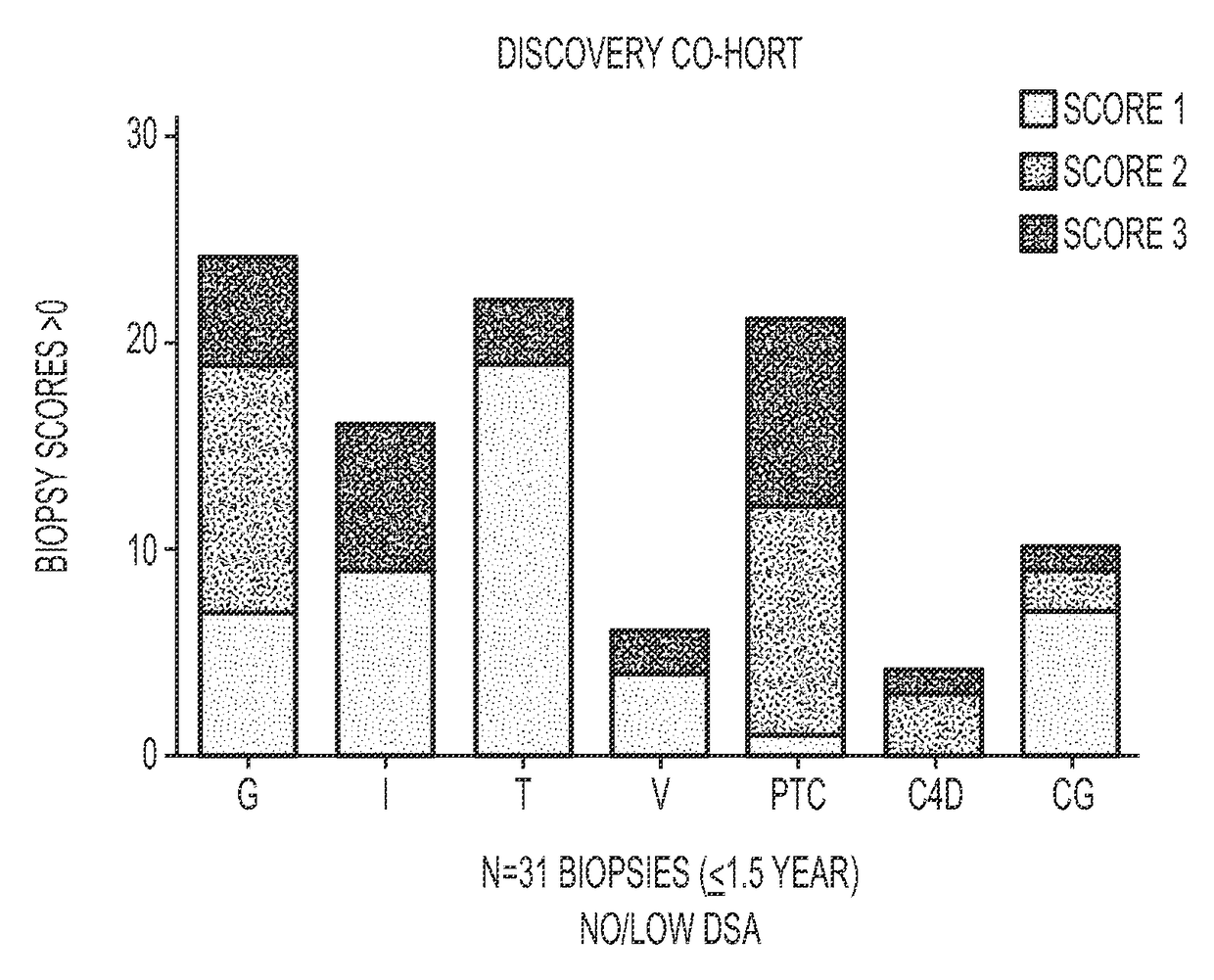
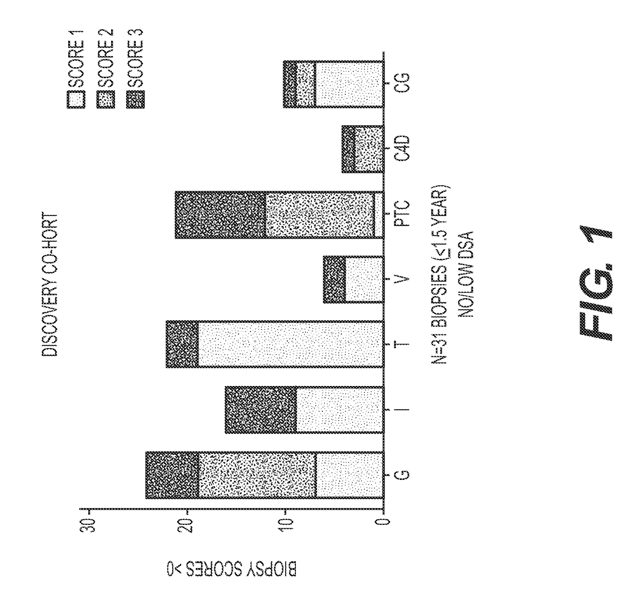
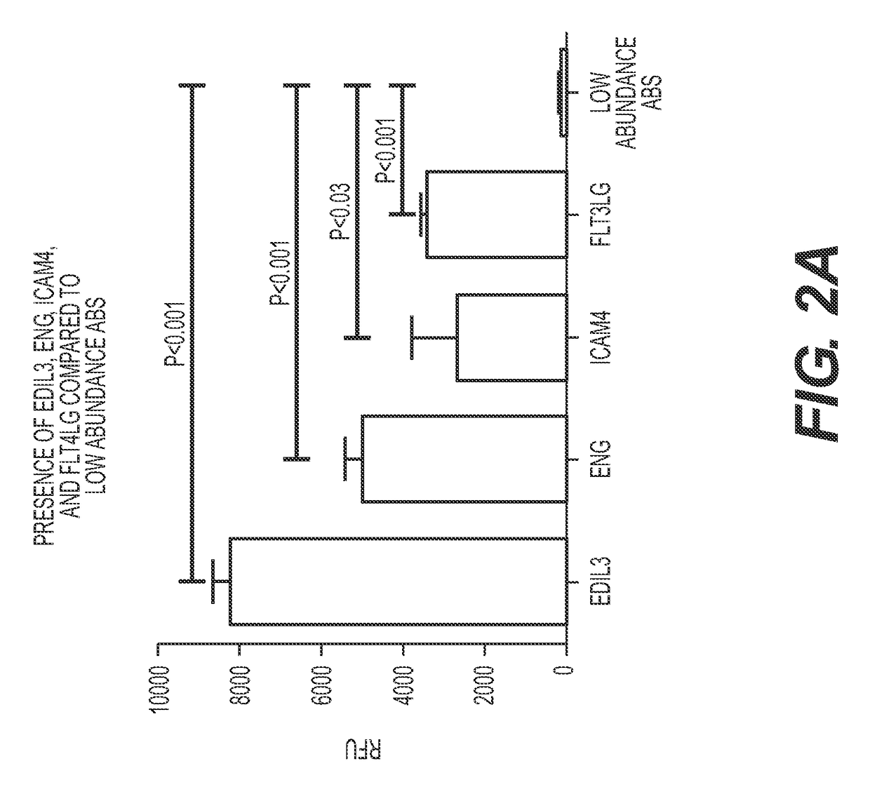
[0139]AECAs were isolated from a Discovery Cohort of ten renal transplant recipients whose demographics are provided in Table 1. The majority (9 of 10) was sensitized to HLA and all tested positive for AECAs in pre-transplant endothelial cell crossmatch tests. Nine experienced allograft dysfunction and biopsy proven rejection with noted glomerulitis and peritubular capillaritis (FIG. 1). Only one recipient had antibody to donor-HLA (DR52) at the time of rejection. To focus our analyses on AECA target antigens, antibody eluates were generated using ECPs derived from blood. In brief, each serum was incubated with ECPs and, following wash steps, the bound antibodies were acid eluted and neutralized. Using a high-density protein platform, we profiled AECA eluates from the ten Discovery Cohort recipients against approximately 9500 human proteins. Four proteins, expressed on vascular endothelium, endoglin, EDIL3, ICAM4 ...
example 2
Novel Antigenic Targets of Anti-Endothelial Cell Antibodies
[0140]Anti-endothelial cell antibody target antigens identified from antibody eluates from ECPs in human protein arrays are listed in Table 2 below. Each anti-endothelial cell antibody formed an antibody / antigen complex and was identified at a threshold cut off RFU>2,000.
TABLE 2Anti-endothelial cell antibody target proteins (antigens)Protein Name (corresponding to arrayAveragepeptide / antigen)Gene SymbolGene IDRFURecombinant human CTLA-4 / FcCTLA4149358077.0tripartite motif-containing 21 (TRIM21)TRIM21673739835.7hematopoietic SH2 domain containing (HSH2D)HSH2D8494128572.5interferon, alpha-inducible protein 6 (IFI6)IFI6253715892.2APEX nuclease (apurinic / apyrimidinic endonuclease)APEX22730112493.32 (APEX2), nuclear gene encoding mitochondrialproteinCAP-GLY domain containing linker protein family,CLIP47974510955.7member 4 (CLIP4)UBX domain containing 8 (UBXD8 / FAF2)UBXD8231978636.5zinc finger, MYM-type 5 (ZMYM5)ZMYM592058260.8EGF-l...
example 3
n of Target Antigens on Endothelial Cells
[0141]Cell phenotype analysis using flow cytometry confirmed surface expression of endoglin, EDIL3, ICAM4 and FLT3 (receptor for FLT3 ligand) on blood derived ECPs when compared to an isotype control antibody (data not shown). Analysis of two EC lines, derived from iliac artery and human umbilical vein, yielded positive staining for endoglin, but negative staining for EDIL3, ICAM4, and FLT3, even following TNFα stimulation. To investigate expression of these antigenic targets in renal tissue, immunohistochemistry was performed on rejection biopsies obtained from the ten Discovery Cohort recipients. FIG. 3 illustrates representative staining for endoglin and FLT3, which were expressed on arterial endothelium and glomerular and peritubular capillaries. Concomitant staining of biopsy tissue for FLT3LG, EDIL3 and ICAM4 yielded negative results.
PUM
| Property | Measurement | Unit |
|---|---|---|
| affinity | aaaaa | aaaaa |
| length | aaaaa | aaaaa |
| light microscopy | aaaaa | aaaaa |
Abstract
Description
Claims
Application Information
 - R&D
- Intellectual Property
- Life Sciences
- Materials
- Tech Scout
- Unparalleled Data Quality
- Higher Quality Content
- 60% Fewer Hallucinations
Browse by: Latest US Patents, China's latest patents, Technical Efficacy Thesaurus, Application Domain, Technology Topic, Popular Technical Reports.
© 2025 PatSnap. All rights reserved.Legal|Privacy policy|Modern Slavery Act Transparency Statement|Sitemap|About US| Contact US: help@patsnap.com


