Compositions and methods for visualization of the vitreous

a technology of vitreous and compositions, applied in drug compositions, animal/human proteins, immunological disorders, etc., can solve the problems of vitrectomy surgery itself, retinal detachment, retinal hole, etc., to improve the safety and efficacy of the surgical procedure, and improve the effect of safety and efficacy

Active Publication Date: 2017-07-20
ALCON INC
View PDF1 Cites 0 Cited by
  • Summary
  • Abstract
  • Description
  • Claims
  • Application Information

AI Technical Summary

Benefits of technology

[0006]The invention is directed to a method to alleviate a structural disorder of an eye. In certain aspects, the method comprises injecting a vitreous-delineating composition into the eye in an effective amount to allow the vitreous to be visible to a surgeon while simultaneously maintaining the view of intraocular structures such as the retina, thereby enabling the surgeon to alleviate the disorder in a safer and more efficacious manner.

Problems solved by technology

The fibrous membranes in proliferative vitreoretinopathy can contract and cause retinal tears, retinal holes, and retinal detachments, all of which can lead to blindness.
However, the surgical procedure of vitrectomy itself can produce complications.
The transparency of vitreous makes it challenging for the surgeon to visualize and hence completely remove the vitreous.
While the surgeon attempts a complete removal of the vitreous near the retina, he or she may inadvertently cut into the retina and thereby create iatrogenic retinal defects or holes or possibly remove irreplaceable neural elements essential for vision.
None of these is completely satisfactory.
For example, the instillation of blood into the vitreous has several drawbacks: blood disperses into the vitreous cavity and is likely to obscure visualization of the retina during vitrectomy, and it has the potential of causing postoperative inflammation and proliferative vitreoretinopathy.

Method used


Image

  • Compositions and methods for visualization of the vitreous
  • Compositions and methods for visualization of the vitreous
  • Compositions and methods for visualization of the vitreous

Examples


example 1

Chromophore Visualization of the Vitreous

[0073]A FITC linked HA-binding protein was generated as follows.

Expression and Purification:

[0074]An HA-binding peptide (Protein 6 in Table 1) was transiently expressed in HEK293F cells using polyethylenimine (PEI) as transfection agent with ratio of 1:3 of plasmid DNA to PEI and grown for five days at 37° C. with 5% CO2. The culture was harvested and the supernatant filtered. The conditioned medium was loaded onto nickel resin column, washed with 50 mM phosphate buffer containing 20 mM immidazole, 500 mM sodium chloride, pH 8 to remove unbound proteins, and gradient eluted with 50 mM phosphate buffer containing 500 mM imidazole, 500 mM sodium chloride, pH 8.

[0075]The purified Protein 6 was site-specifically labeled with fluorescein isothiocyanate on the N-terminus using Sortase-A mediated reaction (see Mao et al., 2004, J Am Chem Soc., March 10; 126(9):2670-1). The fluorophore (fluorescein isothiocyanate)-conjugated HA-binding peptide was pu...

example 2

rgery

[0088]The composition of the invention is placed in a formulation suitable for intravitreal injection (i.e., with the appropriate pH, osmolarity, low endotoxin level, etc., that is customarily achievable by those skilled in the art of formulating drugs for intravitreal injection and demonstrated in the previously mentioned pilot research studies in rabbit eyes.) A few hours prior to surgery or even one to three days prior to surgery, the formulation containing the composition is injected into the vitreous. The volume of the dose would be in the range of 1 microliter to 100 microliters. As is standard for intravitreal injections, the injection site is a few mm posterior to the limbus so that the needle enters the eye at the pars plana. A vitreous delineating composition of the invention is injected about 5-10 mm deep into the vitreous.

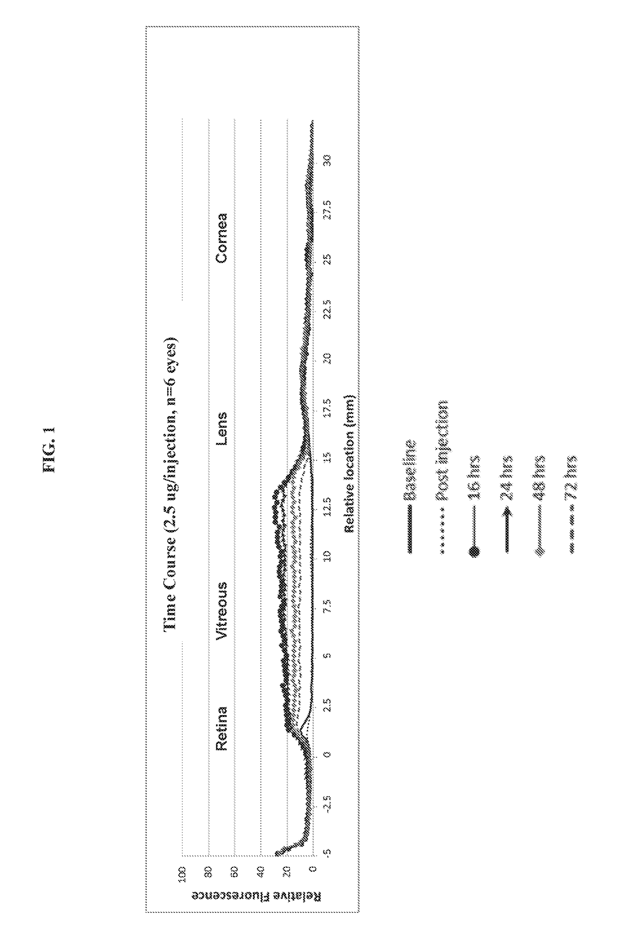
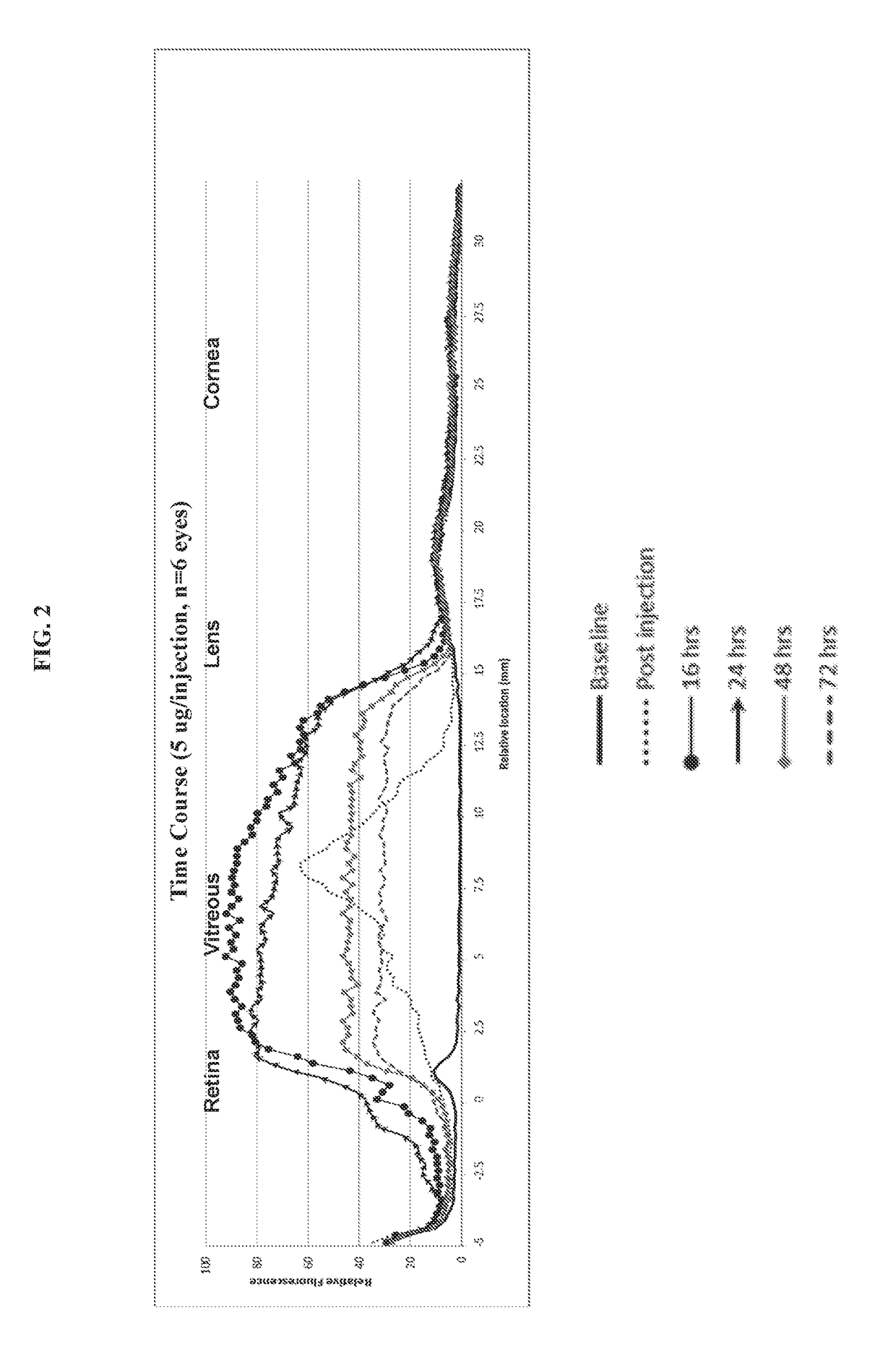
[0089]As demonstrated in the pilot studies in Example 1 above, the composition diffuses throughout the vitreous within hours so that by 16 hours (...


PUM

PropertyMeasurementUnit
concentrationaaaaaaaaaa
volumeaaaaaaaaaa
volumeaaaaaaaaaa

Abstract

The invention provides compositions for rendering a vitreous cavity visible during a surgical procedure to alleviate a structural disorder caused by the vitreous in an eye, and methods of using the compositions. The compositions are vitreous delineating agents that comprise a hyaluronan binding peptide linked to an optically detectable moiety. Such compositions can be in a formulation that may be a solution, a suspension, or an emulsion, and would be injected into the vitreous shortly prior to use. The composition may additionally contain a therapeutic agent, a diagnostic agent, or a chemosensing material, in use, the composition marks or delineates the vitreous by binding preferentially to the hyaluronan that permeates the vitreous humor and binding little or not at all to surrounding tissues such as the retina. The interface between the labelled vitreous humor and the non-labelled surrounding vital tissues produces a visible signal, thereby allowing a surgeon to clearly visualize the entire vitreous cavity and distinguish it from vital ocular structures. Use of the method improves the accuracy and safety of a vitrectomy and thus prevents suboptimal outcomes or the need for repeated procedures. The compositions comprising chemosensing material are useful as long-lasting biosensors, which when used in the vitreous enable repetitive, non-invasive, in vivo measurements of metabolites or pharmacologic agents in the vitreous of animals or humans.

Description

FIELD OF THE INVENTION[0001]The invention relates to compositions, methods, and uses for protein-based, long-lasting agents for injection into the vitreous humor in mammalian eyes. The invention also relates to the use of such compositions during ophthalmic surgery, for example, during vitrectomy surgery, and their use as biosensors.BACKGROUND OF THE INVENTION[0002]In the eye, the cavity between the lens and the retina is filled with a clear, jelly-like substance termed the vitreous body, vitreous humor, or vitreous. Its volume is fixed and is relatively permanent. The vitreous humor is 99% water, with most of the remaining 1% composed of salts (sodium, chloride, bicarbonate, etc.), collagen, and hyaluronan (hyaluronic acid). The collagen and hyaluronan give the vitreous is gel-like consistency and high viscosity. The outer surface of the vitreous is normally weakly adherent to the retina and ciliary body, with a somewhat tighter attachment at the macula, at the far periphery of the...

Claims


Application Information

Patent Timeline
Patent Type & Authority Applications(United States)
IPC IPC(8): A61K49/00A61K9/00
CPCA61K9/0051A61K49/0056A61K45/00A61K49/0021A61K49/0043A61K49/0084A61K49/0091C07K14/4718A61P17/02A61P27/02A61P31/00A61P35/00A61P37/06A61P41/00A61P43/00
Inventor GHOSH, JOYDRYJA, THADDEUS PETERROGUSKA, MICHAELCARLSON, ERIC C.
Owner ALCON INC
Why Eureka
Try Eureka