Method for Detecting SMN Protein Expression

a technology protein, which is applied in the field of detecting the expression of survival motor neuron (smn) protein, can solve the problem of no reliable method for detecting smn protein expression by using blood cell samples, and achieve the effect of reliable method of detection

Inactive Publication Date: 2017-04-27
MICROBIAL CHEM RES FOUND +1
View PDF1 Cites 3 Cited by
  • Summary
  • Abstract
  • Description
  • Claims
  • Application Information

AI Technical Summary

Benefits of technology

[0015]An object of the present invention is to provide a reliable method for detecting SMN protein expression by using blood cell samples, the method being capable of detecting a difference in the level of the SMN protein expressed between a childhood SMA patient and a healthy person and / or between a childhood SMA patient and a carrier thereof.
[0016]The present inventors have conducted earnest studies and as a result, the inventors have newly found out that the sensitivity of detecting the level of the SMN protein expressed is enhanced, by selecting a particular cell population among blood cells in which SMN protein expression is to be detected and detecting the SMN protein expression in the cell population.
[0032]According to one aspect of the present invention, it is possible to detect a difference in the amount of the SMN proteins expressed between a childhood SMA patient and a healthy person and / or a carrier thereof. In other words, the present invention makes it possible to provide a reliable method for detecting SMN protein expression, the method being capable of detecting a difference in the level of the SMN protein expressed between a childhood SMA patient and a healthy person and / or between a childhood SMA patient and a carrier thereof.
[0033]Additionally, one aspect of the present invention makes it possible to detect SMN protein expression using approximately 500 μL of blood, and to provide a method useful when SMN protein serves as a biomarker in testing a drug or a drug candidate against SMA, particularly when blood is collected from a child patient.

Problems solved by technology

Heretofore, no reliable method for detecting SMN protein expression by using blood cell samples, which is capable of detecting a difference in the level of the SMN protein expressed between a childhood SMA patient and a healthy person and / or between a childhood SMA patient and a carrier thereof, has been found.

Method used


Image

  • Method for Detecting SMN Protein Expression
  • Method for Detecting SMN Protein Expression
  • Method for Detecting SMN Protein Expression

Examples


example 1

Detection of SMN Protein Expression Using Lymphoblasts

(1) Lymphoblast Transformation Treatment

[0096]A blood sample obtained from a healthy person was collected into a heparin tube, then overlaid on a Ficoll solution, and centrifuged at 2000 rpm for 15 minutes. Thereby, peripheral blood mononuclear cells were separated and collected. Subsequently, a lymphoblast transformation treatment was performed by a transformation method using EB virus.

[0097]A blood sample obtained from a SMA type I patient was also subjected to the same treatment performed on the blood sample obtained from the healthy person.

(2) Measurement of SMN Protein Expression

[0098]The lymphoblasts derived from the peripheral blood mononuclear cells of the healthy person obtained in (1) above were collected into a centrifuge tube. The cells were fixed with a 4% paraformaldehyde-phosphate buffer solution. Then, the cells were washed with a phosphate buffer solution and subsequently centrifuged at 500×g for 5 minutes to rem...

example 2

Detection of SMN Protein Expression Using Lysis Method

(1) Lysis Treatment

[0109]2 mL of blood from a healthy person was collected into a heparin tube, which was inverted to mix well the blood. Then, 0.5 mL of the blood was collected into a centrifuge tube. BD Phosflow™ Lyse / Fix Buffer (BD Biosciences) was added thereto as a Lysing / Fix solution and left standing at 37° C. for 10 minutes, followed by centrifugation at 2300 rpm for 8 minutes. After the supernatant was removed, the cells were washed with a phosphate buffer solution. A sample containing nucleated cells derived from the blood was thus obtained.

[0110]Samples containing nucleated cells derived from blood of a carrier and a SMA type I patient were also prepared by the lysis method as in the case of the healthy person.

(2) Measurement of SMN Protein Expression

[0111]BD Phosflow™ Perm Buffer II (BD Biosciences) was added as a cell membrane permeation reagent to the nucleated cells of the healthy person obtained in (1) above, and ...

example 3

Detection of SMN Protein Expression in Each Cluster Classified Using Surface Antigen Markers

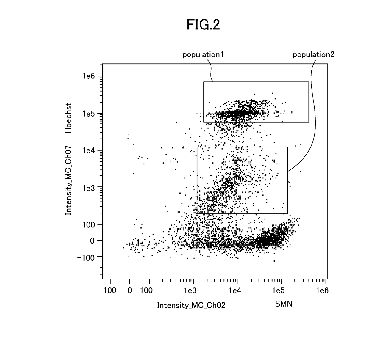
[0117]Samples were prepared according to the following staining protocol.[0118]1. Collect 2 mL of peripheral blood obtained from a healthy person into a tube. Add reagents thereto such that 2 mL of the peripheral blood contains 80 μL of an Fc receptor blocking reagent (Clear Back, MTG-001, MBL) and 100 μL of Hoechst 33342 (Molecular Probes) which has been diluted to 5 μg / mL with PBS.[0119]2. Seal the tube, and slowly rotate it at room temperature for 15 minutes in the dark.[0120]3. Place 500 μL of the peripheral blood thus treated into each of four 15-mL conical tubes.[0121]4. Add 5 μL of fluorochrome-labeled antibodies (purchased from BioLegend, Inc., BD Biosciences, and MERCK KGaA) into each of the tubes in accordance with Table 5 below.

TABLE 5FluorochrometubePEPE-Cy5BV510BV65QBV785Monoclonal#1CD66a / c / eCD33CD45CD11cCD14antibodyCD66b#2HLA-DRCD123CD45CD11cCD14#3CD34CD123CD45CD19CD14#4CD25CD3C...


PUM

PropertyMeasurementUnit
fluorescence intensityaaaaaaaaaa
fluorescentaaaaaaaaaa
fluorescence intensitiesaaaaaaaaaa

Abstract

Provided is a method for detecting the expression of SMN protein, said method comprises:a step for labeling SMN protein in a sample, said sample containing nucleated cells derived from blood;a step for labeling the nuclei of the nucleated cells in the sample;a step for selecting a cell population of the nucleated cells in which nuclei and SMN protein are labeled; anda step for detecting the expression of the SMN protein on the basis of the label for the SMN protein in the selected cell population.

Description

TECHNICAL FIELD[0001]The present invention relates to a method for detecting the expression of survival motor neuron (SMN) protein.BACKGROUND ART[0002]Spinal muscular atrophy (SMA) is a muscular atrophy caused by a lesion of anterior horn cells of the spinal cord, and shows a lower motor neuron sign characterized by muscle weakness and muscle atrophy in the trunk and limbs. SMA is classified into type I to type IV according to the age of onset and severity: type I which is severe type (also referred to as Werdnig-Hoffmann disease) and occurs by the age of six months after birth; type II which is intermediate type (also referred to as Dubowitz disease) and occurs by the age of one year and six months; and type III which is mild type (also referred to as Kugelberg-Welander disease) and occurs after the age of one year and six months. These are childhood SMA. On the other hand, type IV which occurs at the age of 20 years or older is adult SMA.[0003]SMA type I accounts for approximately...

Claims


Application Information

Patent Timeline
Patent Type & Authority Applications(United States)
IPC IPC(8): G01N33/58G01N21/64
CPCG01N33/582G01N21/6428G01N2333/47G01N2800/2878G01N2021/6439G01N33/48G01N33/56972G01N33/6875G01N33/6896C07K14/435
Inventor SAITO, KAYOKOARAKAWA, REIKOARAKAWA, MASAYUKIAKIO
Owner MICROBIAL CHEM RES FOUND
Why Eureka
Try Eureka