
Magnetic Resonance Imaging Method with Asymmetric Radial Acquisition of K-Space Data

a magnetic resonance imaging and k-space data technology, applied in the field of magnetic resonance imaging (mri), can solve the problems of prone to any source of imperfection in balanced ssfp imaging, high susceptibility variation in imaging of tissues, and remains challenging, and achieve the effect of robust radial acquisition of k-space data

Inactive Publication Date: 2016-11-17
UNIVSSPITAL BASEL
View PDF5 Cites 18 Cited by
  • Summary
  • Abstract
  • Description
  • Claims
  • Application Information

AI Technical Summary

Benefits of technology

The present invention provides a magnetic resonance imaging (MRI) method that allows for a robust radial acquisition of k-space data without being prone to imperfections of the MRI-system. By using a balanced SSFP sequence and a centered-in asymmetrical radial acquisition scheme, the trajectory fidelity is improved in the region of the k-space center, leading to an overall improved signal homogeneity in the final image. This results in less pronounced background noise and less pronounced artifacts, particularly in the form of miscalibrations of the data acquisition trajectories. The use of a balanced SSFP sequence as the gradient echo sequence is particularly advantageous because it maximizes the contribution of the remaining transverse magnetization of previous RF excitations to the MR signal. This results in a high signal intensity towards the end of the acquisition period in each basic sequence element, which is not decreased during the acquisition period.

Problems solved by technology

Over the last two decades, balanced SSFP imaging has become increasingly popular for applications where rapid data acquisition is needed, e.g., for cardiac imaging, native magnetic resonance angiography, or thoracic imaging, but remains challenging even on current state-of-the-art MRI systems, since for constant phase accruals, all gradient moments have to be perfectly balanced within every TR.
As a result, balanced SSFP imaging is prone to any source of imperfection that perturbs the perfectly balanced gradient scheme, such as eddy-currents.
Hence, imaging of tissues with high susceptibility variations (e.g., lung parenchyma, bone tissue, cartilage), becomes challenging and requires proper shimming to achieve an excellent main magnetic field homogeneity.
One of the major limitations of contemporary Cartesian sampling schemes, however, is its inherent sensitivity to motion, resulting in shifted copies (“ghosting artifacts”) of the object along the phase-encoding direction.
Similar to Cartesian MRI, the sampling density of a fully sampled radial dataset is determined by the Nyquist limit and undersampling will thus results in streaking artifacts in the image.
Nevertheless, radial trajectories have higher sampling requirements than Cartesian methods as a result of a less efficient k-space coverage.
Although conceptually, radial data acquisition is known since the early days of MRI, its application in the clinical setting is still limited due to technical challenges.
A major issue represents proper data reconstruction in the presence of even tiny deviations from the nominal k-space trajectory, e.g., caused by gradient system imperfections, such as eddy currents or heating effects and internal synchronization errors.
In general, any discrepancy between the nominal and actual k-space trajectory results in data inconsistencies, i.e., perpendicular and parallel offset of the central k-space point with respect to the k-space center emanating in the image as increased diffuse background noise (‘smearing’ artifacts or ‘halo-effect’).
Unfortunately, centered-out half-radial sampling in combination with balanced SSFP imaging is particularly affected by the aforementioned MRI-system imperfections.

Method used


Image

  • Magnetic Resonance Imaging Method with Asymmetric Radial Acquisition of K-Space Data
  • Magnetic Resonance Imaging Method with Asymmetric Radial Acquisition of K-Space Data
  • Magnetic Resonance Imaging Method with Asymmetric Radial Acquisition of K-Space Data

Examples


Embodiment Construction

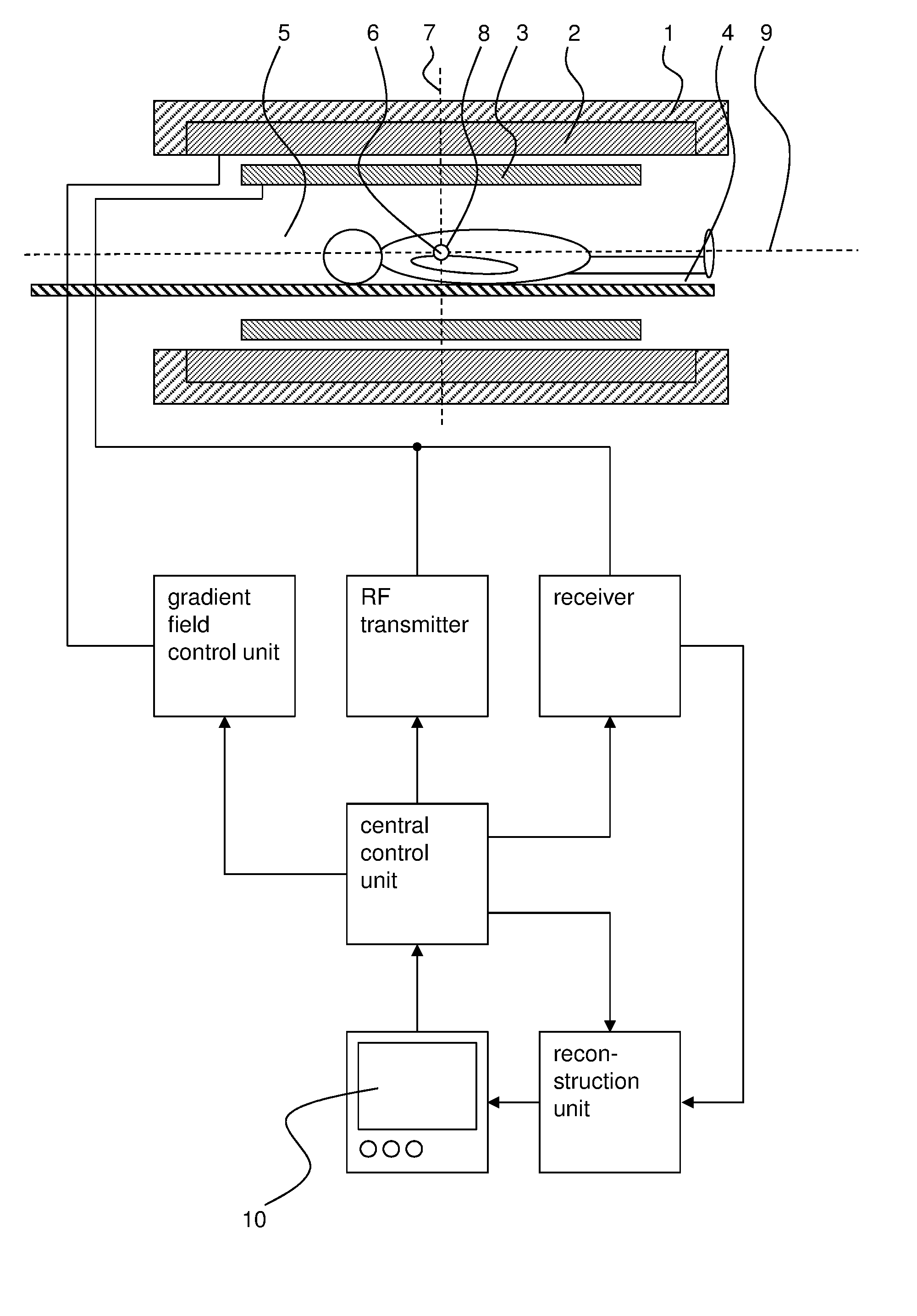
[0048]In FIG. 1, an exemplary MRI system is shown which serves to carry out the inventive method in which a sample 5, such as a living or dead human or animal body, is subjected to a gradient echo imaging sequence with asymmetric radial acquisition of k-space data.

[0049]The MRI system comprises a main magnet 1 for producing a main magnetic field B0. The main magnet 1 usually has the essential shape of a hollow cylinder with a horizontal bore. Inside the bore of the main magnet 1 a magnetic field is present, which is essentially uniform at least in the region of the isocenter 6 of the main magnet 1. The main magnet 1 serves to at least partly align the nuclear spins of a sample 5 arranged in the bore. Of course, the magnet 1 does not necessarily be cylinder-shaped, but could for example also be C-shaped.

[0050]The sample 5 is arranged in such a way on a moving table 4 in the bore of the main magnet 1, that the part of the sample 5, which is to be imaged by the inventive method, is arr...


PUM


Abstract

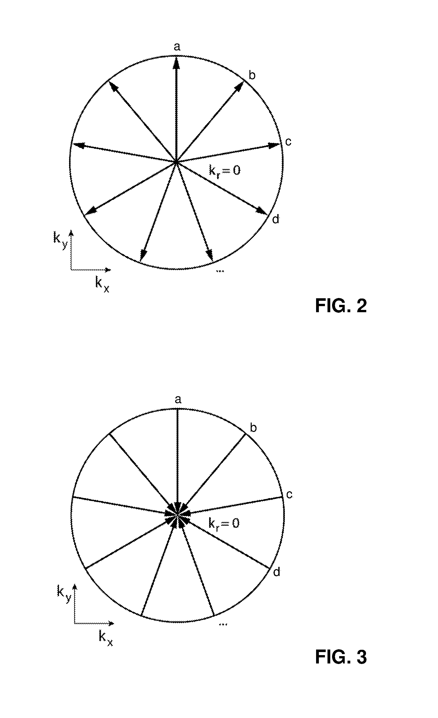
A magnetic resonance imaging (MRI) method is provided in which a sample is subjected to a gradient echo imaging sequence having a plurality of basic sequence elements each of which includes a radiofrequency (RF) pulse, at least one frequency encoding gradient moment kx for generating a magnetic resonance (MR) signal, at least one first phase encoding gradient moment ky for phase encoding the MR signal and a data acquisition period during which k-space data reflecting the MR signal are acquired. The frequency encoding gradient moment kx and the first phase encoding gradient moment ky are applied such during the data acquisition period of each basic sequence element, that the k-space data are acquired in a radial direction and asymmetrically with respect to the center of k-space in the direction from the periphery towards the center of k-space.

Description

CROSS-REFERENCE TO RELATED APPLICATION[0001]This application claims priority to German Patent Application No. 10 2015 107 347.1 filed May 11, 2015, the disclosure of which is hereby incorporated in its entirety by reference.TECHNICAL FIELD[0002]The present invention concerns a magnetic resonance imaging (MRI) method in which a sample is subjected to a gradient echo imaging sequence with asymmetric radial acquisition of k-space data. The present invention also concerns a MRI system configured to carry out such a MRI method as well as a computer program for controlling a MRI system, the computer program comprising executable instructions to carry out such a MRI method.BACKGROUND OF THE INVENTION[0003]Magnetic resonance imaging (MRI) is a well-known medical imaging technique that allows visualizing internal structures of the human (and the animal) body in great detail.[0004]With gradient echo sequences, which are widely used in MRI, the nuclear spins of a sample are excited by less tha...

Claims


Application Information

Patent Timeline
IPC IPC(8): G01R33/561G01R33/48
CPCG01R33/4818G01R33/5614G01R33/4824G01R33/4826G01R33/565
Inventor BIERI, OLIVERBAUMAN, GRZEGORZ
Owner UNIVSSPITAL BASEL
Learn More