
System and apparatus comprising a multisensor guidewire for use in interventional cardiology

a multisensor guidewire and guidewire technology, applied in the field of system and apparatus comprising a multisensor guidewire for use in interventional cardiology, can solve the problems of increasing the risk of tissue damage, reducing the stiffness of the multisensor guidewire, and difficulty in positioning the sensors, so as to achieve the effect of reducing one or more disadvantages

Inactive Publication Date: 2016-05-12
THREE RIVERS CARDIOVASCULAR SYST
View PDF3 Cites 11 Cited by
  • Summary
  • Abstract
  • Description
  • Claims
  • Application Information

AI Technical Summary

Benefits of technology

The present invention aims to improve the accuracy and minimize the risks associated with measuring cardiovascular parameters and performing interventional cardiac procedures, specifically transcatheter valve therapy (TVT) for heart valve repair or replacement. The invention offers a multisensor guidewire with high stiffness that can directly measure various hemodynamic parameters before and after the valve replacement, and also provide feedback to the interventional cardiologist to avoid tissue trauma or perforation. The guidewire has a grooved core wire with high stiffness, which can provide better support during the procedure. Additionally, the invention provides a fabrication method for improving stiffness and torque characteristics of the guidewire. Overall, the invention offers an improved solution for minimizing risks and improving the efficacy of TVT procedures.

Problems solved by technology

Since the stiffness of a wire varies as the fourth power of its diameter, the small diameter core wire significantly reduces the stiffness of the multisensor guidewire.
Thus, during measurement of a transvalvular pressure gradient, movement of the guidewire may create difficulty in positioning the sensors and the cardiologist may need to readjust the positioning of the guidewire to maintain the pressure sensors each side of the heart valve.
While a larger gauge, stiffer guidewire would be less easily displaced during measurements, for measurement of transvalvular pressure gradients, it would tend to interfere more with normal heart valve operation, and may increase the risk of tissue damage.
When a stiff support guidewire is introduced into the left ventricle of the heart through the aortic valve, if too much force is applied to the guidewire or it is pushed too far, there is some risk that the guidewire could cause damage or trauma to the heart tissues, e.g. damage to the aortic wall or ventricular perforation and pericardial effusion resulting in pericardial tamponade.
Moreover, there is increased risk of trauma or damage to the heart wall in a diseased, weakened or calcified heart.

Method used


Image

  • System and apparatus comprising a multisensor guidewire for use in interventional cardiology
  • System and apparatus comprising a multisensor guidewire for use in interventional cardiology
  • System and apparatus comprising a multisensor guidewire for use in interventional cardiology

Examples


first embodiment

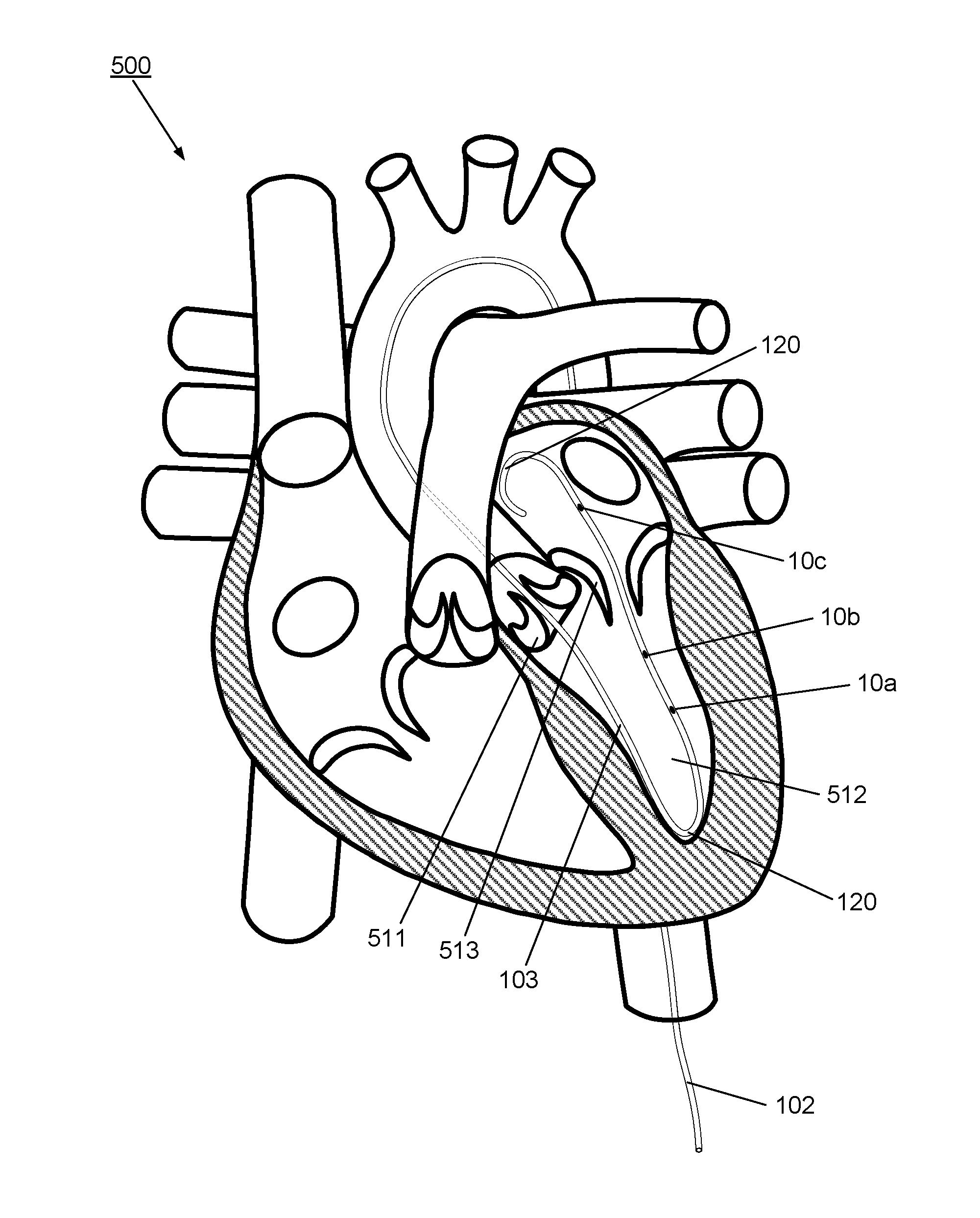
[0090]FIG. 2 illustrates schematically a longitudinal cross-sectional view of the apparatus 100 according to the invention, comprising a multisensor guidewire. The apparatus 100 extends from the optical input / output connector 112 at the proximal end 110 through the proximal part 101 to the distal part 102 which extends to the distal tip 120.

[0091]The distal part 102 takes the form of a multisensor guidewire and comprises components of a conventional guidewire comprising a tubular outer layer in the form of a flexible fine metal coil 35 and an inner mandrel or core wire 31 within the outer coil 35. The outer coil 35 and the core wire 31 each have a diameter and mechanical properties to provide the required flexibility and stiffness to act as a support guidewire for TAVI. Typically, for TAVI, the coil forms a flexible tubular covering layer of the guidewire and has an outside diameter of 0.035 inch or 0.89 mm or less. To provide the appropriate stiffness and other mechanical propertie...

second embodiment

[0104]As illustrated schematically in FIGS. 4B to 4D, assuming the coil 35 has an outside diameter of 0.89 mm (0.035 inch) including any coating, and is formed from 0.002 inch thick coil wire, which would provide a coil with an inside diameter of about 0.787 mm (0.031 inch), then a core wire having a maximum outside diameter of about 0.736 mm (0.029 inch) could be accommodated within, allowing for clearance between the core wire and the coil. Preferably the coil and the core of the guidewire are made from stainless steel having high stiffness and tensile strength, e.g. 304V stainless steel, or other approved types of stainless steel for medical applications. Other biocompatible metal alloys with suitable mechanical characteristics may alternatively be used. Typical medical grade nitinol alloys would not offer sufficient stiffness for a core wire for a multisensor support guidewire for transcatheter valve replacements. On the other hand, medical grade nitinol alloys may provide suffi...


PUM


Abstract

A system and apparatus comprising a multisensor guidewire for use in interventional cardiology, e.g. for Transcatheter Valve Therapies (TVT), comprises a plurality of optical sensors for direct measurement of cardiovascular parameters, e.g. transvalvular blood pressure gradients and flow. A conventional outer coil contains a shaped core wire having a cross-section defining helical grooves extending along its length, which accommodate optical fibers and optical sensors within a diameter Dcore of the core wire. Advantageously, the diameter and material of the core wire provides the guidewire with sufficient stiffness for use as a support guidewire for valve replacement, e.g. Transcatheter Aortic Valve Implantation (TAVI), while accommodating multiple sensors and optical fibers within a guidewire of outside diameter ≦0.89 mm. An optical connector couples the guidewire to a control system. Optionally, the guidewire includes a contact force sensor; a pre-formed tip; and / or a separable micro-connector for proximal mounting of over-the-guidewire components.

Description

CROSS-REFERENCE TO RELATED APPLICATIONS[0001]This application is a Continuation-in-Part of PCT International Application No. PCT / IB2015 / 055240, entitled “System and Apparatus Comprising a Multisensor Guidewire for Use in Interventional Cardiology”, filed Jul. 10, 2015, designating the United States, and claiming priority from U.S. Provisional patent application No. 62 / 023,891, entitled “System And Apparatus Comprising a Multisensor Support Guidewire for Use in Trans-Catheter Heart Valve Therapies”, filed Jul. 13, 2014 and from U.S. Provisional patent application No. 62 / 039,952, entitled “System And Apparatus Comprising a Multisensor Support Guidewire for Use in Trans-Catheter Heart Valve Therapies”, filed Aug. 21, 2014; all said applications are incorporated herein by reference in their entirety.[0002]This application is related to U.S. patent application Ser. No. 14 / 354,624 which is a national stage entry of PCT International Application No. PCT / IB2012 / 055893, entitled “Apparatus, ...

Claims


Application Information

Patent Timeline
IPC IPC(8): A61B5/0215
CPCA61B5/02154A61B2560/066A61B2560/0462A61B5/02158A61F2/24A61B5/6851A61B5/283A61B2562/228A61F2/2418A61F2/2427
Inventor CARON, ERICBILODEAU, LUC
Owner THREE RIVERS CARDIOVASCULAR SYST
Learn More