Cellular transplant site
- Summary
- Abstract
- Description
- Claims
- Application Information
AI Technical Summary
Benefits of technology
Problems solved by technology
Method used
Image
Examples
example 1
Preparation of the Subcutaneous Space
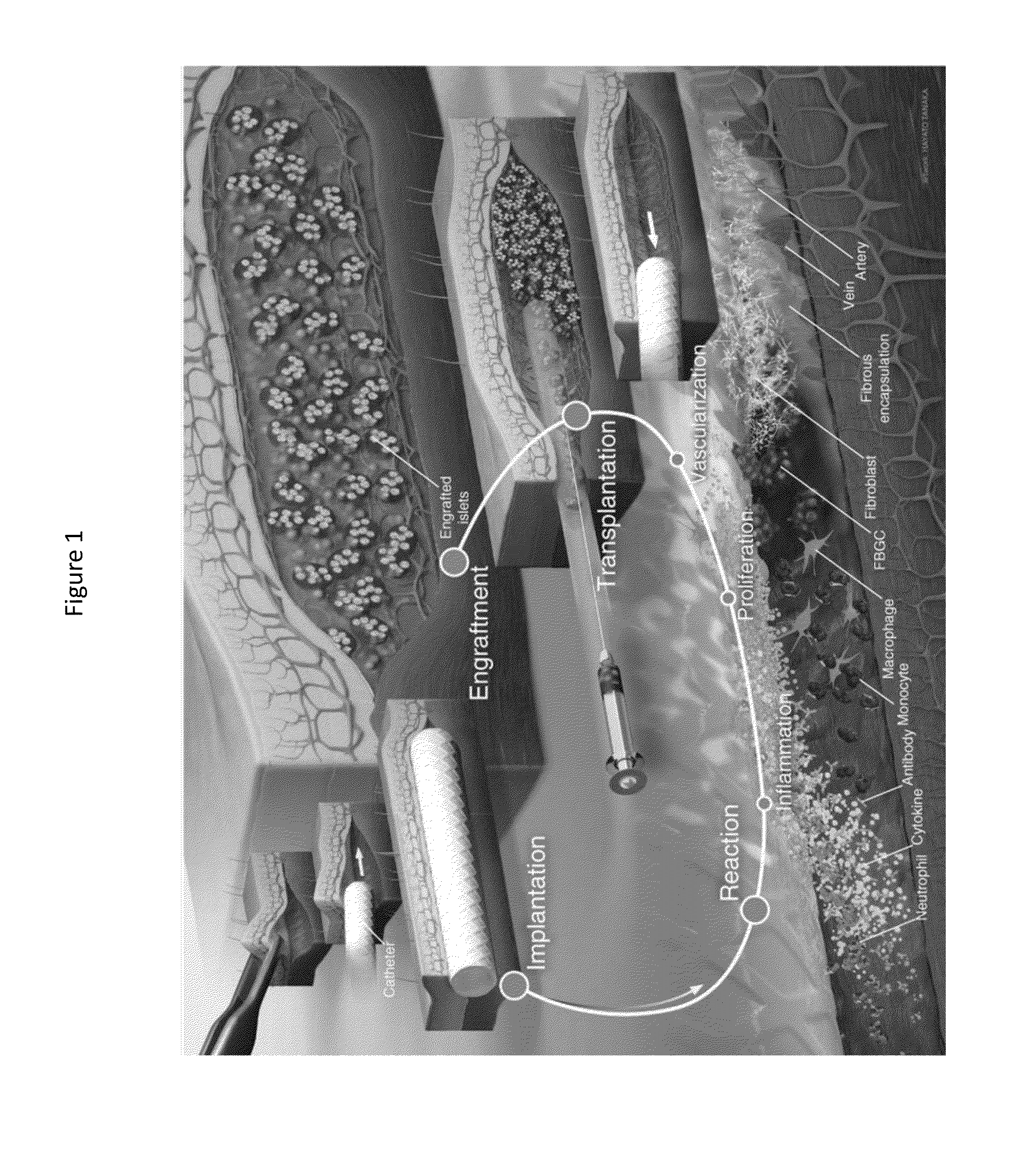
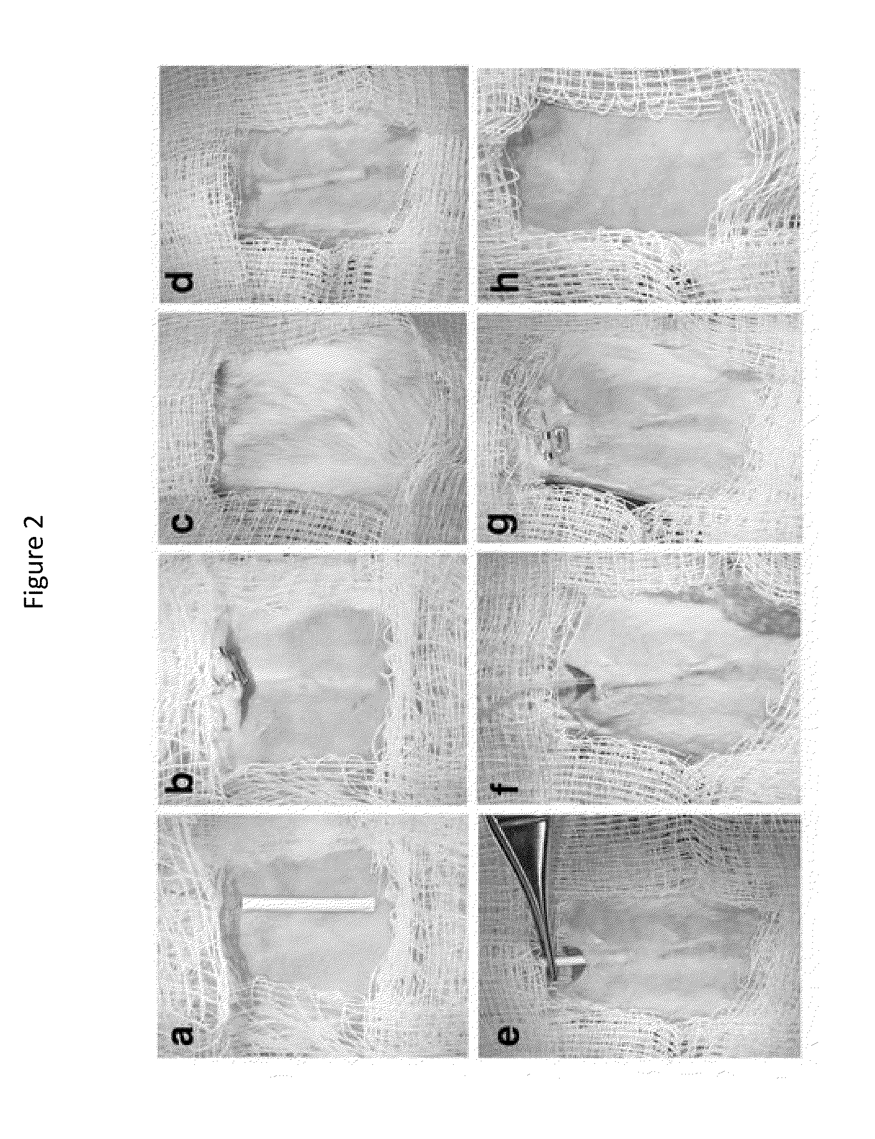
[0063]Three to six weeks prior to islet transplant, 2 cm segments of a 5 French textured nylon radiopaque angiographic catheter (Cook Medical, Indiana, USA) or a 6.5 French smooth silicone catheter (Cook Medical, Indiana, USA) was implanted subcutaneously into the lower left quadrant of 20-25 g male BALB / c mice (Jackson Laboratories, Canada) for mouse syngeneic islet transplant (FIG. 2(a)) or B6.129S7-Rag1tm1Mom immunodeficient mice for human islet transplants. A b)). Once implanted, the catheter became engrossed with blood proteins, leading to the formation of densely vascularized tissue, which exhibited a minimally visible profile (FIG. 2(c-d)). Removal of the implant revealed a vascularized lumen allowing for cellular transplant infusion (FIG. 1 and FIG. 10(a)).
example 2
Proinflammatory Cytokine and Chemokines Measurements
[0064]A one-centimeter segment from both nylon and silicon catheter material was placed subcutaneously into the left lower quadrant of 20-25 g male BALB / c mice. Implanted catheters with surrounding skin and muscle tissue margins were explanted 24 hours, 1 week and 2 weeks post-implantation. Similarly, tissue dissections were retrieved from the abdomen of non-implanted mice, serving as background cytokine and chemokine control specimens. The respective catheter segments were carefully removed from the surrounding tissue, yielding a hollow void encompassed by a vascularized matrix. Tissue samples were immediately placed in pre-weighed microcentrifuge tubes. The tissue weights were recorded, then subsequently flash frozen with liquid nitrogen and stored at −80° C. prior to conducting the cytokine and chemokine proinflammatory analysis. Once all tissue samples from respective implantation period were collected and frozen, 1 mL of lysis...
example 3
Mouse Pancreatectomy and Islet Isolation
[0072]Pancreatic islets were isolated from 8 to 12 week old male BALB / c mice. Animals were housed under conventional conditions having access to food and water ad libitum. The care for all mice within the study was in accordance with the guidelines approved by the Canadian Council on Animal Care. Prior to pancreatectomy, the common bile duct was cannulated with a 27 G needle and the pancreas was distended with 0.125 mg / mL cold Liberase TL Research Grade enzyme (Roche Diagnostics, Laval, QC, Canada) in Hanks' balanced salt solution (Sigma-Aldrich Canada Co., Oakville, ON, Canada). Islets were isolated by digesting the pancreata in a 50 mL tube placed in a 37° C. water bath for 14 minutes with light shaking. Subsequent to the digestion phase, islets were purified from the pancreatic digests using histopaque-density gradients (1.108, 1.083 and 1.069 g / mL, Sigma-Aldrich Canada Co., Oakville, ON, Canada).
PUM
 Abstract
Description
Claims
Application Information
 - R&D
- Intellectual Property
- Life Sciences
- Materials
- Tech Scout
- Unparalleled Data Quality
- Higher Quality Content
- 60% Fewer Hallucinations
Browse by: Latest US Patents, China's latest patents, Technical Efficacy Thesaurus, Application Domain, Technology Topic, Popular Technical Reports.
© 2025 PatSnap. All rights reserved.Legal|Privacy policy|Modern Slavery Act Transparency Statement|Sitemap|About US| Contact US: help@patsnap.com


