Eureka AIR delivers breakthrough ideas for toughest innovation challenges, trusted by R&D personnel around the world.

QUANTITATIVE MULTIPLEX METHYLATION SPECIFIC PCR METHOD- cMethDNA, REAGENTS, AND ITS USE

Active Publication Date: 2015-04-02
THE JOHN HOPKINS UNIV SCHOOL OF MEDICINE
View PDF8 Cites 4 Cited by
  • Summary
  • Abstract
  • Description
  • Claims
  • Application Information

AI Technical Summary

Benefits of technology

The present invention is a new assay called cMethDNA that uses a panel of genes to detect methylated DNA in cancer and normal serum with high sensitivity and specificity. The assay can monitor treatment response and reflect the methylation pattern of the primary tumor and its metastatic lesions.

Problems solved by technology

However, reproducible detection of methylated DNA from serum / plasma proved to be more difficult.
However, no clinical validation studies have followed these preliminary findings, suggesting that there were technical difficulties, yet to be resolved, in this analysis: 1) First, the amount of methylated tumor DNA shed in the serum is a very small fraction of the total unmethylated DNA (either matching gene-specific DNA or a reference DNA such as actin) shed by normal cells.
This leads to lack of robustness of the assay due to masking of methylated signals by the more abundant species; 2) The extent of shedding of normal unmethylated DNA fluctuates daily and with various clinical states, such as surgery or infection; if the reference fluctuates, then inaccuracies occur in interpretation of % methylation of a target gene of interest; 3) The most sensitive assays available utilize nested PCR (with or without multiplex) to pre-amplify amplicons of target and reference DNA using external primers outside the region of interest prior to round two quantitative PCR.
But there are technical limitations such that the target and reference DNA regions need to be co-amplified in the pre-amplification PCR reaction with the same efficiency, ideally using the same pair of external primers in order to maintain the accurate starting ratio of target to reference DNA.
In nested PCR Actin or other genes cannot accurately serve as a reference for the gene of interest because of differences in efficiencies of primer hybridization between the non-identical genes.
Since the level of total DNA in serum of cancer patients is low [median 65.4 ng / ml; range 6.3-268.6 ng / ml; Holdenrieder et al., Ann. N.Y. Acad. Sci. 1137:162-170 (2008)] assays of several genes require a significant amount of serum, making repeat studies problematic and clinical validation difficult because archival sera from existing clinical studies are available only in small volumes.

Method used


Image

  • QUANTITATIVE MULTIPLEX METHYLATION SPECIFIC PCR METHOD- cMethDNA, REAGENTS, AND ITS USE
  • QUANTITATIVE MULTIPLEX METHYLATION SPECIFIC PCR METHOD- cMethDNA, REAGENTS, AND ITS USE
  • QUANTITATIVE MULTIPLEX METHYLATION SPECIFIC PCR METHOD- cMethDNA, REAGENTS, AND ITS USE

Examples


example 1

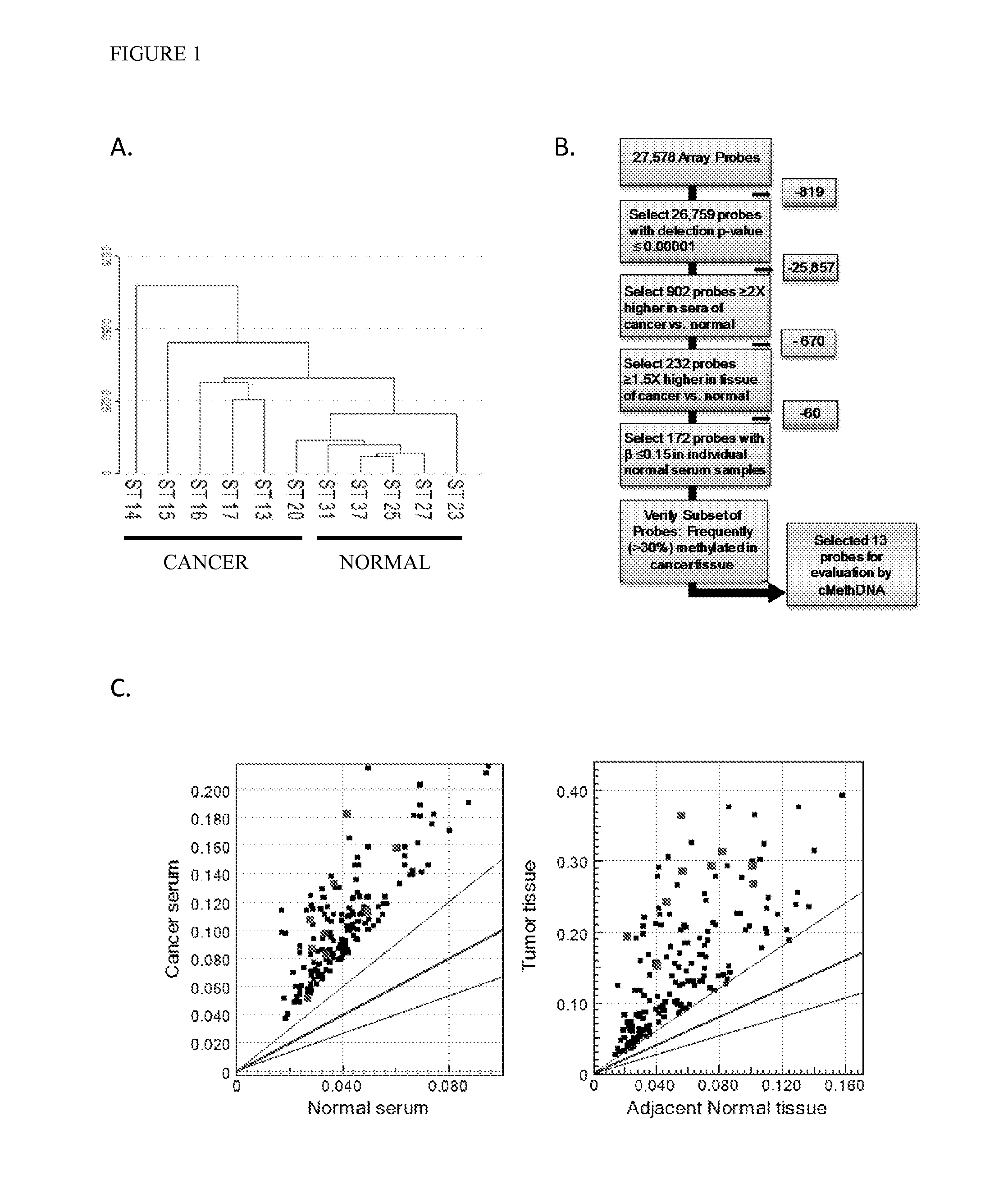
[0084]Recently, genome wide-DNA methylation arrays and bisulfite treated DNA sequencing of primary breast tumors have resulted in the identification of novel subgroups from the “intrinsic subtypes” identified by gene expression profiling (Breast Cancer Res. 2007; 9:R20; Pathobiology. 2008; 75:149-52; Anal Cell Pathol (Amst). 2010; 33:133-41; Cancer Epidemiol Biomarkers Prev. 2007; 16:1812-21; J Epidemiol. 2012; 22:384-94). As a first step to comprehensively analyze the methylation profile of circulating DNA in breast cancer patients, a methylation array analysis was conducted using the Infinium HumanMethylation27 BeadChip Array (Illumina, Inc.) on a discovery set of six recurrent metastatic breast cancer sera, five normal sera, and twenty normal leukocyte (four pools of 5 individuals each) (Table 1). An unsupervised hierarchical cluster analysis performed using 26759 probes (97% of the array; probe detection p-values <0.0001) found that cancer and normal sera clustered into two sepa...

example 2

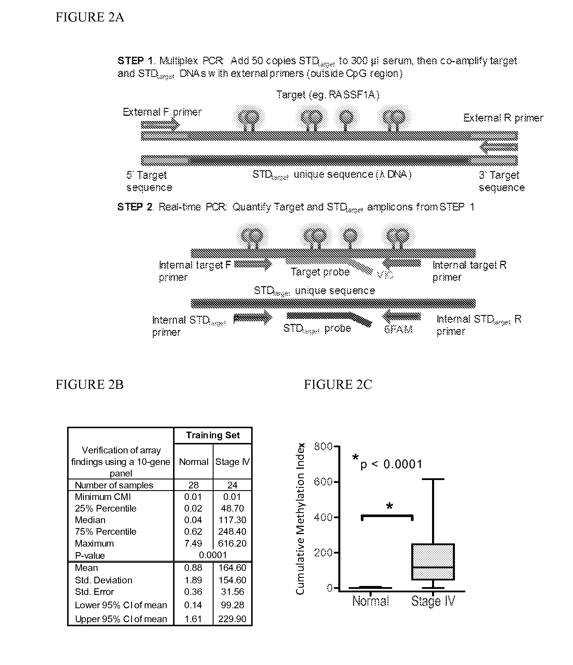
[0086]Marker detection frequency in circulating tumor DNA. In this example, fourteen genes hypermethylated in cancer sera compared to normal were tested-thirteen novel genes (HOXB4 miRNA, RASGRF2, ARHGEF7, AKR1B1, TMEFF2, TM6SF1, COL6A2, GPX7, HIST1H3C, ALX1, DAB1, DABIP2, and GAS7) identified by array analysis and RASSF1A, based upon our prior experience. Methylation frequency of the 14 genes was then verified in the patient and normal serum DNA samples used for arraying with the cMethDNA assay. The gene panel was refined by removing genes from the panel with high background or low frequency (FIG. 2B), specifically eliminating ALX1, DABIP2, and DAB1 which displayed levels of methylation of above 7 units (the set threshold for “normal” in normal control serum), and eliminating GAS7 based on its low methylation frequency (less than 20%) in cancer sera. Next, the finalized ten-gene panel was evaluated in a training set of samples (n=52, Table S5). As shown in FIG. 2C, statistical eval...

example 3

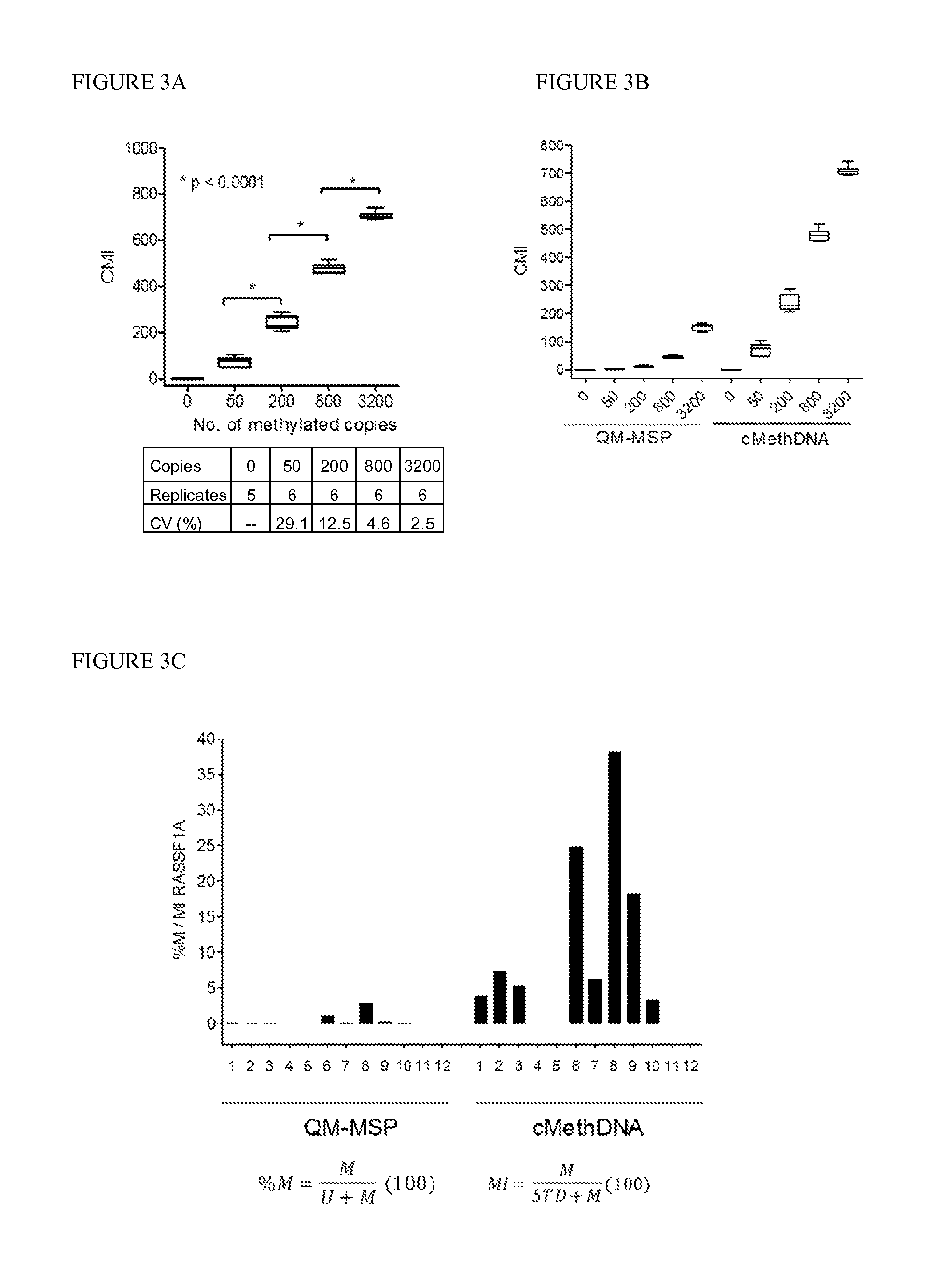
[0087]Technical evaluation of the cMethDNA assay, Analytical linearity and sensitivity. To establish assay linearity and sensitivity, a series of experiments were performed. An optimal procedure was established for isolating minute amounts of circulating DNA by testing three different serum DNA extraction methods (UltraSens Virus Kit, Qiagen; MiniElute Virus Kit, Qiagen, and Quick-gDNA Prep Zymo Research). A master mix of serum from a single normal donor (300 μl serum per assay point) was spiked with a physiological range (0, 50, 200, 800 or 3200 copies) of fully methylated DNA extracted from the MDA-MB-453 breast cancer cell line. The coefficient of variation for each replicate (5-6 replicates) was calculated for each method (FIG. 3A). The lowest level of 50 copies of spiked genomic DNA was detectable by all three methods, and negligible levels of methylated DNA were observed in the normal sera. However, of the three kits tested (FIG. 3A), the MiniElute Virus Spin Kit method displa...


PUM

PropertyMeasurementUnit
Ratioaaaaaaaaaa
Fluorescenceaaaaaaaaaa

Abstract

The cMethDNA method of the present invention is a novel modification of the QM-MSP method (U.S. Pat. No. 8,062,849), specifically intended to quantitatively detect tumor DNA (or other circulating DNAs) in fluids such as serum or plasma at the lowest copy number yet reported. Unique compared to any other PCR-based assay, a small number of copies of a synthetic polynucleotide standard (STDgene) is added to an aliquot of patient serum. In a standard procedure, a cocktail of standards for a plurality of genes of interest (TARGETgene) is added to a sample of serum. Once total DNA is purified and processed, a PCR (multiplex step) is performed wherein the STDgene and the TARGETgene are co-amplified with the same external primer set. In the N second nested PCR step, amplicons present in a dilution of the first PCR reaction are subjected to real time PCR, and quantified for each gene in one well by two-color real-time PCR. Products are calculated by absolute quantitation with internal primer sets specific for the methylated TARGETgene and associated STDgene. Methods of making the STDgene standards and the use of the cMethDNA methods and kits containing the same are disclosed.

Description

REFERENCE TO RELATED APPLICATIONS[0001]This application claims the benefit of U.S. Provisional Patent Application No. 61 / 650,083, filed on May 22, 2012, which is hereby incorporated by reference for all purposes as if fully set forth herein.INCORPORATION-BY-REFERENCE OF MATERIAL SUBMITTED ELECTRONICALLY[0002]The instant application contains a Sequence Listing which has been submitted in ASCII format via EFS-Web and is hereby incorporated by reference in its entirety. Said ASCII copy, created on May 13, 2013, is named P12014-02_SL.txt and is 58,416 bytes in size.BACKGROUND OF THE INVENTION[0003]It is now clear that epigenetic alterations including hypermethylation of gene promoters and some regions internal to a gene are consistent and early events in neoplastic progression. Such alterations are thought to contribute to the neoplastic process by transcriptional silencing of tumor suppressor gene expression, and by increasing the rate of genetic mutation. DNA methylation is reversible...

Claims


Application Information

Patent Timeline
IPC IPC(8): C12Q1/68
CPCC12Q1/6883C12Q2600/16C12Q2600/118C12Q2600/154C12Q1/6851C12Q1/6886C12Q2523/125C12Q2545/114C12Q2563/107C12Q2565/1015C12Q1/686C12Q2525/301C12Q2525/204C12Q2525/185C12P19/34C12Q2525/161
Inventor SUKUMAR, SARASWATIFACKLER, MARY JOTEO, WEI WENLOPEZ BUJANDA, ZOILA ARELI
Owner THE JOHN HOPKINS UNIV SCHOOL OF MEDICINE
Why Eureka
Eureka Blog
Learn More