Naphthalene-based two-photon fluorescent probes, preparation method and use thereof
a fluorescent probe and two-photon technology, applied in the field of naphthalene-based two-photon fluorescent probes, can solve the problems of inability to diagnose cancer by independent labeling, lack of specific imaging, and inability to deep imaging for tumors
- Summary
- Abstract
- Description
- Claims
- Application Information
AI Technical Summary
Benefits of technology
Problems solved by technology
Method used
Image
Examples
example 1
[0090]The synthesis of fluorescent probe A1:
[0091](1) The Synthesis of Intermediate 1:
[0092]20 mmol of 4-bromo-1,8-naphthalic anhydride and 25 mmol of methylamine were added into a round-bottom flask containing 10 mL acetic acid under nitrogen protection, the mixture was heated to reflux for 2 h at 100° C. Then the solution was poured into cooled water and filtrated. The white solid powder was collected to obtain the intermediate 1 in a yield of 96%.
[0093](2) The Synthesis of Intermediate 2:
[0094]20 mmol of intermediate 1 and 30 mmol of hexamethylenediamine were added into a round-bottom flask containing 20 ml ethylene glycol monomethylether under nitrogen protection, the mixture was heated to reflux for 5 h at 125° C., then the solution was poured into cooled water and filtrated. The residue was collected and purified by silica gel column chromatography, affording the intermediate 2 as a yellow solid powder in a yield of 55%.
[0095](3) The Synthesis of A1
[0096]20 mmol intermediate ...
example 2
The Labeling Experiment of Probe Compound A1 for Cancer Cells and Non-Cancer Cells
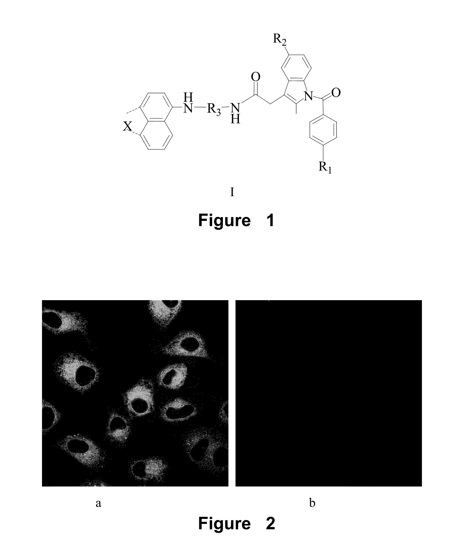
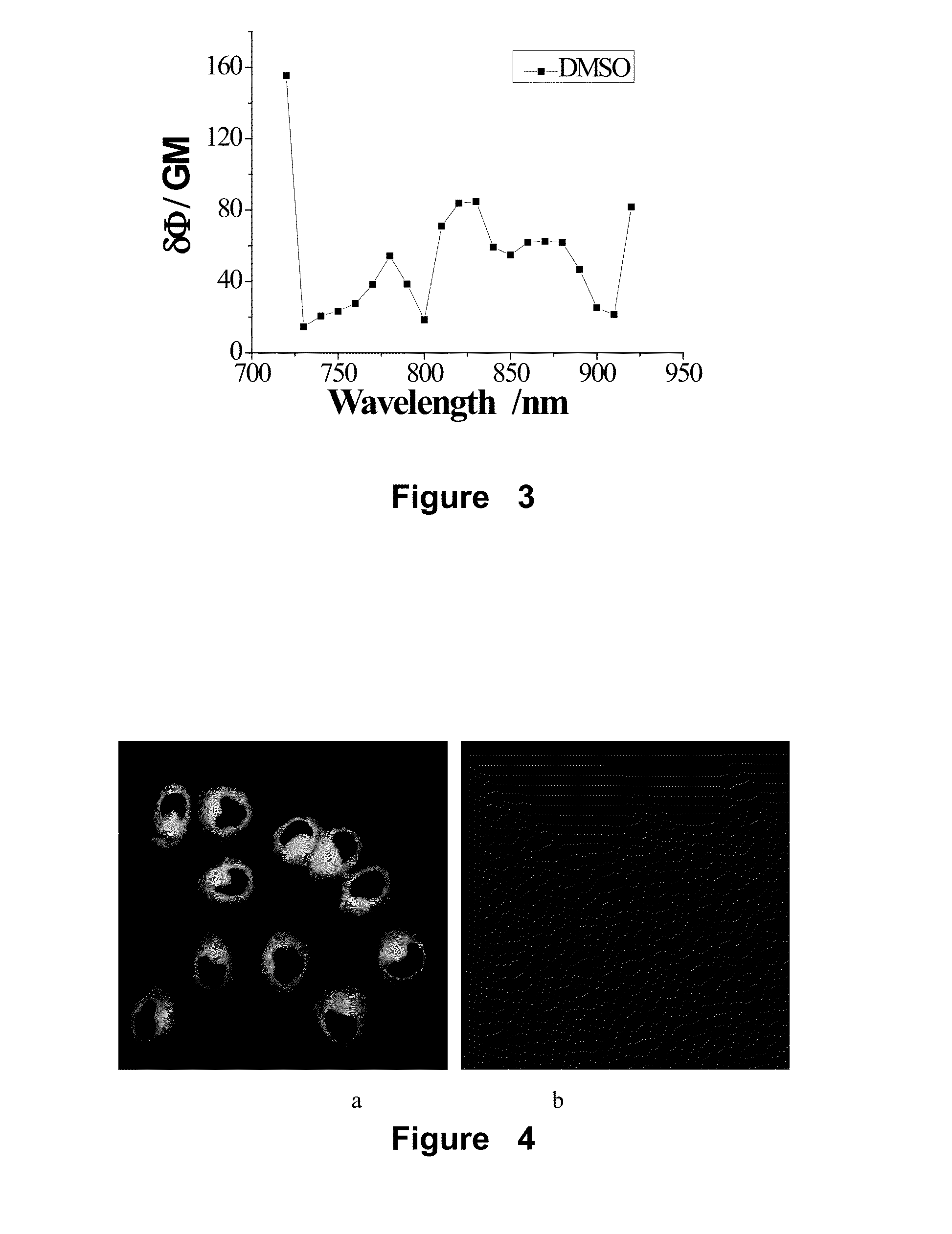
[0097]Compound A1 was used, which was synthesized in the example 1. 4 μL of compound A1-DMSO solution (4 μM) was added into HeLa and HEK 293 cells, respectively. HeLa and HEK 293 cells with probe A1 were cultured for 60 min in 5% CO2 at 37° C. Then, they were washed with phosphate-buffered saline 5 min×3. After that, the fresh medium was added into every cell. The fluorescence imaging was obtained with a two-photon spectral confocal multiphoton microscope. The representative areas were selected and imaged three times with oil-immersion objective lens (100×). The imaging results indicated that there were strong fluorescence signals in HeLa cells, but there were no any fluorescence signal in HEK 293 cells.FIG. 2(a) is confocal image of HeLa cell after adding probe A1, FIG. 2(b) is confocal image of HeLa cell after adding probe A1. The images were recorded with the emission in the range of 500-550 nm.
example 3
The Detection Experiment of the Effective Two-Photon Cross Section (δ) of A1
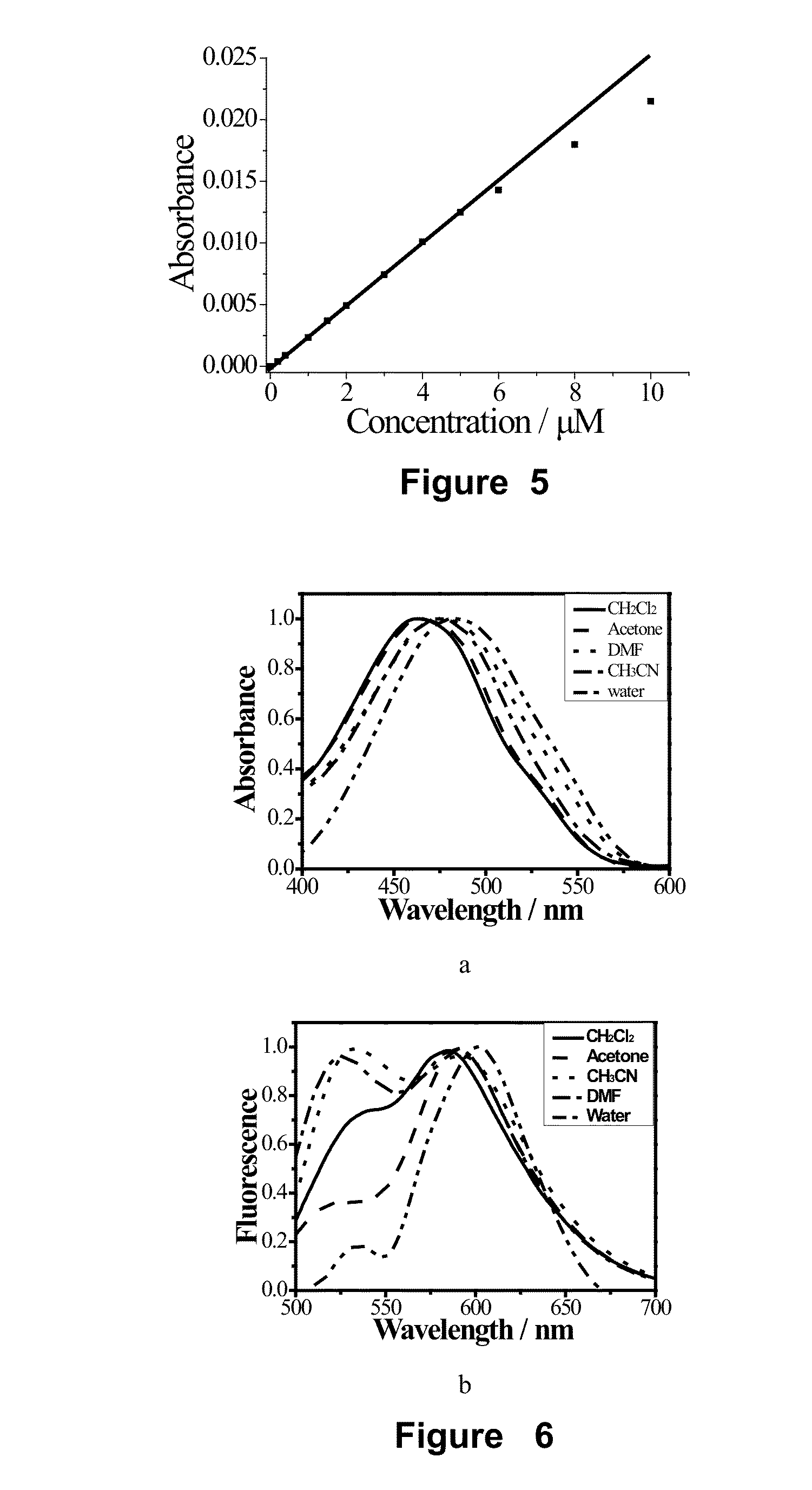
[0098]The two-photon cross section (δ) was determined by the femtosecond two-photon induced fluorescence method The compound A1, which was synthesized in the example 1, was dissolved in methanol, ethanol, acetone, acetonitrile, dioxane, dimethyl sulfoxide, tetrahydrofuran, N,N-dimethyl formamide, water and so on, respectively, at concentration of 1.0×10⁻⁴ M and then the two-photon cross section (δ) was measured by using fluorescein-sodium hydroxide solution (pH=11) as reference solution. The calculated equation was as follow:
δs=δrCrCsnrnsFsFrΦrΦs
[0099]In this equation, the concentration of solutions was denoted as c, the refractive index was denoted as n which was found in common data table. The upconversion fluorescence intensity was denoted as F, which was obtained by experiment. δ is the two-photon cross section. The reference solution was subscripted r.
[0100]The effective two-photon cross section (δΦ) i...
PUM
 Abstract
Description
Claims
Application Information
 - R&D
- Intellectual Property
- Life Sciences
- Materials
- Tech Scout
- Unparalleled Data Quality
- Higher Quality Content
- 60% Fewer Hallucinations
Browse by: Latest US Patents, China's latest patents, Technical Efficacy Thesaurus, Application Domain, Technology Topic, Popular Technical Reports.
© 2025 PatSnap. All rights reserved.Legal|Privacy policy|Modern Slavery Act Transparency Statement|Sitemap|About US| Contact US: help@patsnap.com


