Nonwoven membrane as a drug delivery system
a drug delivery and non-woven technology, applied in the field of non-woven membranes, can solve the problems of difficult processing into a macroscopic structure, difficult to obtain a sustained release kinetics of these small fibers, and lack of control over the arrangement of fibers
- Summary
- Abstract
- Description
- Claims
- Application Information
AI Technical Summary
Benefits of technology
Problems solved by technology
Method used
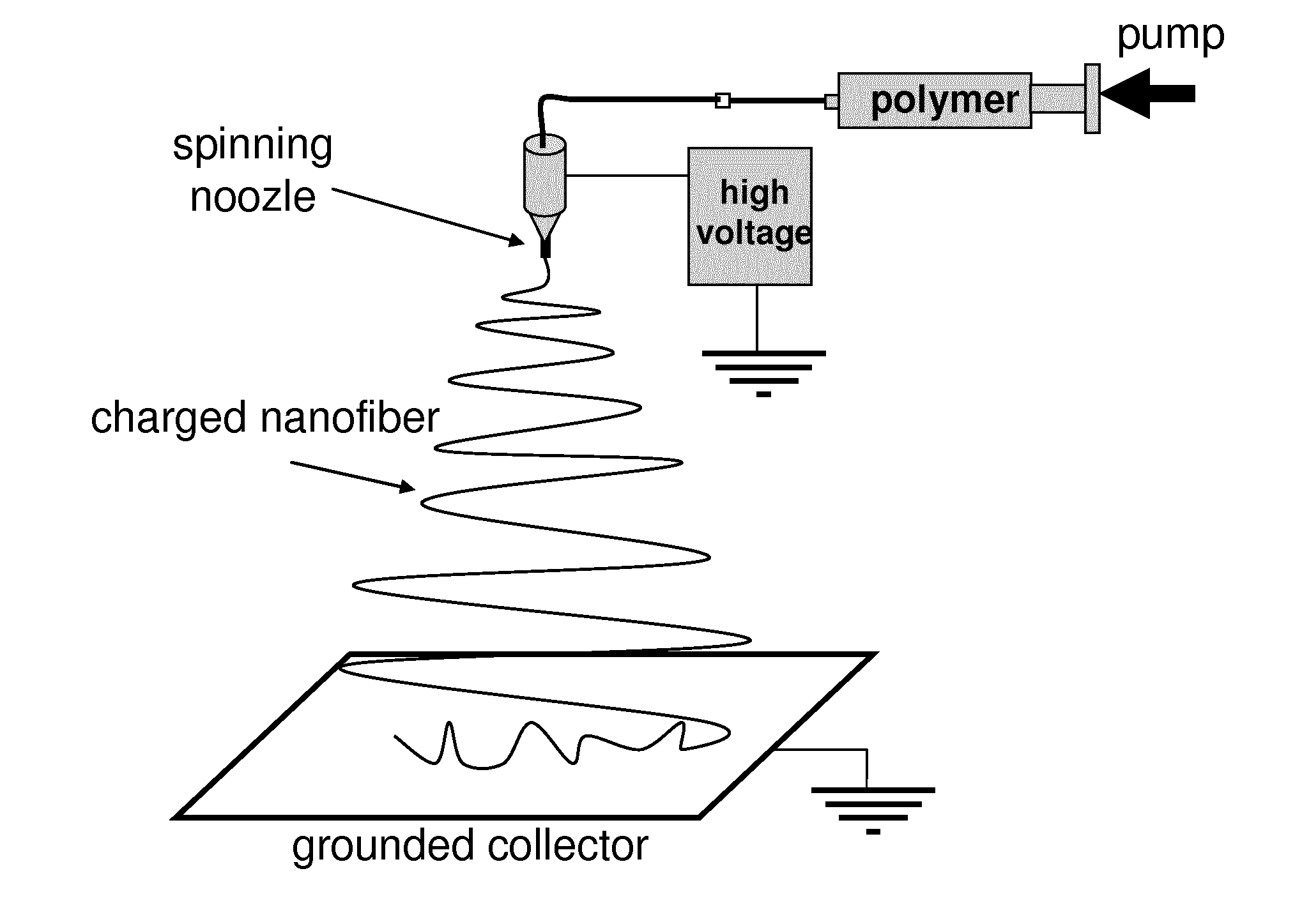
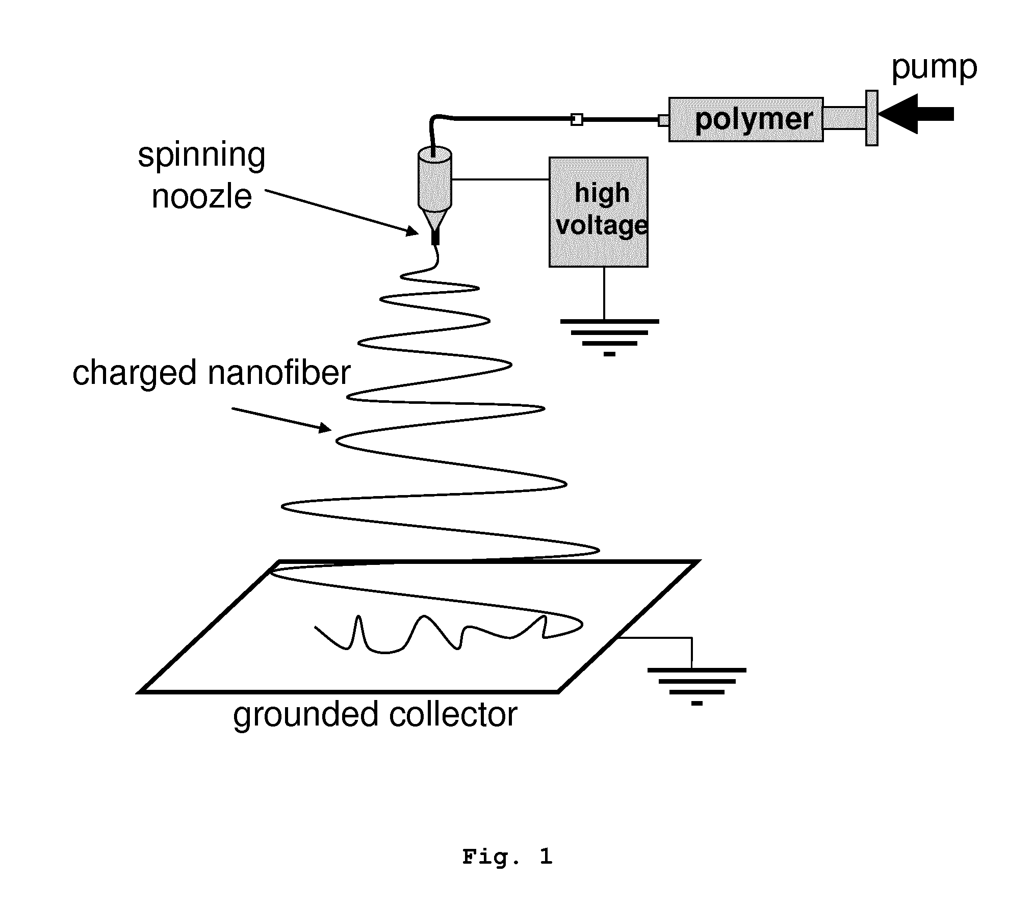
Image

Examples
example 1
Preparation of a Nonwoven Membrane Comprising Electrospun PLA Nanofibers and SN-38 Microcrystals
1.1 Preparation of a Suspension of SN-38 Microcrystals.
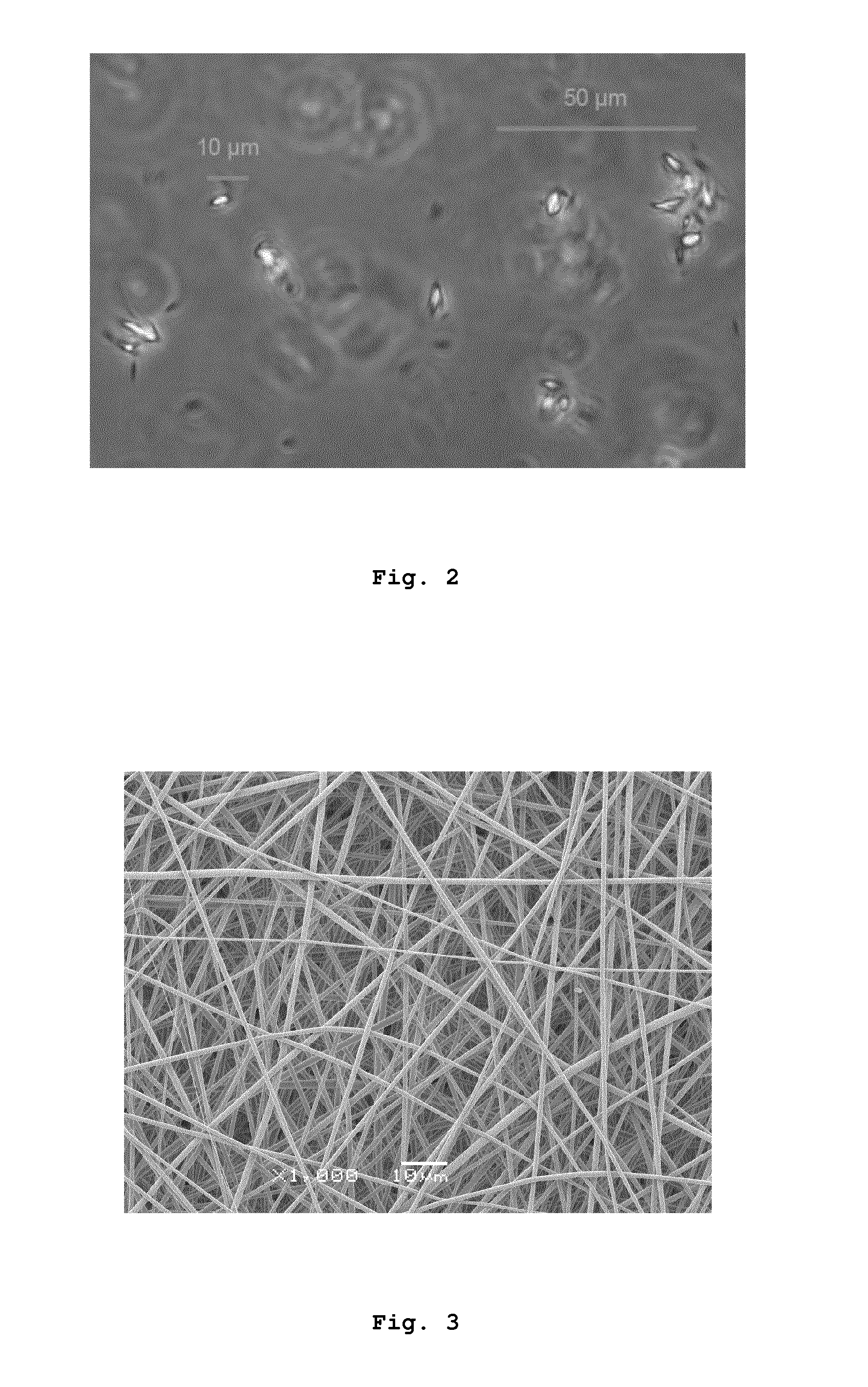
[0138]To a 50 mL pH 5.0 acetate buffer was added 2% Pluronic F68. Then SN-38 was dissolved (4 mg / mL) in NaOH 0.1 N, ratio 9:1. The acetate buffer (450 μL) was poured on the SN-38 solution (50 μL, in an eppendorf) with agitation at room temperature. No turbidity was observed during the first hour at room temperature. After 1 hour, small crystals appeared. Samples were stored at 4° C. for 24 h with hourly agitation. After that no aggregates were observed as the suspension was very stable. The mean diameter of the microcrystals in the suspension was determined by optical microscopy, being of 0.5-5 μm. FIG. 2 shows an optical microscopy image of the suspension of SN-38 microcrystals so prepared.
1.2 Preparation of a Nonwoven Membrane Comprising Electrospun PLA Nanofibers and SN-38 Microcrystals Entangled Between the PLA Nanofibers.
[0139]A ...
example 2
In Vitro Characterization of the Nonwoven Membrane Obtained in Example 1
[0146]Assays were carried out in order to characterize the in vitro release pattern and the activity in vitro of the prepared membrane.
[0147]To study the release profile of the drug entrapped in the nonwoven membrane of the invention in vitro experiments were performed using 5 mm diameter membranes containing 5 pg of SN-38 crystals. Membranes were introduced in 24 well plates containing 400 μL of cell culture medium (RPMI) each, at 37° C. The complete volume was removed at established time points (8, 24, 48 and 96 hours) and renewed with fresh medium. Released drug was quantified with a high performance liquid chromatographer (Shimadzu) with a fluorescence detector. Drug released at each time point is represented in FIG. 6, and accumulated release is represented in FIG. 7.
[0148]FIG. 6 shows the in vitro release profile of the 5 mm diameter membranes prepared. Individual dots represent dru...
experiment a
Conservation of Antitumor Activity of the Active Agent after Manufacturing the Nonwoven Membrane of the Invention
[0150]To determine whether the antitumor activity of the drug was conserved after the manufacturing process of the membranes, the present inventors co-incubated the membranes with neuroblastoma cell lines. In a previous experiment, the present inventors determined the extent to which SN-38 inhibits neuroblastoma. Such experiment was performed by exposing LAN-1, SK-N-BE(2)c and SK-N-AS cell lines to a fresh solution of SN-38 in RPMI, prepared from a stock solution of SN-38, 1 mg / mL in DMSO. Briefly, cells were plated in 96 well plates, 3000 cells per well, in RPMI medium. 24 hours later, the cells were exposed to SN-38 (concentration range 10-0.000001 μM). After 4 hours, the drug was withdrawn and fresh medium was added. The viability of the cell culture was quantified 4 days later, by using the MTS cell viability assay (Promega). The established IC50 values were in the na...
PUM
| Property | Measurement | Unit |
|---|---|---|
| Percent by mass | aaaaa | aaaaa |
| Diameter | aaaaa | aaaaa |
| Density | aaaaa | aaaaa |
Abstract
Description
Claims
Application Information
 - R&D
- Intellectual Property
- Life Sciences
- Materials
- Tech Scout
- Unparalleled Data Quality
- Higher Quality Content
- 60% Fewer Hallucinations
Browse by: Latest US Patents, China's latest patents, Technical Efficacy Thesaurus, Application Domain, Technology Topic, Popular Technical Reports.
© 2025 PatSnap. All rights reserved.Legal|Privacy policy|Modern Slavery Act Transparency Statement|Sitemap|About US| Contact US: help@patsnap.com