
Cytosolically-active peroxidases as reporters for microscopy

a technology of cytosolic active peroxidases and reporters, applied in microorganisms, organic chemistry, enzymology, etc., can solve the problems of inactivation of other reporters, limited use of minisog as an em reporter, lack of fluorescent protein equivalent, etc., and achieve the effect of enhancing enzymatic activity

Active Publication Date: 2015-02-05
MASSACHUSETTS INST OF TECH
View PDF1 Cites 2 Cited by
  • Summary
  • Abstract
  • Description
  • Claims
  • Application Information

AI Technical Summary

Benefits of technology

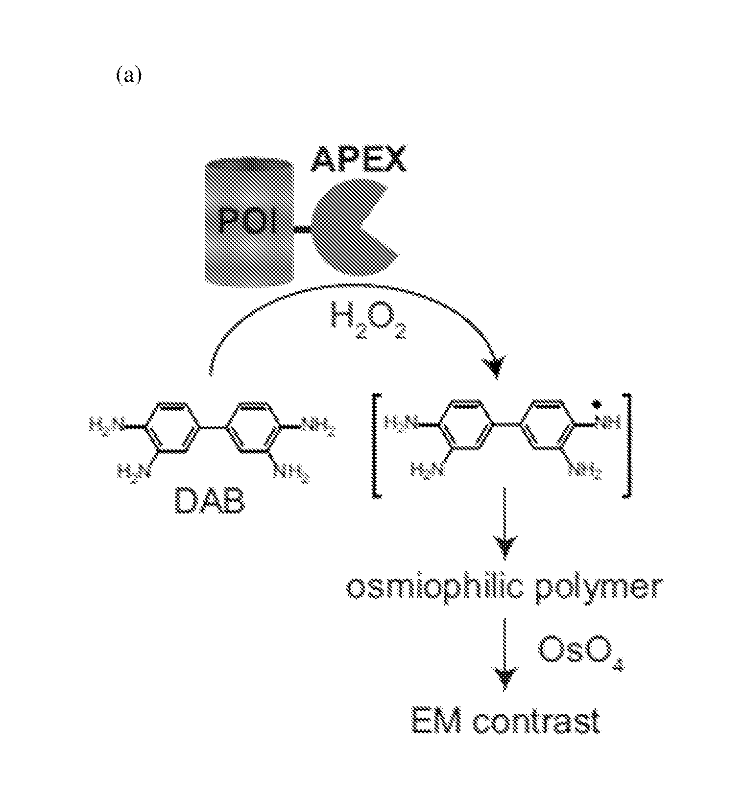
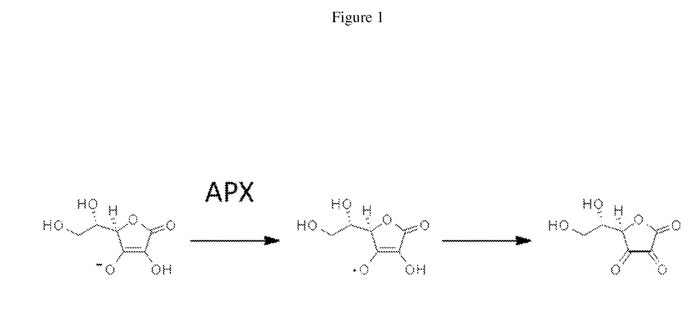
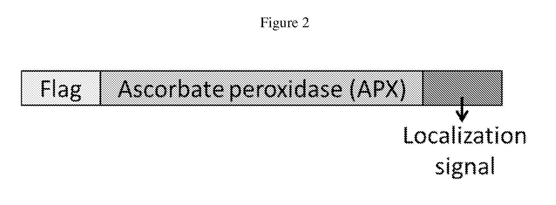
The present patent is based on the discovery of the cytosolic activity of ascorbate peroxidase (APX) and its use in microscopy imaging, particularly in EM imaging. The patent describes the use of monomeric mutants of APX that have elevated enzymatic activity and can be used in imaging methods to detect signals released from the substrates of APX. The patent also describes the development of fusion proteins containing APX and other proteins or cellular localization signals for use in imaging specific subcellular compartments. The technical effects of the patent include the discovery of the cytosolic activity of APX and its use in microscopy imaging, the development of monomeric mutants of APX with elevated enzymatic activity, and the development of fusion proteins containing APX and other proteins or cellular localization signals for use in imaging specific subcellular compartments.

Problems solved by technology

However, a major limitation of EM is the lack of a fluorescent protein equivalent for highlighting specific proteins of interest.
As such, use of miniSOG as an EM reporter is limited to small fields of view.
Other reporters are prone to inactivation due to the strong fixation typically employed in EM.

Method used


Image

  • Cytosolically-active peroxidases as reporters for microscopy
  • Cytosolically-active peroxidases as reporters for microscopy
  • Cytosolically-active peroxidases as reporters for microscopy

Examples


example 1

Generation and Characterization of Ascorbate Peroxidase Mutants

Materials and Methods

Cloning and Mutagenesis of APX Plasmids

[0081]The pTRC99A expression vector encoding pea cytosolic APX with a His6-tag appended to its N-terminus has been described previously (Cheek, et. al., Journal of Biological Inorganic Chemistry 4, 64-72. (1999)). This gene was originally derived from a pea leaf (Pisum sativum L.) cDNA library (Patterson, et. al., Journal of Biological Chemistry 269, 17020-17024 (1994)) and encodes the following amino acid sequence for APX:

(SEQ ID NO: 22)RGKSYPTVSPDYQKAIEKAKRKLRGFIAEKKCAPLILRLAWHSAGTFDSKTKTGGPFGTIKHQAELAHGANNGLDIAVRLLEPIKEQFPIVSYADFYQLAGVVAVEITGGPEVPFHPGREDKPEPPPEGRLPDATKGSDHLRDVFGKAMGLSDQDIVALSGGHTIGAAHKERSGFEGPWTSNPLIFDNSYFTELLTGEKDGLLQLPSDKALLTDSVFRPLVEKYAADEDVFFADYAEAHLKLSELGFAEA

[0082]Mutants of pea APX, including K14D, E112K, E228K, D229K, K31S, A233D, I185K, A28K, A28K / E112K, K14D / E112K, K14D / E228K, K14D / D229K, E112K / E228K, E17N / K20A / R21L, W41F, G69F, G174...

example 2

Use of Ascorbate Peroxidase Mutants in Microscopy

Materials and Methods

Genetic Constructs

[0099]The genetic constructs used in this example consist of at least those found in Example 1, as exampled in Table 3.

Mammalian Cell Culture and Transfection

[0100]Culture and transfection methods were employed as described in Example 1.

Fixation and Staining with DAB, Osmium Tetroxide (OsO4), and Uranyl Acetate

[0101]A solution of 4% formaldehyde (freshly depolymerized from paraformaldehyde) with 2% glutaraldehyde in 10 mM phosphate buffered saline (PBS) was added to cells 24 hours after transfection. Cells were treated for 3 min at room temperature, then 30 min on ice. Cells were then washed 5×2 min using ice cold 10 mM PBS. Cells were blocked with 20 mM glycine for 5 min on ice. Cells were then washed 5×2 min with ice-cold 10 mM PBS. Cells were then treated with an ice-cold solution of DAB (0.5 mg / mL) and H2O2(0.03%) in 10 mM PBS for 20 min. Cells were then washed 5×2 min with ice-cold 10 mM PBS...


PUM

PropertyMeasurementUnit
temperatureaaaaaaaaaa
flow rateaaaaaaaaaa
thicknessaaaaaaaaaa

Abstract

An imaging method comprising expressing in cells a Class I heme peroxidase, which optionally is fused with a protein of interest or a cellular localization signal peptide, and contacting the cells with a substrate of the Class I heme peroxidase to allow conversion of the substrate into a product via an oxidation reaction catalyzed by the Class I heme peroxidase, wherein the product releases a signal detectable by a microscope such as an electron microscope. Also disclosed herein are monomeric mutants of a Class I heme peroxidase and mutants of the enzyme that exhibit elevated enzymatic activity as compared to the corresponding wild-type counterpart.

Description

RELATED APPLICATIONS[0001]This application claims the benefit under 35 U.S.C. §119(e) of U.S. Provisional Application Ser. No. 61 / 535,839, entitled “Cytosolically-Active Peroxidase as Reporters for Microscopy,” filed on Sep. 16, 2011, which is herein incorporated by reference in its entirety.BACKGROUND OF THE INVENTION[0002]Electron microscopy (EM) offers far better spatial resolution than fluorescence microscopy and therefore is a very important tool for cell biology. For example, mitochondria are just a few pixels wide by fluorescence but details and subcompartments can be seen by EM. However, a major limitation of EM is the lack of a fluorescent protein equivalent for highlighting specific proteins of interest.[0003]Two existing genetically encoded reporters for EM are Horse Radish Peroxidase (HRP) and mini-singlet oxygen generator (SOG), both generating contrast by catalyzing the polymerization of a diaminobenzidine (DAB) into an osmiophilic polymer. Photo oxidation of miniSOG r...

Claims


Application Information

Patent Timeline
Patent Type & Authority Applications(United States)
IPC IPC(8): C12Q1/28G01N23/04
CPCC12Q1/28C12Y111/01011G01N23/04
Inventor TING, ALICE Y.MARTELL, JEFFREY DANIEL
Owner MASSACHUSETTS INST OF TECH
Learn More