
Endodontic imaging

a technology for endodontics and endoscopy, applied in the field of endodontic imaging, can solve the problems of pulpitis process, inconvenient preparation, and inability to accurately diagnose the disease, and achieve the effects of minimizing the risk of re-infection, and reducing the risk of infection

Active Publication Date: 2014-10-16
FOSCHI FEDERICO +2
View PDF2 Cites 2 Cited by
  • Summary
  • Abstract
  • Description
  • Claims
  • Application Information

AI Technical Summary

Benefits of technology

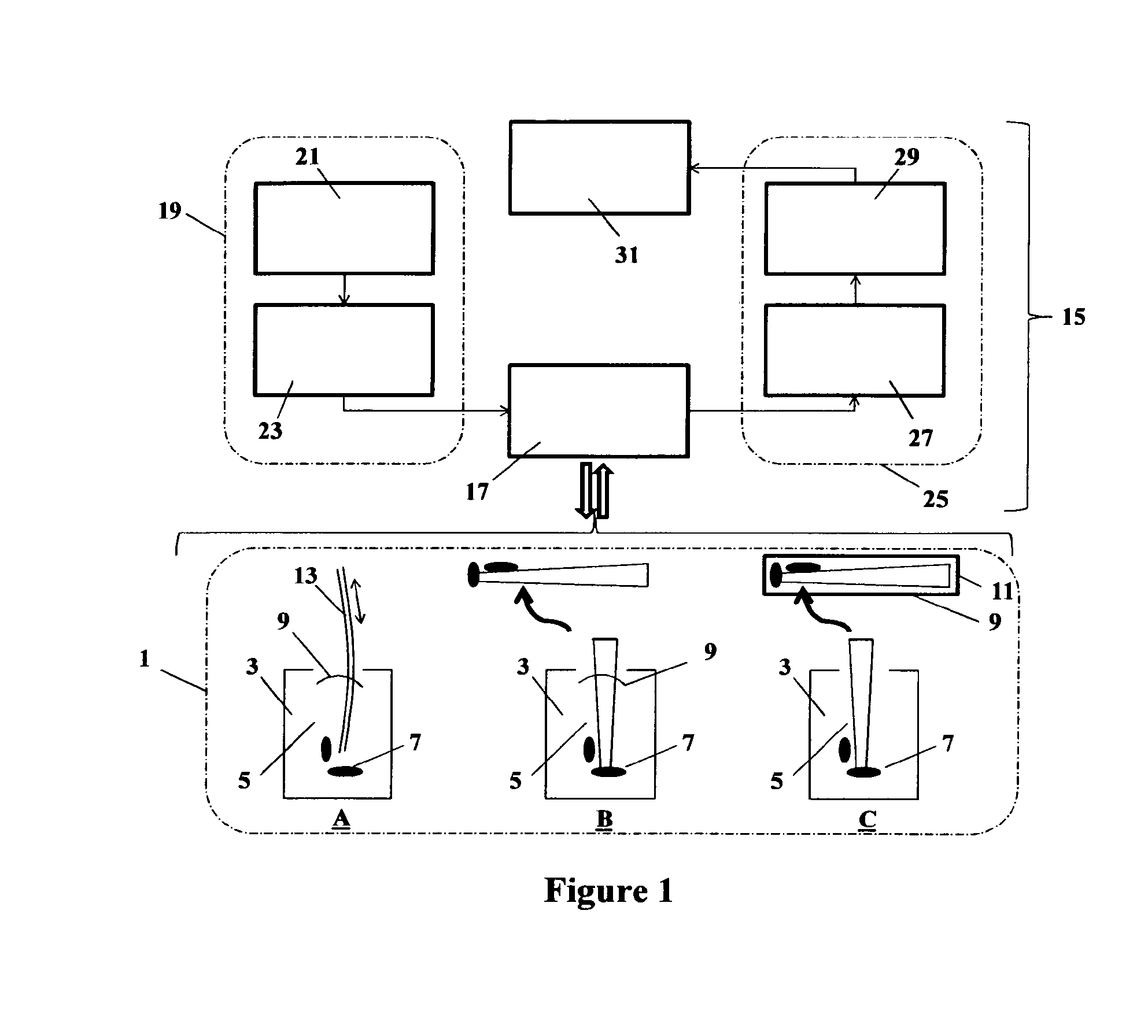
The invention provides a method for detecting cells or cellular material in an endodontic sample using a cell indicator. This method involves incubating the cell indicator with the sample for a specific period and measuring or detecting a change in a parameter associated with the cell indicator to get a result indicative of cells or cellular material in the sample. This invention also includes the use of a cell indicator for diagnosis, prevention, and treatment of endodontic infection or recurrence.

Problems solved by technology

Bacterial infection in the root canal or tooth, typically affecting the pulp material, causes a process of pulpitis, an often painful process affecting the pulp chamber or endodontic chamber and root canal structure.
This results in an infection of the tooth and often results in an extending lesion beyond the apex or other canal connecting the nerve space to the adjacent soft and hard tissues of a tooth and may present as an abscess within these surrounding tissues.
Infected and inflammatory bacterial fluids emanating from within the tooth structure and apical portion of the tooth escape through the apex of the root into the surrounding periodontal tissue, causing substantial inflammation, with associated tissue changes.
Without intervention, the tooth will die and may require extraction and the infection may spread into the surrounding tissues causing potentially life-threatening abscesses in the head and neck region.
Furthermore, roots may have a complex morphology in terms of overall structure including curvature and accessory canals.
The most common cause of failure of root canal treatment, aside from poor technique (e.g. missed root canal or improperly sealed access cavity) or iatrogenic contamination, is residual bacteria in the root canal space and at the apex of the tooth, which can arise due to difficulty in visualising or accessing all aspects of the inside of the roots, particularly in the apical region, of the root canal which may harbour remaining tissue or bacteria.
As a result, dental practitioners may over-prepare the canals to remove the infected dentine, at potentially unnecessary time and cost, or may underprepare leaving a substantial bio-burden within the tooth structure with concomitant risk of re-infection.
Yet, still, approximately 15-25% of endodontic root canal procedures fail.
Therefore, these procedures often need to be repeated due to persistent infection, thus representing a significant time and cost burden, in addition to often requiring specialist intervention and dramatically reducing the prognosis of the tooth to be treated.
However, this procedure is inherently flawed as it adds several days to the procedure time (since microbial analysis in a cell culture laboratory is required) and furthermore, bacteria cultures will only identify bacteria present in the obtained sample as well as being limited to culturable material and so can result in false negatives.
This leads to canals which ultimately fail as they are still infected at the time of obturation despite negative culture results.
However, the apparatus is expensive and only effective in and insofar as the probe is inserted into the appropriate root canals.
Furthermore, this device will not assess the actual presence of viable bacteria pre- or post-treatment.
A problem with X-ray is that the apical portion of the root canal (the portion where structural complexities are likely to exist) is partially obscured in such images by the jaw bone.

Method used


Image

  • Endodontic imaging
  • Endodontic imaging
  • Endodontic imaging

Examples


example

Example 1

Processing of Extracted Teeth

[0116]Twenty freshly extracted, single rooted teeth were obtained under ethical Approval (South East London ethical approval 10 / H0804 / 056). Each tooth had the crown removed to obtain 15 mm long specimens, using a 0.3 mm thickness diamond wafering blade (Extec, Enfield, Conn., USA) on a 11-1180 Isomet low speed saw (Buehler, Düsseldorf, Germany). Root canal patency was confirmed using an ISO 10 K-file. Canals were instrumented with Gates Glidden (nr. 2-4) for coronal flaring followed by ProTaper F3 instrumentation and apical instrumentation with an ISO 45 K-file 1 mm short of the working length. Irrigation throughout the instrumentation was undertaken with sterile saline. Each root was then sectioned longitudinally through the root canal using the low speed saw. The specimen's halves were then reapproximated and placed in a block of freshly mixed impression material (Aquasil™ Hard Putty / Fast Set, Dentsply) contained in 5 ml bijou vials with the c...


PUM


Abstract

A method and apparatus for detecting the presence of the viable cells in an endodontic sample in or taken from an endodontic cavity in the tooth, such as the root canal, enables the rapid and low cost identification of the presence or absence of bacteria or other viable cell tissue in the endodontic space by first incubating a viable cell indicator with an endodontic sample for a pre-determined period suitable for chair-side test and then measuring and / or detecting a change in a parameter (e.g. fluorescence) associated with the viable cell indicator. Thereby, the required level of root canal treatment can be reached which minimizes the risk of re-infection without the time, cost and potentially harm of over-preparation and over treatment by the dental surgeon or of a re-treatment.

Description

FIELD OF THE INVENTION[0001]This invention pertains generally to the field of endodontic imaging. More particularly, the invention relates to a method and apparatus for imaging, quantitative measurement and / or data collection inside a tooth, and more particularly the root canal.BACKGROUND OF THE INVENTION[0002]Endodontic therapy or root canal treatment is the treatment of choice for prevention or recovery of a tooth from infection, or where it is otherwise threatened by infection, cracking or decay, extending (into or near) to or approximating the pulp chamber.[0003]Bacterial infection in the root canal or tooth, typically affecting the pulp material, causes a process of pulpitis, an often painful process affecting the pulp chamber or endodontic chamber and root canal structure. This results in an infection of the tooth and often results in an extending lesion beyond the apex or other canal connecting the nerve space to the adjacent soft and hard tissues of a tooth and may present a...

Claims


Application Information

Patent Timeline
IPC IPC(8): C12Q1/04C12Q1/37
CPCC12Q1/37C12Q1/04A61B1/0638A61B1/0684A61B1/24A61B1/00165A61B1/043
Inventor COOK, RICHARDKOLLER, GARRITWATSON, TIMOTHYFOSCHI, FEDERICOMANNOCCI, FRANCESCOFESTY, FREDERIC
Owner FOSCHI FEDERICO
Learn More