
Methods and devices for occluding blood flow to an organ

a technology of occluding blood flow and organs, applied in the field of surgical implantation of blood occluding devices, can solve the problems of sterility and/or premature menopause, chemotherapy drugs that are toxic to reproductive cells and organs, and drugs that might have undesired side effects on organs and tissues, so as to reduce the exposure of in vivo organs and prevent blood flow.

Inactive Publication Date: 2014-09-04
A A CASH TECH
View PDF0 Cites 767 Cited by
  • Summary
  • Abstract
  • Description
  • Claims
  • Application Information

AI Technical Summary

Benefits of technology

This patent relates to a device that can protect an organ, like a gonad, from exposure to drugs that are in a patient's blood system. The device can prevent blood flow to the organ for a short time, reducing the risk of damage. It can also be implanted and removed using a laparoscopy. This technology may be useful for medical procedures where there is a risk of harm to organs, such as ovarian surgery.

Problems solved by technology

Some of these organs and tissues may be targets of treatment, while others might not, and the drugs might have undesired side effects on organs and tissues which are not intended for treatment.
For example, some chemotherapy drugs are toxic to reproductive cells and organs.
Known side effects of chemotherapy in young women include sterility and / or premature menopause.
Similarly, men undergoing chemotherapy often suffer damage to their natural fertility, for example chemotherapy induced oligospermia.
Both techniques require a delay in cancer treatment for at least one month, which is not an option for some patients.

Method used


Image

  • Methods and devices for occluding blood flow to an organ
  • Methods and devices for occluding blood flow to an organ
  • Methods and devices for occluding blood flow to an organ

Examples


example 1

[0085]The following experiment was designed to demonstrate ovary resistance to prolonged blood occlusion.

[0086]The ovaries of three 5 month old female sheep were exposed by laparoscopy and blood flow to and from the ovaries was occluded for an occlusion time interval of 24.5 or 27.5 hours. In one ovary of each sheep blood flow was occluded (full block) at the ovarian artery and ovarian vein, while in the other ovary, all of the ovarian artery and vein and the uterine artery and vein were occluded.

[0087]After the occlusion time interval the ovaries were removed and fixed in Buyen solution for histology evaluation. The results are depicted in Table 1 below. In addition, the ovaries were observed for coloration changes. The results are summarized in Table 1.

[0088]Firstly, it was noted that ovaries where all four arteries and veins were occluded, maintained a natural appearance in size and coloration, wherein ovaries with blood occlusion only at the ovarian artery and vein appeared to b...

example 2

[0089]The following experiment shows an example for reducing gonadotoxic damage according to some embodiments of the invention.

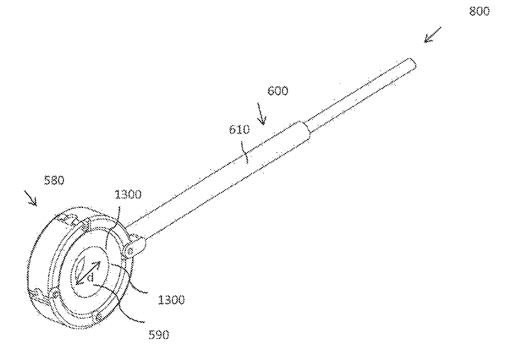
[0090]The protocol and design of all parts of this study were approved by the Animal Research Ethics Committee of Israel. Three female pigs (The Institute of Animal Research, Kibbutz Lahav, Israel) weighing 60 Kg each, were anesthetized with 10 mg / kg intramuscular ketamine hydrochloride and 4 mg / kg xylazine hydrochloride (Vetmarket, Israel). One ovary of each pig was exposed by a longitudinal midline incision. A conventional gastric band (Lap-band; Allergen, Irvine, Calif., USA) was modified according to some embodiments of the invention, to occlude blood flow to the ovaries. The band was wound around the ovarian hilum, and a zip tie was placed on the center of the band ring to reduce the diameter of the band's aperture. 10 ml saline was injected via the band's port thus completely blocking blood flow through the ovarian and uterine arteries and veins. The c...


PUM


Abstract

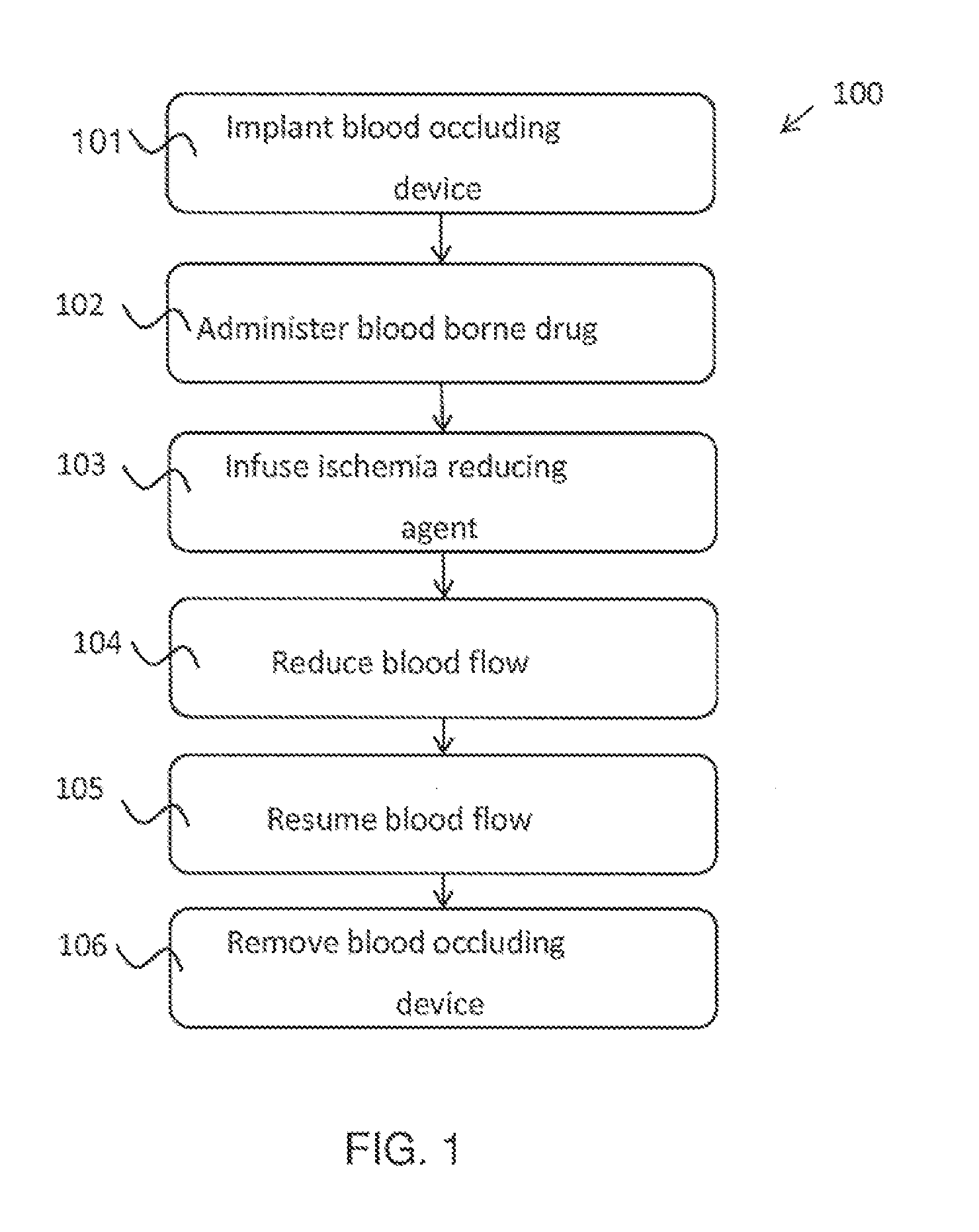
A method for protecting at least one gonad from a blood borne cytotoxic drug, the method comprising reducing blood flow to at least one gonad of a patient undergoing cytotoxic treatment for an occlusion time interval, and allowing blood flow to the at least one gonad to resume after the occlusion time interval. Optionally the method is performed to protect an ovary during chemotherapy treatment.

Description

FIELD OF THE INVENTION[0001]The invention, in some aspects thereof, relates to the field of surgical implantation of blood occluding devices.BACKGROUND OF THE INVENTION[0002]Many drugs are carried by a patient's blood system from a site of administration to organs and tissues throughout the body. Some of these organs and tissues may be targets of treatment, while others might not, and the drugs might have undesired side effects on organs and tissues which are not intended for treatment. For example, some chemotherapy drugs are toxic to reproductive cells and organs. Known side effects of chemotherapy in young women include sterility and / or premature menopause. Similarly, men undergoing chemotherapy often suffer damage to their natural fertility, for example chemotherapy induced oligospermia.[0003]Men undergoing chemotherapeutic treatments often preserve their fertility through sperm banking. To date, the solutions offered to women include cryopreservation of embryos or, albeit still...

Claims


Application Information

Patent Timeline
Patent Type & Authority Applications(United States)
IPC IPC(8): A61B17/12
CPCA61B17/12009A61B17/12013A61B17/12036A61B17/1204A61B17/12109A61B17/12136A61B17/1285A61B2017/00022A61B17/42A61B2017/00557
Inventor ARAV, AMIR
Owner A A CASH TECH
Learn More