
Methods and tools for the diagnosis and prognosis of urogenital cancers

a technology for urogenital cancer and diagnosis, applied in the field of cancer, can solve the problems of poor disease outcome, over-expression of prostate cancer cells, and major health risks of genital cancer types for the public, and achieve the effects of improving the diagnosis and treatment process

Inactive Publication Date: 2014-08-21
CANCER GENETICS
View PDF0 Cites 4 Cited by
  • Summary
  • Abstract
  • Description
  • Claims
  • Application Information

AI Technical Summary

Benefits of technology

The present invention provides methods and tools for the diagnosis and prognosis of cancer and pre-cancer in the prostate, renal, and bladder regions using genomic analysis. The invention involves the use of microarrays, such as array-based comparative genomic hybridization (CGH) or whole genome array CGH, to analyze genetic material from samples obtained from humans. The invention can also involve the use of other technologies, such as labeling the genetic material and hybridizing it with the microarrays. The methods can be used to diagnose and predict the recurrence of prostate, renal, and bladder cancers, as well as to determine the metastasis potential of bladder cancers. The invention also provides a tool for identifying the subtype of renal cancer and predicting the outcome of patients with low risk insignificant prostate cancers. Overall, the invention provides a valuable resource for the diagnosis and treatment of cancer and pre-cancer in these regions.

Problems solved by technology

(2007) Oncologist 12:20-37), these three types of genitourinary cancers still represent major health risk and substantial medical cost burden to the public with their high rates of incidence.
However, for a disease with a potentially long progression time, the challenge arises in determining the most appropriate and cost-effective treatment for each patient after the initial diagnosis.
Putting expression of ERG under androgen regulation results in its over-expression in prostate cancer cells.
The presence of the TMPRSS2:ERG fusion and the loss of the PTEN protein are associated with poorer disease outcome (Yoshimoto et al.
The widespread of modern imaging techniques has led to an increased detection of incidental and smaller kidney masses, leading to the difficult question of when and to what extent the physician should intervene.
Although the procedure of kidney biopsy is associated with little complications and demonstrable clinical significance, the challenge lies in the accurate diagnosis to distinguish between the malignant tumors (mostly RCCs) from the benign ones, most frequently the renal oncocytoma, using such minimal amount of material.
Often, small renal mass biopsies could not be diagnosed due to either the failure to obtain biopsy tissue of adequate quantity and / or quality for pathological examination or the inability to distinguish RCC subtypes pathologically.
(2009) BMC Cancer 9:152) but due to sample size limitations and differences in cohort population, there is no standard consensus in the diagnostic scheme that has been rigorously validated and translated to clinical practice.
The probability of progression varies with initial stage, grade, and size of the tumor and currently, there is no reliable biomarker that can distinguish tumors with the more aggressive characteristics from the ones that have low reoccurring potential.
In many cases, intravescial therapy may be overly used if the prognosis is good and the chance of reoccurrence is low [85].
(2011) Genet Med 13:676-79) have been suggested for the performance of array-CGH as a replacement for (or as an adjunct to) standard cytogenetic techniques (e.g., karyotyping, FISH) as commercially available FDA-approved devices, as commercially available Investigational Use Only (IUO) devices requiring validation, or as “home-brew” or in-house developed and validated devices; however, they have not yet been adopted.

Method used


Image

  • Methods and tools for the diagnosis and prognosis of urogenital cancers
  • Methods and tools for the diagnosis and prognosis of urogenital cancers
  • Methods and tools for the diagnosis and prognosis of urogenital cancers

Examples


example 1

Development of a Microarray for Diagnosis and Prognosis of Urogenital Cancers

[0114]A literature survey was performed to facilitate development of tools (particularly the microarray—e.g., the urogenital cancer array, also known as the UroGenRA® urogenital cancer array as described herein) for the use in the diagnosis and prognosis of specific urogenital cancers including prostate, kidney, and bladder cancers. Initially, an evaluation of the literature was performed for molecular genetic and cytogenetic applications in the study of gynecological cancers. These studies utilized chromosomal CGH, FISH, array CGH (BAC and oligonucleotide), SNP-array, ROMA, and / or PCR-based assessment of single gene copy numbers. Across these studies vastly differing technologies were utilized and even within technologies, different platforms were used, making direct comparisons and extraction of data difficult. In addition, different cut-offs and algorithms were used for data analysis again complicating c...

example 2

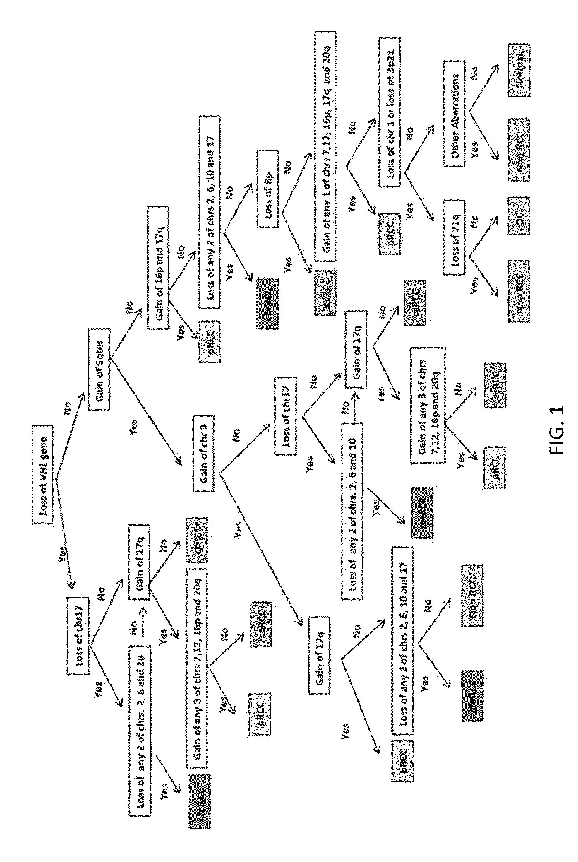
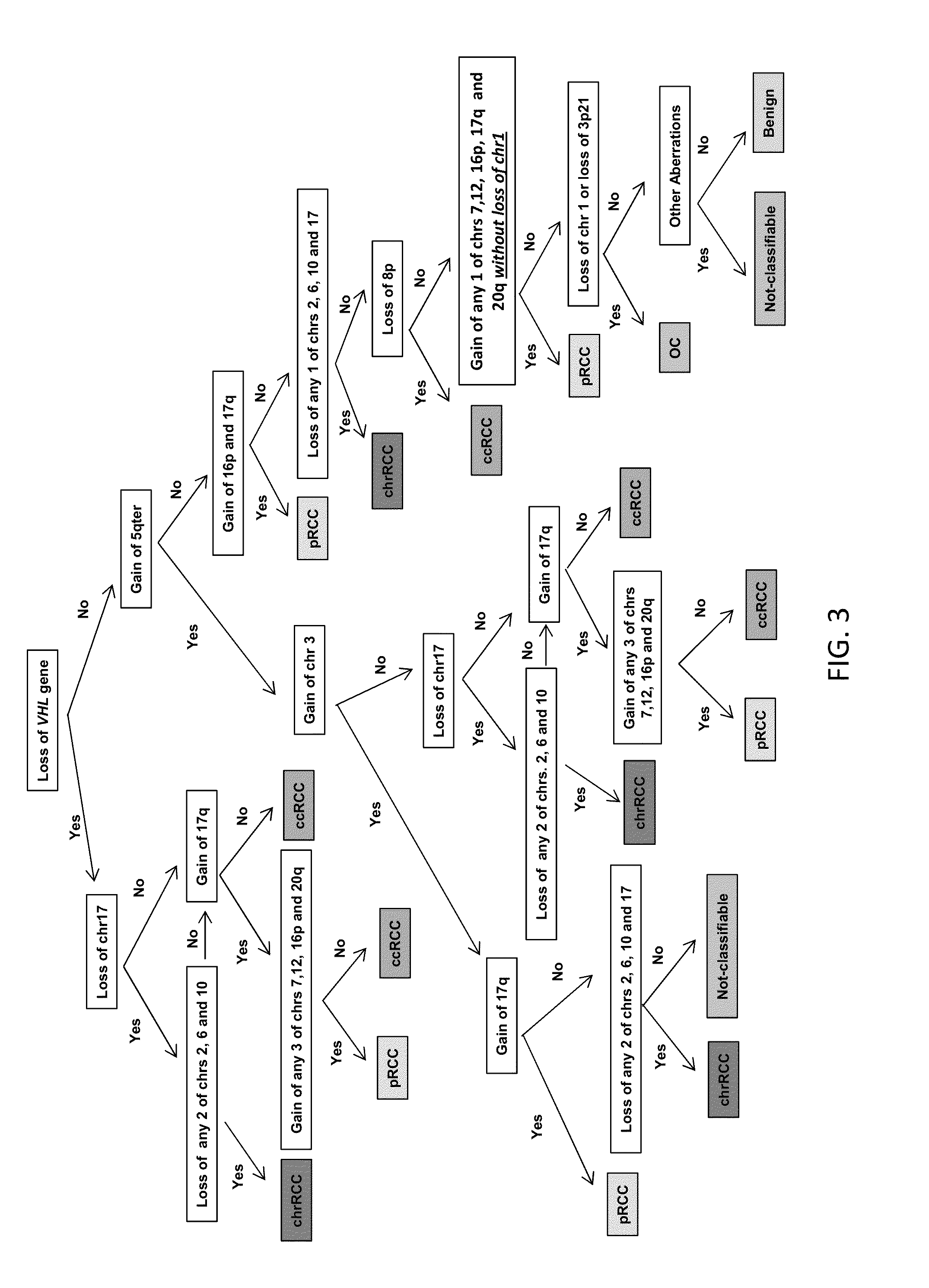
A Preliminary Decision Tree for the Diagnosis of Renal Cortical Neoplasms

Description of the Disease

[0123]In 2012, nearly 64,770 new cases of kidney cancer are estimated in United States and about 13,570 deaths from the disease are predicted to occur [1]. Renal cell carcinoma (RCC) is the most abundant form of kidney cancer, and despite being the most lethal can be cured by surgery if diagnosed at an early stage. RCC arises in the renal cortex and is the predominant malignant type of renal cortical neoplasm as shown in Table 9.

TABLE 9Renal Cortical Neoplasm Histologic Subtype and FrequencyRenal Cortical Neoplasm Histologic SubtypeFrequencyBenignOncocytoma (OC)6-9%Papillary adenomaMetanephric adenomaNephrogenic adenofibromaMalignantClear cell (conventional) RCC (ccRCC)60-65%Papillary RCC (pRCC)13-15%Chromophobe RCC (chrRCC) 6%Collecting duct carcinomaMedullary carcinomaTubulocystic RCCRCC, unclassified 7%Mucinous tubular and spindle cell carcinomaTranslocation-associated carcinomasTum...

example 3

Diagnostic Classification of Renal Tumors Using a Copy Number-Based Decision Tree Algorithm

Abstract

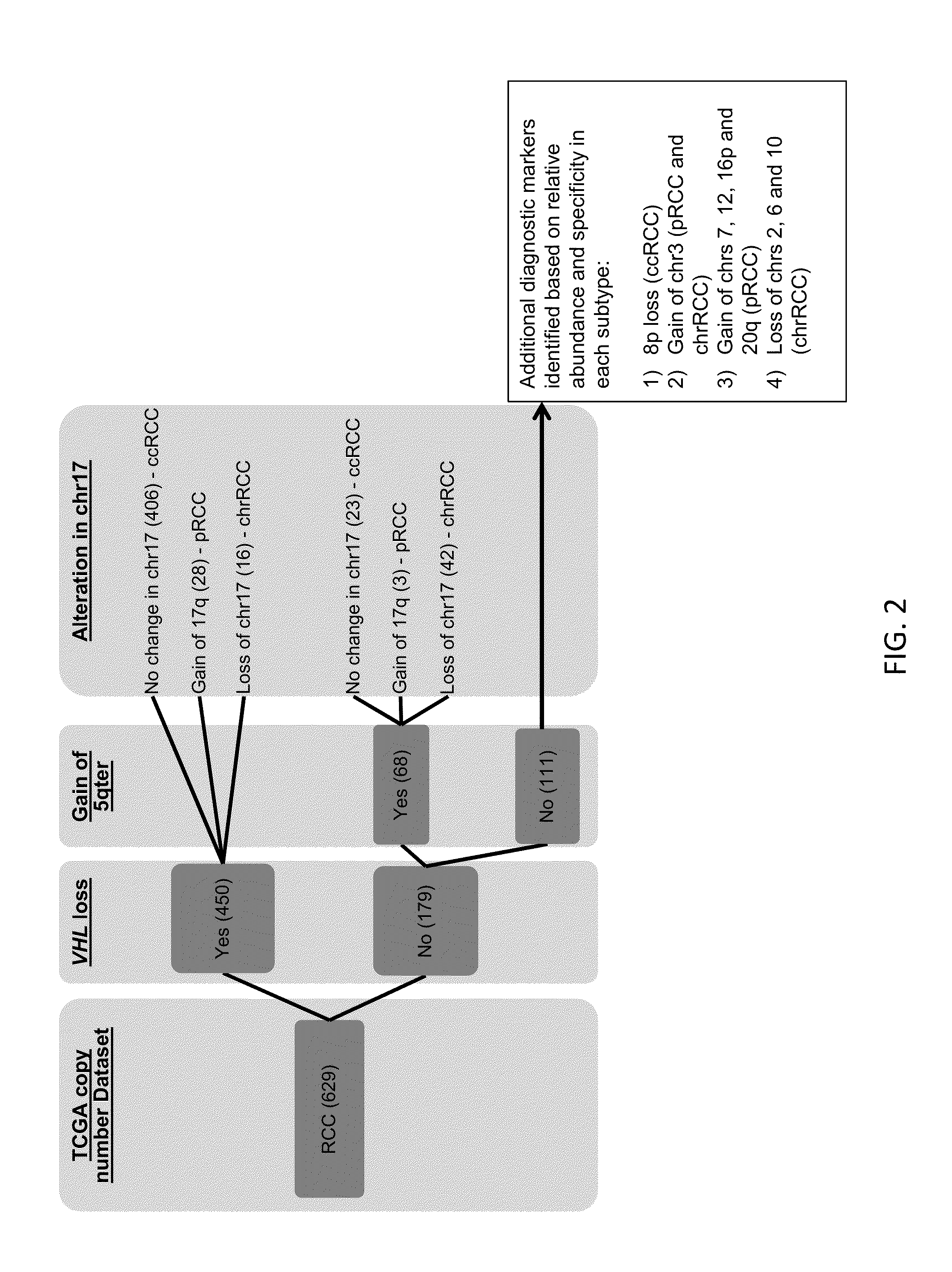
[0181]Accurate diagnostic discrimination of major renal cortical neoplasms (clear cell, papillary, chromophobe and oncocytoma subtypes) is not only useful for predicting prognosis of the disease but also to plan appropriate treatment strategies which include active surveillance, cryoablation, partial and radical nephrectomy followed by with or without systemic therapy. Diagnosis solely by histopathology could often be challenging both in percutaneous needle biopsies as well as in surgically resected specimens. The aim of this study is to utilize the genomic imbalance associated with the major renal tumor subtypes to achieve correct diagnosis. Using TCGA (The Cancer Genome Atlas) copy number dataset, prior FISH study and published literature, a total of 15 genomic markers were identified. A decision tree algorithm was developed based on these genomic markers to assist in renal tumor sub...


PUM

PropertyMeasurementUnit
Massaaaaaaaaaa

Abstract

The present invention provides a microarray useful as a tool in the diagnosis and / or prognosis of certain types of cancers, particularly urogenital cancers. The microarray can include a plurality of genomic regions represented thereon, the genomic regions corresponding to regions wherein alterations, such as copy number aberrations, at such locations correlate to specific, identifiable cancers, particularly prostate, renal, or bladder tumors. The invention further provides methods of diagnosing certain types of cancers, particularly urogenital cancers, more particularly renal cortical cancers. The methods can comprise analyzing genetic material from a human individual to determine the presence or presence of certain aberrations and using a decision tree to classify the subtype of renal cortical neoplasm present in the sample.

Description

CROSS-REFERENCE TO RELATED APPLICATION[0001]This application claims the benefit of U.S. Provisional Application No. 61,765,678, filed Feb. 15, 2013, which is hereby incorporated herein in its entirety by reference.FIELD OF THE INVENTION[0002]The present invention relates to cancer and in particular to urogenital cancers. In particular, the present invention relates to methods and tools for the diagnosis and prognosis of prostate, kidney, and bladder cancers. The invention also provides methods for the diagnosis and prognosis of such malignancies using alternative platforms or technologies, preferentially with minimal invasiveness.BACKGROUND OF THE INVENTION[0003]The reproductive organs (prostate, testis, penis, cervix, uterine, ovary, vulva, and vagina) and the urinary system (two kidneys, bladder, combined with the urine transporting ureters and urethra) are commonly grouped together as the genitourinary system due to their physiological and developmental proximity and sharing of t...

Claims


Application Information

Patent Timeline
IPC IPC(8): C12Q1/68
CPCC12Q1/6886C12Q2600/112C12Q2600/118C12Q2600/156
Inventor HOULDSWORTH, JANE C.CHAGANTI, RAJU S.K.
Owner CANCER GENETICS
Learn More