
Oligonucleotide-based probes for detection of bacterial nucleases

a technology of oligonucleotide-based probes and bacterial nucleases, which is applied in the direction of instruments, diagnostic recording/measuring, ultrasonic/sonic/infrasonic diagnostics, etc., can solve the problems of affecting the development of assays, affecting the detection accuracy of fluorophores, and complicated probe design

Active Publication Date: 2014-07-17
UNIV OF IOWA RES FOUND +1
View PDF8 Cites 16 Cited by
  • Summary
  • Abstract
  • Description
  • Claims
  • Application Information

AI Technical Summary

Benefits of technology

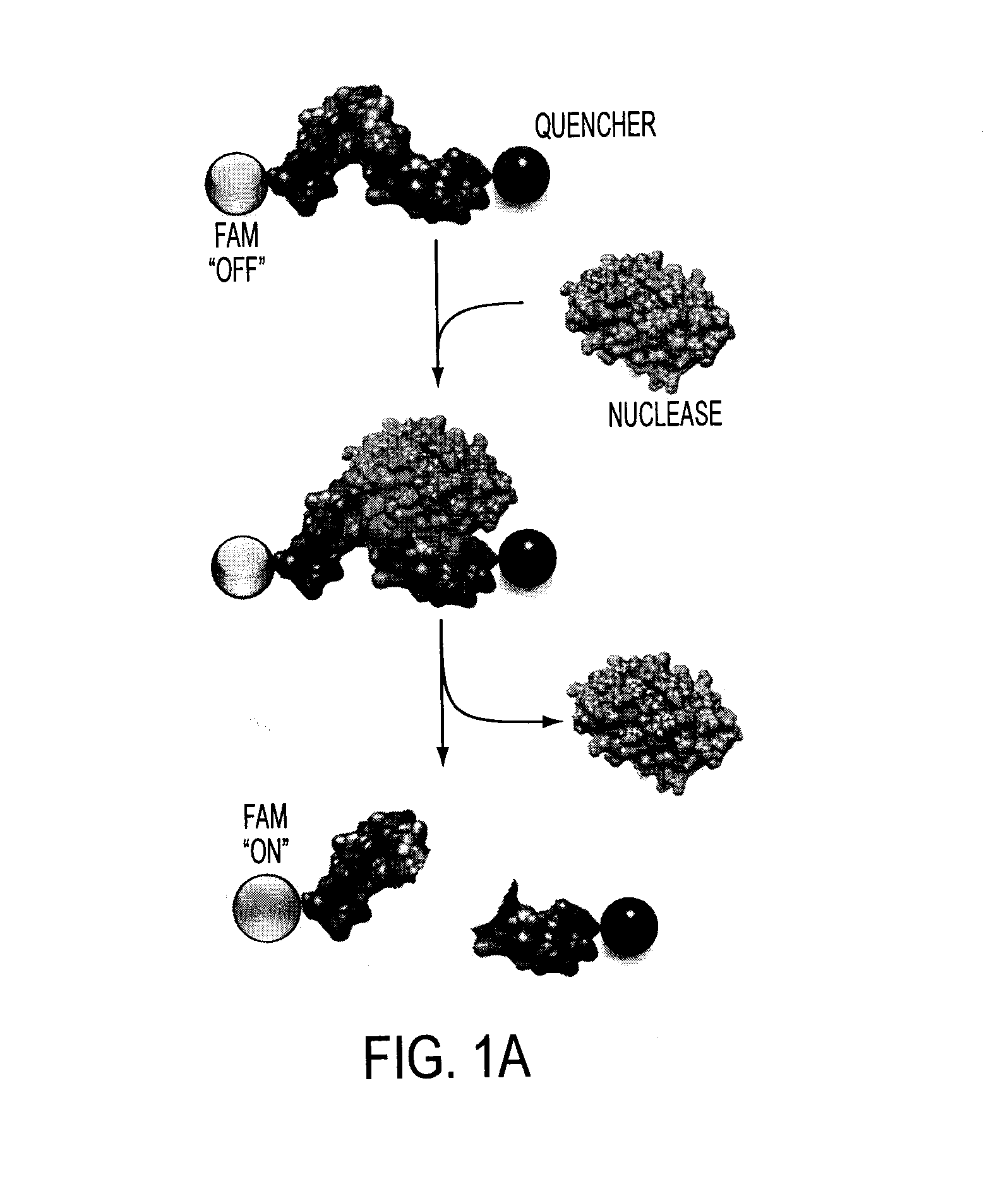
The present invention provides a probe for detecting microbial endonuclease activity using a substrate oligonucleotide of 2-30 nucleotides in length, a fluorescence-reporter group, and a fluorescence-quencher group. The fluorescence-reporter group and the fluorescence-quencher group are separated by at least one RNAse-cleavable residue or DNAse-cleavable residue. The oligonucleotide can be a DNA or RNA molecule, and can be modified with fluorescences such as FAM, TET, HEX, JOE, MAX, Cy3, or TAMRA. The invention also provides methods for detecting microbial infections in samples using the probe, as well as methods for evaluating the activity of microbial nucleases on various nucleic acid substrates.

Problems solved by technology

This complicates the design of probes because not all potential quencher / donor pairs can be used.
The use of varied quenchers complicates assay development because the purification of a given probe can vary greatly depending on the nature of the quencher attached.
Such quenchers interfere with the use of fluorophores that fluoresce at similar wavelength ranges.
This limits the number of fluorophores that can be used with such quenchers thereby limiting their usefulness for multiplexed assays which rely on the use of distinct fluorophores in distinct probes that all contain a single quencher.
RNases are extremely common, resulting in very short lifespans for any RNA that is not in a protected environment.
Present technologies for detection of bacterial pathogens are time-consuming and expensive because they usually require the isolation and culturing of the bacteria.
Also, many of the existing technologies are toxic and / or use radioactive tracers.
Further, technologies for imaging bacterial colonization in humans lack sensitivity.

Method used


Image

  • Oligonucleotide-based probes for detection of bacterial nucleases
  • Oligonucleotide-based probes for detection of bacterial nucleases
  • Oligonucleotide-based probes for detection of bacterial nucleases

Examples


example 1

[0134]Degradation of Nuclease-Stabilized RNA Oligonucleotides in Mycoplasma-Contaminated Cell Culture Media

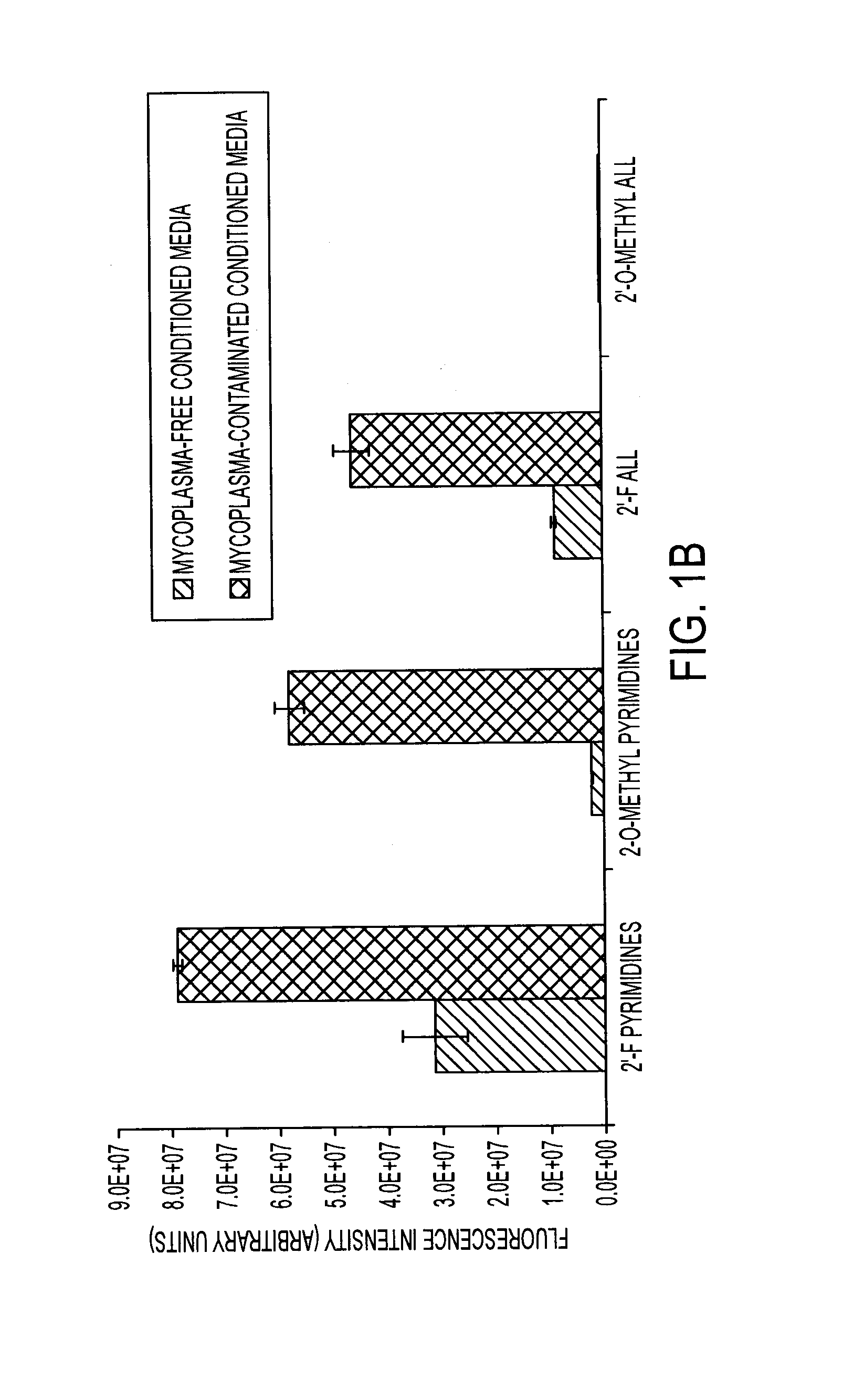
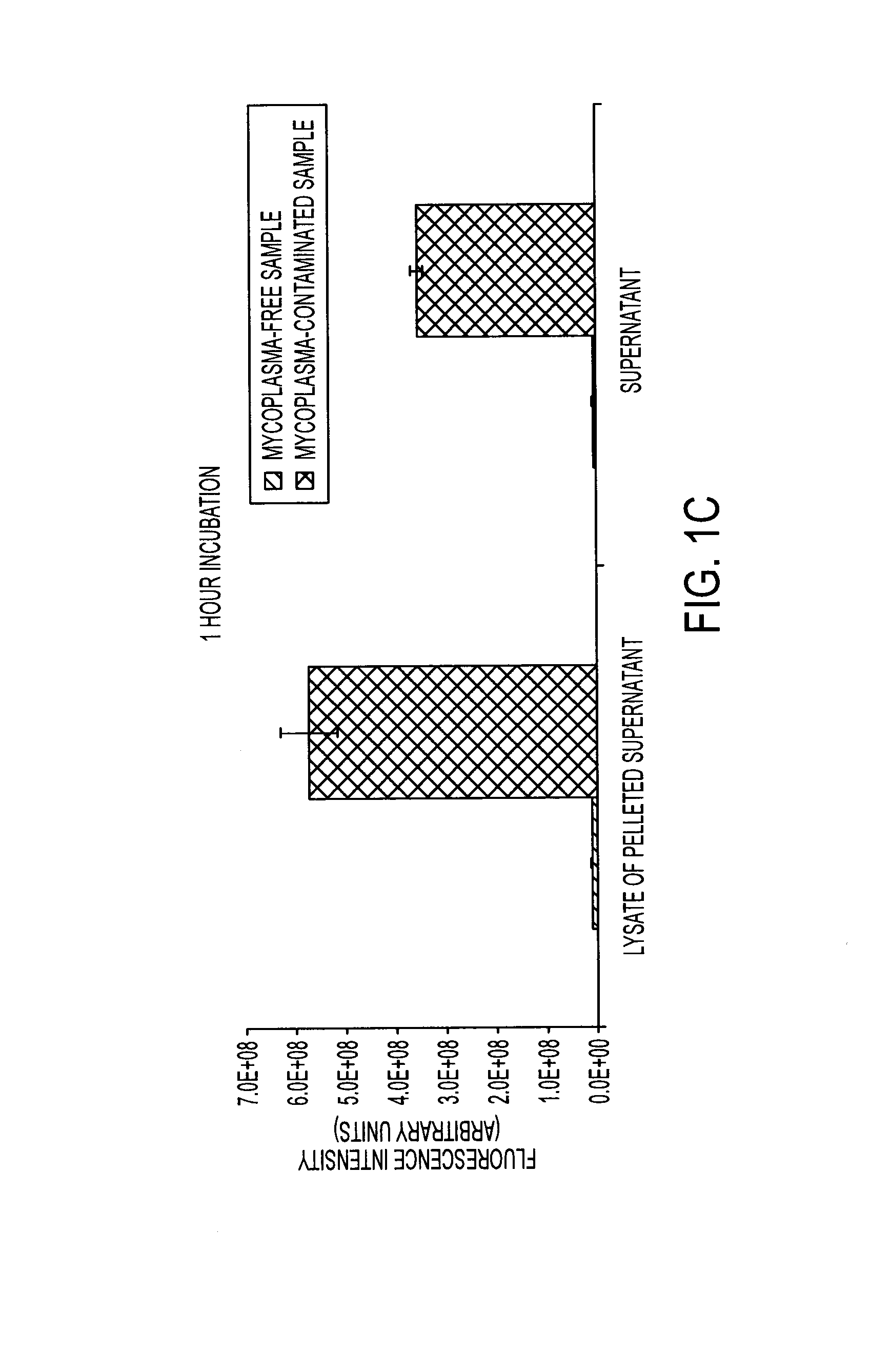
[0135]Artificial RNA reagents such as siRNAs and aptamers often must be chemically modified for optimal effectiveness in environments that include ribonucleases. Mycoplasmas are common bacterial contaminants of mammalian cell cultures that are known to produce ribonucleases. Here, the inventors describe the rapid degradation of nuclease-stabilized RNA oligonucleotides in an HEK cell culture contaminated with Mycoplasma fermentans, a common species of mycoplasma. RNA with 2′-fluoro- or 2′-O-methyl-modified pyrimidines was readily degraded in conditioned media from this culture, but was stable in conditioned media from uncontaminated HEK cells. RNA completely modified with 2′-O-methyls was not degraded in the mycoplasma contaminated media. RNA zymogram analysis of conditioned culture media and material centrifuged from the media revealed several distinct protein bands (ranging fr...

example 2

In Vivo Detection

[0180]The cleavage of oligonucleotides can be visualized in various ways, but as discussed above, the inventors favor flanking the sequences with a fluorophore and a quencher for rapid detection of the cleavage activity with a fluorometer. Advancing one step further, the inventors envision injecting the fluorescent probe into patients and using fluorescent detection to localize sights of infection (in addition to specific pathogen data) clearly within the patient. Preliminary data of this type has been established in mouse models. Briefly, mice were injected with micrococcal nuclease (purified Staph. aureus nuclease) in leg and injected with probe in tail vein. This procedure resulted in fluorescence being seen at site of nuclease and subsequently in liver.

example 3

In Vitro Detection

[0181]Nuclease Probe Plate-Reader Assay:

[0182]The nuclease probes were synthesized by Integrated DNA Technologies (IDT; Coralville, Iowa). These probes consist of a 12 nucleotide long RNA oligo, 5′-UCUCGUACGUUC-3′, with the chemical modifications, flanked by a FAM (5′-modification) and a pair of fluorescence quenchers, “ZEN” and “Iowa Black” (3′-modifications. Three samples were assayed for degradation. PBS, 1 μl of each probe (50 picomoles) was combined with 9 μl of PBS. RNase A, 1 μl of each probe was combined with 8 μl of PBS and 1 μl RNase A (˜50 U / μl). Micrococcal Nuclease (MN), 1 μl of each probe was combined with 8 μl of PBS and 1 μl of MN (10 U / μl). All the samples were incubated at 37° C. for 4 hours. After the incubation period, 290 μl of PBS supplemented with 10 mM EDTA and 10 mM EGTA was added to each sample and 95 μl of each sample was loaded in triplicate into a 96-well plate (96F non-treated black microwell plate (NUNC)). Fluorescence levels are show...


PUM

PropertyMeasurementUnit
depthaaaaaaaaaa
wavelengthaaaaaaaaaa

Abstract

The present invention relates to a rapid detection of microbial-associated nuclease activity with chemically modified nuclease (e.g., ribonuclease) substrates, and probes and compositions useful in detection assays. Accordingly, in certain embodiments, the present invention provides a probe for detecting a microbial endonuclease comprising a substrate oligonucleotide of 2-30 nucleotides in length, a fluorescence-reporter group operably linked to the oligonucleotide, and a fluorescence-quencher group operably linked to the oligonucleotide. The fluorescence-reporter group and the fluorescence-quencher group are separated by at least one RNAse-cleavable residue, e.g., RNA base.

Description

RELATED APPLICATION[0001]This application claims priority under 35 U.S.C. 119(e) to provisional application U.S. Ser. No. 61 / 530,246 filed Sep. 1, 2011 and to provisional application U.S. Ser. No. 61 / 593,595 filed Feb. 1, 2012, which applications are incorporated hereby by reference.STATEMENT REGARDING FEDERALLY SPONSORED RESEARCH[0002]This invention was made with government support under AI083211 awarded by the National Institutes of Health. The government has certain rights in the invention.BACKGROUND OF THE INVENTION[0003]Chemical moieties that quench fluorescent light operate through a variety of mechanisms, including fluorescence resonance energy transfer (FRET) processes and ground state quenching. FRET is one of the most common mechanisms of fluorescent quenching and can occur when the emission spectrum of the fluorescent donor overlaps the absorbance spectrum of the quencher and when the donor and quencher are within a sufficient distance known as the Forster distance. The e...

Claims


Application Information

Patent Timeline
Patent Type & Authority Applications(United States)
IPC IPC(8): A61K49/00C12Q1/68
CPCC12Q1/689A61K49/0052G01N33/569C12Q1/44G01N33/542A61K49/0054C12Q1/6818C12Q1/6823G01N2333/922
Inventor MCNAMARA, II, JAMES O.FLENKER, KATIE R.HUANG, LINGYANHORSWILL, ALEXANDER R.BEHLKE, MARK A.HERNANDEZ, FRANK J.
Owner UNIV OF IOWA RES FOUND
Learn More