The importance of
early detection and treatment of colorectal diseases is generally recognised, but the range of existing non-invasive screening and diagnostic tests remains strictly limited.
Unfortunately it is often diagnosed late (Richards, 2009) because in many cases clinical manifestations do not appear until advanced CRC stages.
Flexible
colonoscopy is currently regarded as the most sensitive and specific diagnostic procedure for colorectal tumour detection, but it is an invasive investigation requiring
bowel preparation and occasionally causing serious complications (Ransohoff, 2009).
It is also important that
colonoscopy is an expensive procedure, which should be performed only by highly qualified and specially trained medical professionals.
Although flexible
colonoscopy is correctly defined as the final common pathway of every colorectal screening program (Lieberman, 2009), the idea of introducing it as the only valid approach to CRC screening advocated by some US experts does not constitute a viable option for most countries (Hoff et al., 2010).
The necessity of a simple, non-invasive and inexpensive test, which could be used as the “first line” of CRC screening is a long-standing problem in clinical
medicine.
U.S. Pat. Nos. 3,252,762; 3,996,006; 4,092,120; 4,199,550; 4,333,734; 4,562,043; 4,939,097; 5,391,498; 5,563,071 describe different versions of the conventional guaiac-based test detecting the presence of haemoglobin in faeces, but these tests may give false-positive results due to the presence of residual haemoglobin in food remnants.
Although FOBT and iFOBT have some attractive characteristics, being non-invasive, simple, cheap and easily repeatable, application of these tests frequently produces false-positive and especially false-negative results (Allison et al, 1996; 2007; Collins et al, 2005; Burch et al, 2007; Duffy et al, 2011) reflecting the fact that the presence or absence of blood in stool is often unrelated to the presence of CRC (Itzkowitz, 2009).
Another minor drawback of this test is related to the necessity of temporarily preserving excreted stool for collecting samples for analysis.
Nevertheless, the lavage-based method of material collection was invasive and unreliable and has never been introduced into clinical practice.
Nevertheless, no cytological evidence of colonocyte presence in preparations made according to the patents has been provided by the authors.
Eventually the approach has never been used for clinical purposes.
U.S. Pat. No. 5,981,651 suggested application of
epithelium-targeting immunomagnetic beads for recovering epithelial cells from small stool samples, but the method appeared to be complex.
This requirement made its wide application for clinical purposes impossible.
Nevertheless, no single
genetic change is known to be universally present in all colorectal cancers, therefore it became evident that only complex and expensive assays targeting a panel of multiple
mutation markers in stool could detect CRC with the specificity approaching 95%.
Although preliminary investigation of some
methylation-related assays look promising (Glöckner et al, 2009; Hellebrekers et al., 2009; Nagasaka et al, 2009),
DNA methylation assessment methods remain relatively complex and expensive.
Despite growing interest in stool-based assays targeting
nucleic acid-associated biomarkers of neoplasia, faecal material remains a problematic substance for detecting changes in the
human DNA.
The abundance of non-human, bacterial or food-derived
DNA and the presence of substances interfering with PCR amplification are well-known problems related to
stool sample analysis (Nechvatal et al., 2008).
These approaches are not sufficiently developed to be used clinically for CRC diagnosis or screening.
In most cases it is characterized by long remissions and incidental
flare-ups usually requiring treatment.
Unfortunately, repeated diagnostic colonoscopies may be dangerous in IBD patients, and the range of non-invasive tests available for these purposes is very limited and includes only the use of serologic and faecal biomarkers, detection of which requires blood or stool collection and laboratory analysis (Foell et al., 2009).
The use of quantification of
DNA isolated from collected material was successfully employed for CRC detection (Loktionov et al., 2009; Loktionov et al., 2010; Wallin et al., 2010), but this method is unlikely to be suitable for CRC screening purposes due to the proctoscopy requirement making the approach invasive.
Unfortunately efforts of the scientific
community in this direction were predominantly concentrated on stool-based methods with little thought devoted to the development of simple techniques for material self-collection by tested individuals and eventual creation of point-of-care tests or self-testing systems for CRC screening and IBD detection and monitoring.
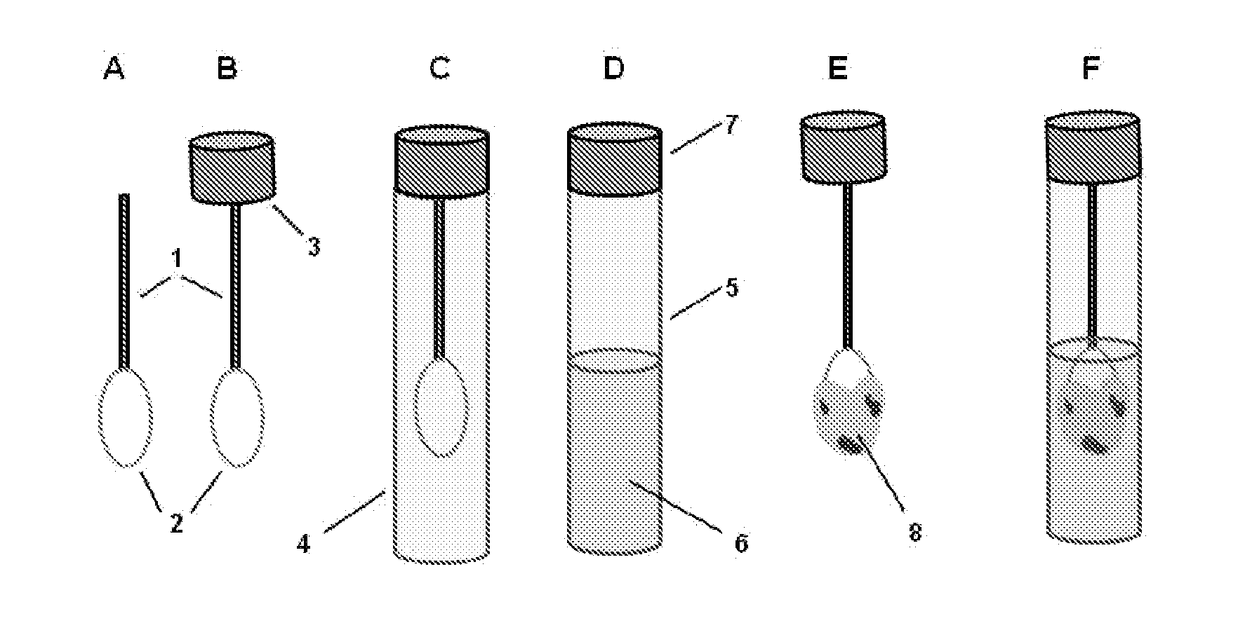
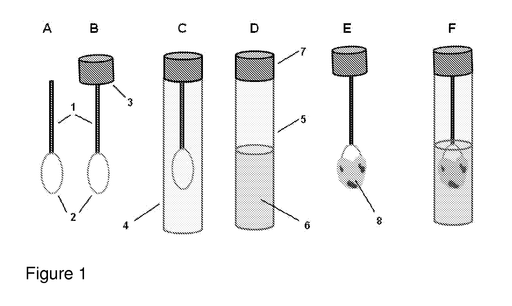
However, as discussed above, there are at present no methods for reliably sampling the mucocellular layer using non-invasive techniques.
Therefore, the most challenging task in testing the mucocellular layer is to devise a non-invasive, simple and inexpensive technique for its sampling and analysis.
