
Angiographic examination method

a technology of angiography and angiography, which is applied in the field of angiographic examination methods, can solve the problems of low contrast attenuation values in the brain tissue, sensitivity to noise, and inability to fast acquire protocols in the vast majority of interventional workplaces, so as to reduce the noise level and reasonable computing time

Inactive Publication Date: 2014-05-08
SIEMENS AG
View PDF1 Cites 12 Cited by
  • Summary
  • Abstract
  • Description
  • Claims
  • Application Information

AI Technical Summary

Benefits of technology

This patent is for a way to make angiographic examinations faster and quieter. The method uses slow-speed rotation and low-noise levels to create better contrast intensity curves. This allows for quicker and more accurate results.

Problems solved by technology

The present problem is the reconstruction of TACs (time attenuation curves), which describe the flow of the contrast medium in the tissue and blood vessels of the brain, from acquisitions with a slowly rotating C-arm angiograph system.
Moreover, the contrast attenuation values in the brain tissue are low and are therefore sensitive to noise.
However, such fast acquisition protocols are not possible in the vast majority of interventional workplaces.
Also, the adoption of constant hemodynamic behavior between the interleaved acquisitions is, however, not guaranteed in reality.
Although these algorithms can be implemented for similarly classical algebraic reconstruction techniques, the use of basis functions with non-compact support renders additional, expensive, CPU-intensive steps necessary.
However, the accuracy of the interpolation is limited by the low number of sampling points that are acquired during a perfusion scanning sequence with the C-arm.
This is imaged on the basis of a rear-projection regulation that is dependent on the projection regulation, such that defective voxel data is generated.

Method used


Image

  • Angiographic examination method
  • Angiographic examination method
  • Angiographic examination method

Examples


Embodiment Construction

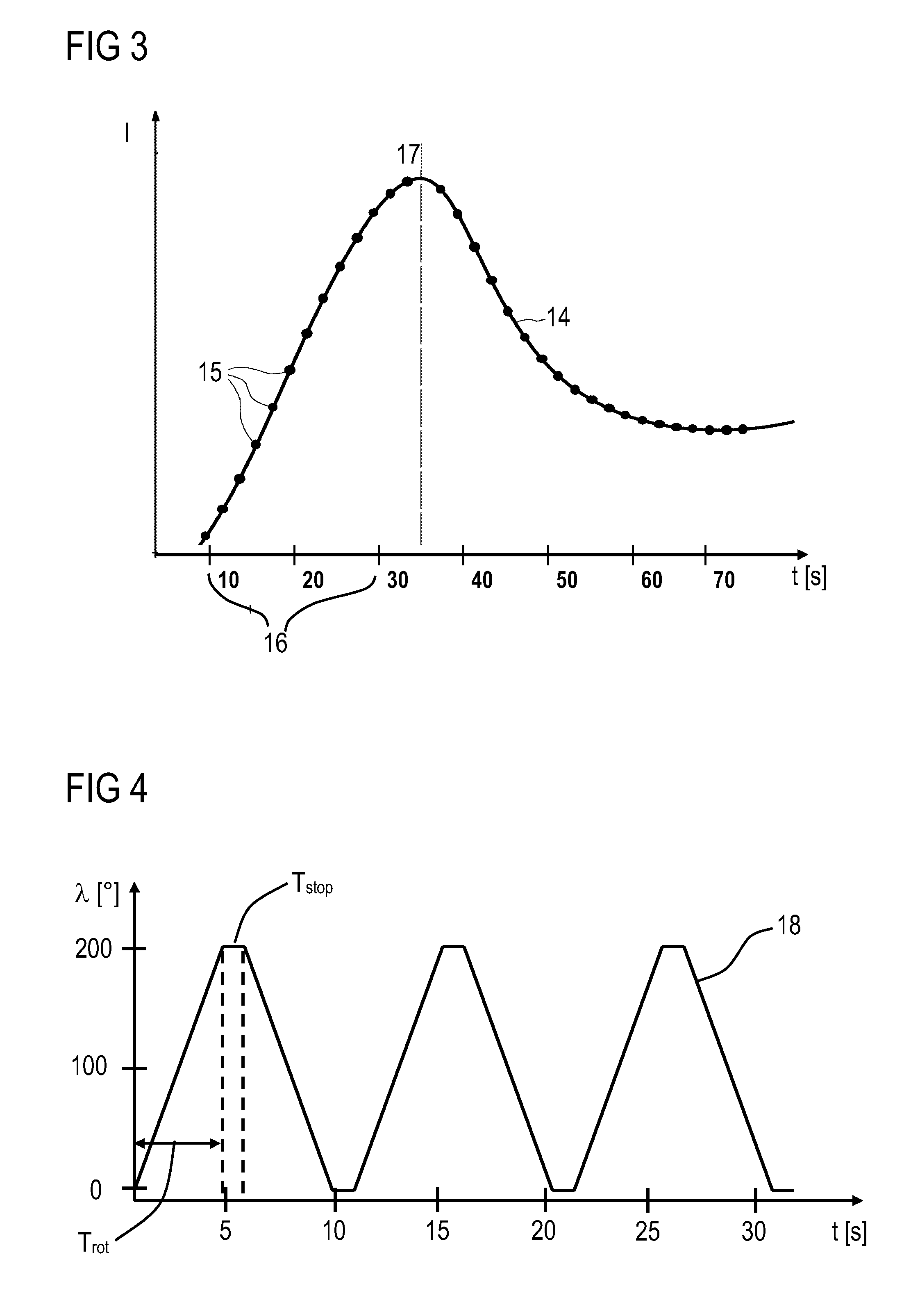
[0043]An acquisition protocol 18 for perfusion by means of a C-arm is described in FIG. 4, which can serve for data acquisition. Since the known C-arm systems enable continuous rotation in only one direction, the C-arm is rotated bidirectionally forwards and backwards. The first C-arm rotation in a forward and backward direction acquires basis projections with the static, anatomical structures—a so-called mask. During each rotation, Nproj=248 projections are acquired along an angular region of approx. 200°. After a contrast agent has been injected, the C-arm is rotated approx. Nrot=7 times bidirectionally, as is shown in FIG. 4. Each rotation lasts Trot=4.3 seconds, with a pause of Tstop=1.2 seconds between two successive rotations.

[0044]Then a direct reconstruction of the rotations would allow a temporal sampling of TACs with a duration of Ts=Trot+Tstop=5.5 seconds over a total scanning time of Tscan=Nrot*Trot+(Nrot−1)*Tstop=37.3 seconds. The basis projections are subtracted from t...


PUM


Abstract

An angiographic examination method for examining an organ, vascular system or other regions of a patient is proposed. Projection images are acquired by an angiography system having an X-ray tube assembly and an X-ray image detector applied to ends of a C-arm, a patient table having a tabletop for carrying the patient, a system control unit, an imaging system and a monitor. Projection images are generated by rotational angiography from a plurality of projection angles. The projection images are subjected to a pre-processing of an FDK reconstruction, the result of which is filtered by a noise-reduction method. A predetermined number of dynamic, iterative reconstruction steps is carried out. Time attenuation curves are reconstructed, which are modeled with a weighted sum of linear basis functions.

Description

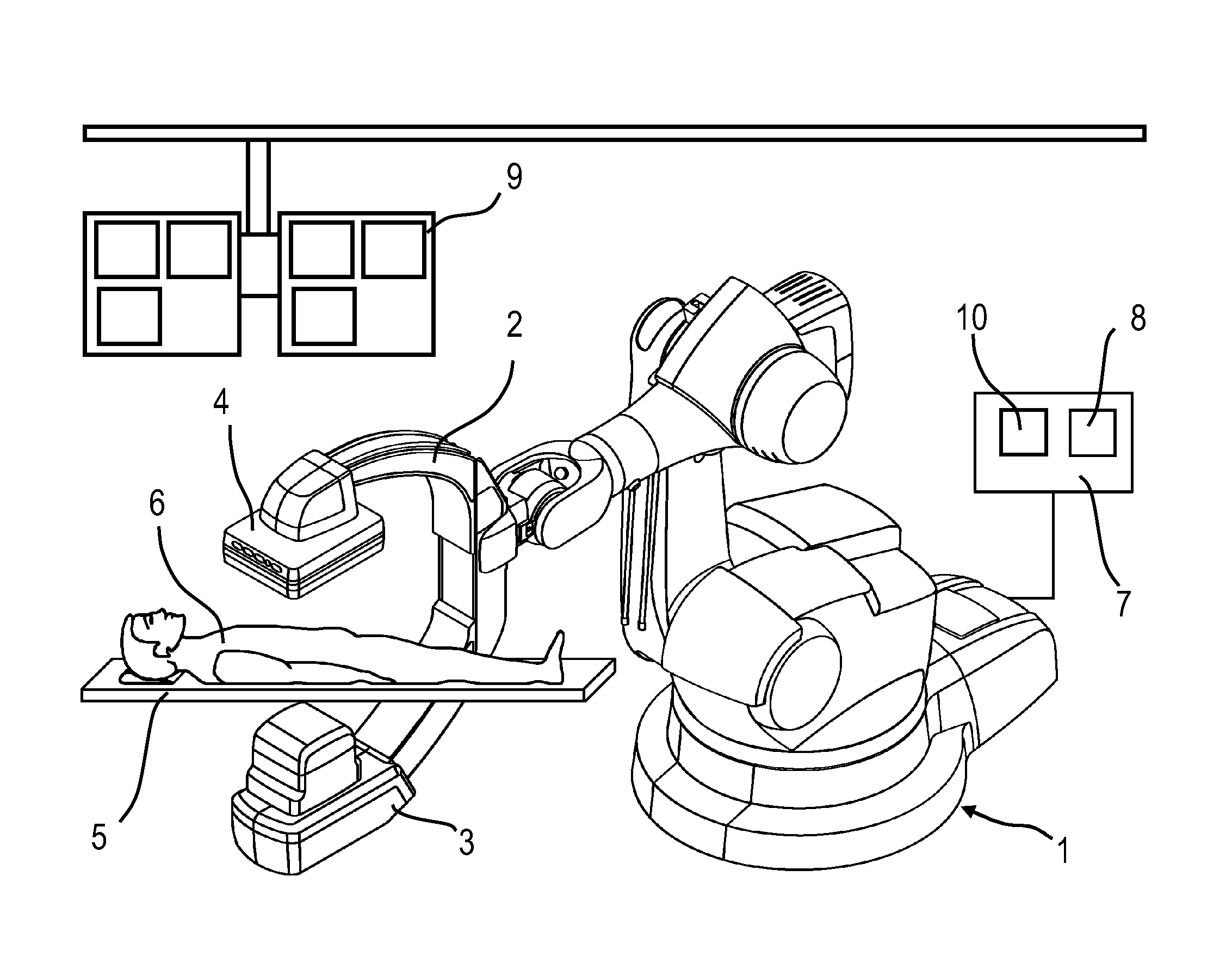
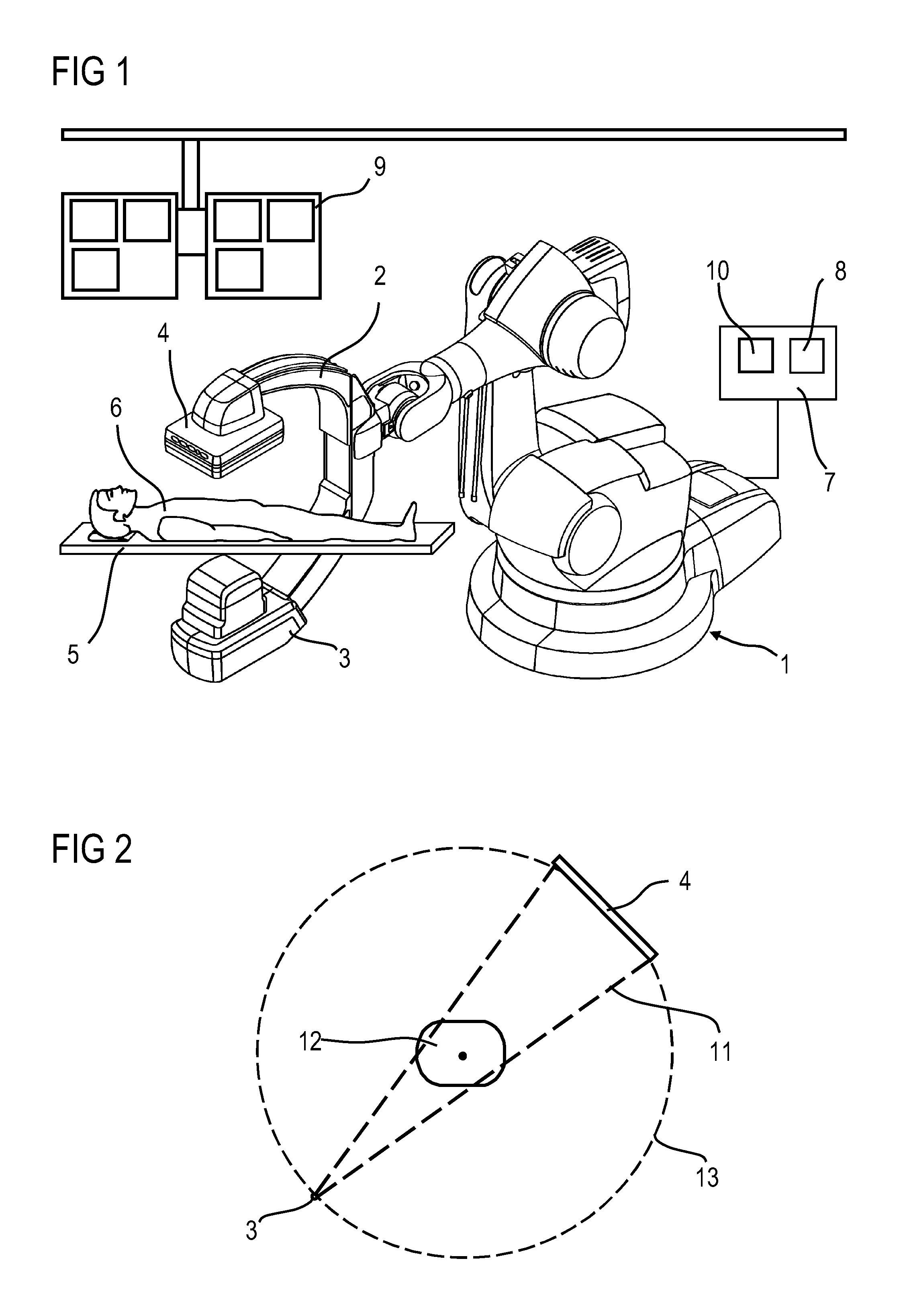
CROSS REFERENCE TO RELATED APPLICATIONS[0001]This application claims benefit of provisional patent application 61 / 722,916 filed on Nov. 6, 2012, the entire content of which is hereby incorporated by reference. This application also claims priority to German application No. 10 2012 220 028.2 DE filed Nov. 2, 2012, the entire content of which is hereby incorporated herein by reference.FIELD OF INVENTION[0002]The invention relates to an angiographic examination method of an organ, vascular system or other regions of the body as an examination object of a patient by means of an angiography system having an X-ray emitter, an X-ray image detector, which are applied to the ends of a C-arm, a patient table having a tabletop for carrying the patient, a system control unit, an imaging system and a monitor, wherein projection images are generated by means of rotational angiography from a plurality of projection angles.BACKGROUND OF INVENTION[0003]An angiography system for carrying out such an ...

Claims


Application Information

Patent Timeline
Patent Type & Authority Applications(United States)
IPC IPC(8): A61B5/00
CPCA61B5/0033A61B6/4441A61B6/4458A61B6/4464A61B6/481A61B6/487A61B6/504A61B6/507G06T11/006G06T2211/404G06T2211/424
Inventor DEUERLING-ZHENG, YUMANHART, MICHAEL
Owner SIEMENS AG
Learn More