
Bioengineered human trabecular meshwork for biological applications

Active Publication Date: 2014-02-06
THE RES FOUND OF STATE UNIV OF NEW YORK
View PDF0 Cites 6 Cited by
  • Summary
  • Abstract
  • Description
  • Claims
  • Application Information

AI Technical Summary

Benefits of technology

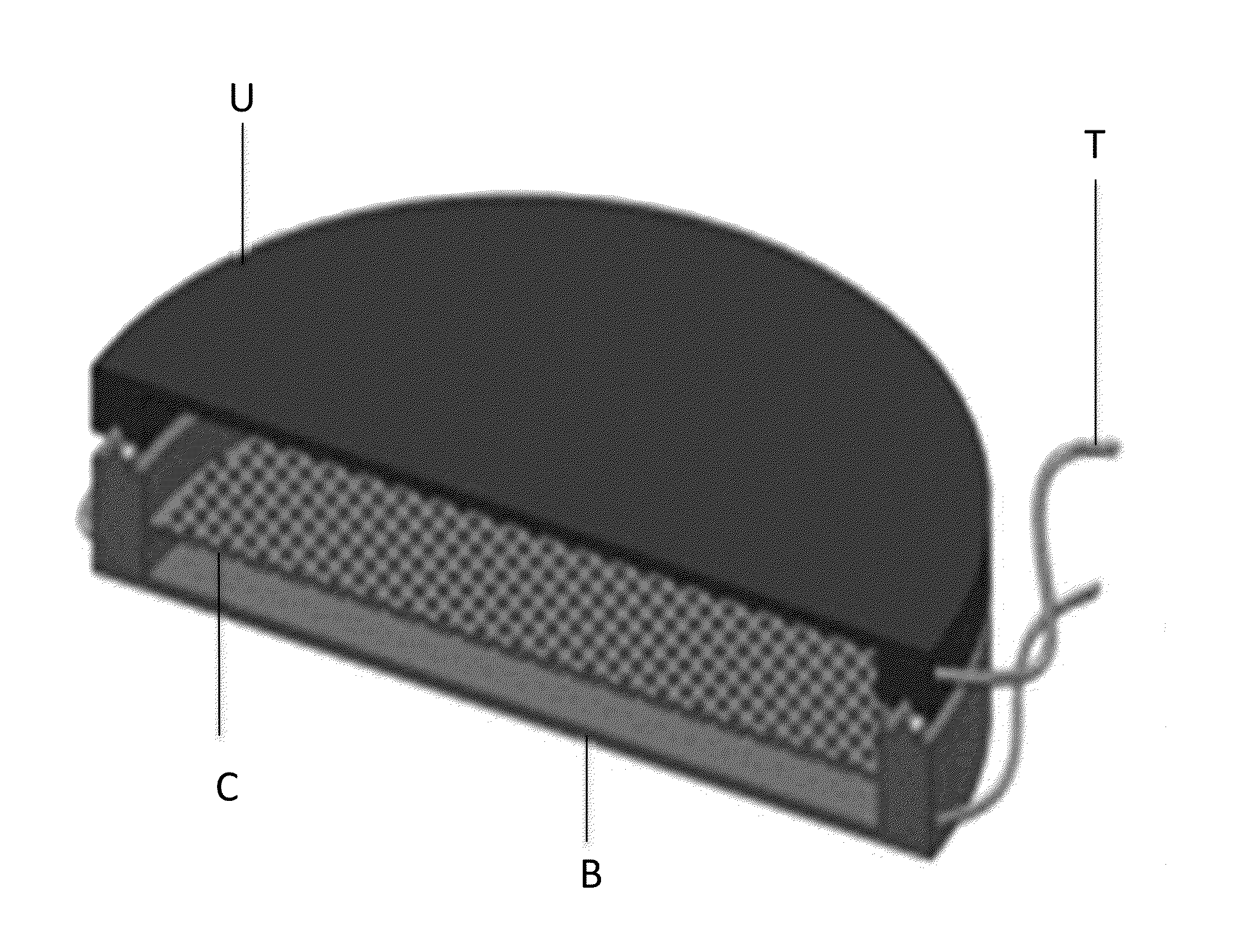
The present invention provides a method of bioengineering a trabecular meshwork by creating a micro- and nanofabricated scaffold with a pore structure that mimics the in vivo structure of the HTM. The scaffold is designed to allow cells to grow and sent out cellular processes in a vertical direction. The scaffold is then coated with a biocompatible coating and used to culture and form a confluent layer of HTM cells. This resulting in a three-dimensional porous trabecular meshwork that can be used for screening and therapy of the outflow facility.

Problems solved by technology

High pressure may damage the sensitive optic nerve and result in vision loss.
In the majority of cases of open angle glaucoma, the eye's TM drainage system becomes “clogged” so the aqueous humor cannot drain efficiently.
As IOP builds up within the eye, it damages the nerve fibers and they begin to die.
If the IOP remains too high for an individual, for too long, then it could lead to optic nerve damage and result in vision loss.
Further advances in pharmacological treatment of glaucoma are currently limited by the lack of proper, efficient, in vitro models for the screening of new potential therapeutics.
While conventional HTM cell cultures may be useful for studying the biology of HTM cells, they are not suitable for evaluating the effects of medications on outflow facility (see, e.g., Koga et al., Exp Eye Res 82:362-70, 2006; incorporated herein in entirety).
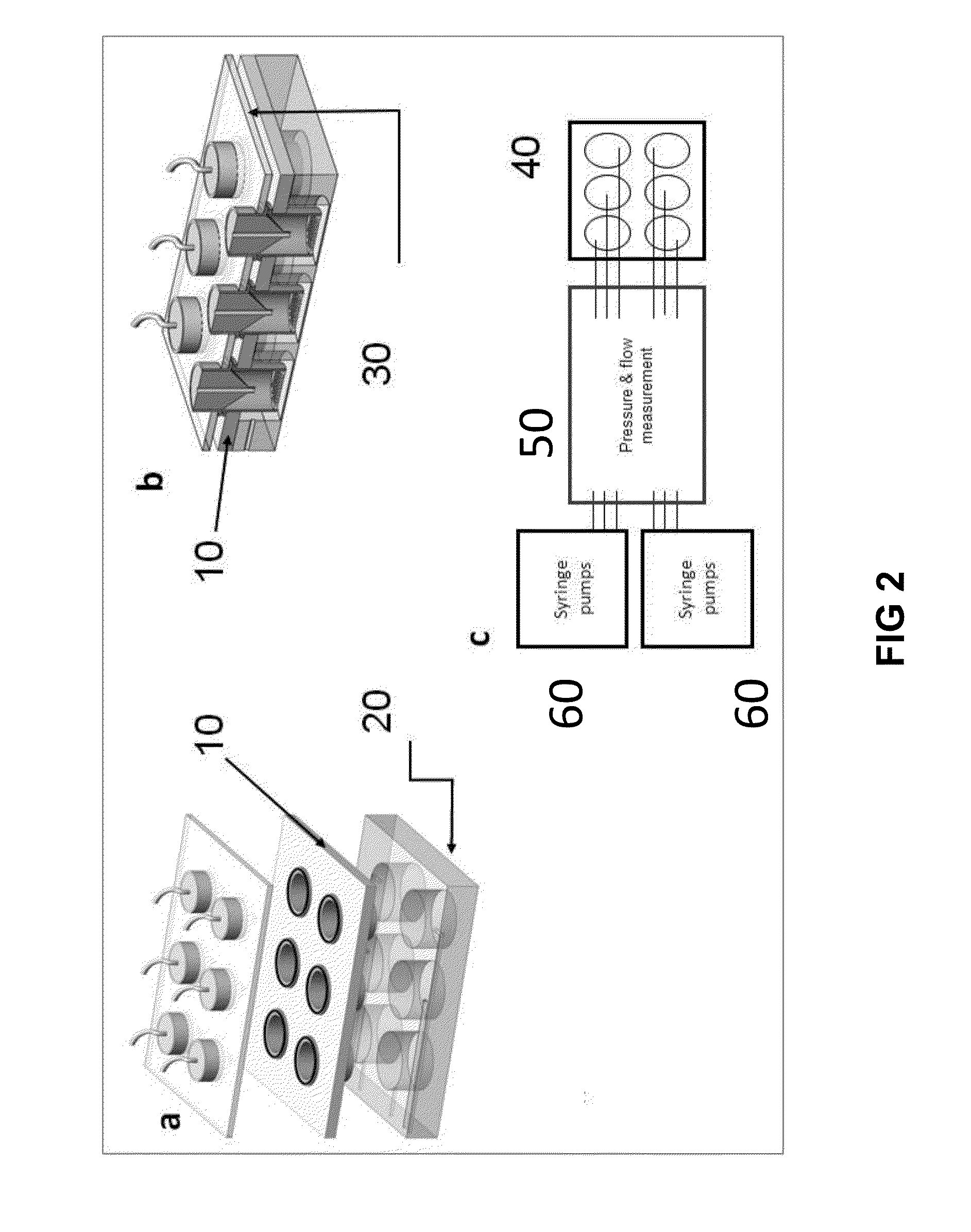
Current outflow facility studies mainly rely on anterior segments of animal or human eyes (see, e.g., Ethier et al., Invest Ophthalmol V is Sci 47:1991-8, 2006; incorporated herein in its entirety); however, the preparation of these perfusion systems is cumbersome, expensive, and not suitable for high-throughput screening.
Commercially available, porous membrane was tried for HTM cell culture, but with limited success (see, e.g., Perkin et al., Invest Ophthalmol V is Sci 29:1836-1846, 1988; incorporated herein in their entirety).
This is because these membranes either possess irregular pore structure or low porosity (e.g., 4-20%), which limits their performance for HTM cell growth and usefulness in perfusion experiments.
In addition these membranes have not been adapted, suggested or used for high throughput screening of medications.
However, this system does not contain HTM cells, has not been used for study of pharmacologic agents, and would be difficult and very expensive to integrate in a high throughput system.
In particular, a hydrogel-based system is not ideal for regulating flow and studying flow physiology at a stable condition.
Since this response is variable, surgical outcomes are often unpredictable.
This remodeling ultimately leads to failure of surgery by limiting the area of diffusion of the aqueous thus increasing resistance to flow.
Its long-term failure is again the result of tissue remodeling of the plate capsule to make it progressively thicker and less permeable.
Attempts to create such bypass with zero resistance to flow have in the long-term generally been unsuccessful to date.
In addition, since such attempts rely on complete bypass of the TM / SC complex, they can cause early hypotony, which can lead to significant vision-threatening complications.
In addition, if the surgery is effective, the regulatory function of the TM / SC complex is lost.
Prior attempts to create a device that would utilize acellular micropatterned structures (see, e.g., Helies et al., J Fr Ophtalmol 21:351-60, 1998; Pan et al., Proc 28th IEEE EMBS Ann Int Conf, 2006) to act as HTM have failed to result in clinically useful devices because they rely on passive regulation of flow afforded by very narrow channels that over time get occluded by cells and debris in the aqueous humor in vivo, as well as by the fact that as with conventional glaucoma surgery, they shunt aqueous humor to the subconjunctival space, thus inducing a fibrotic response.

Method used


Image

  • Bioengineered human trabecular meshwork for biological applications
  • Bioengineered human trabecular meshwork for biological applications
  • Bioengineered human trabecular meshwork for biological applications

Examples


example 1

Human Trabecular Meshwork Cell Culture

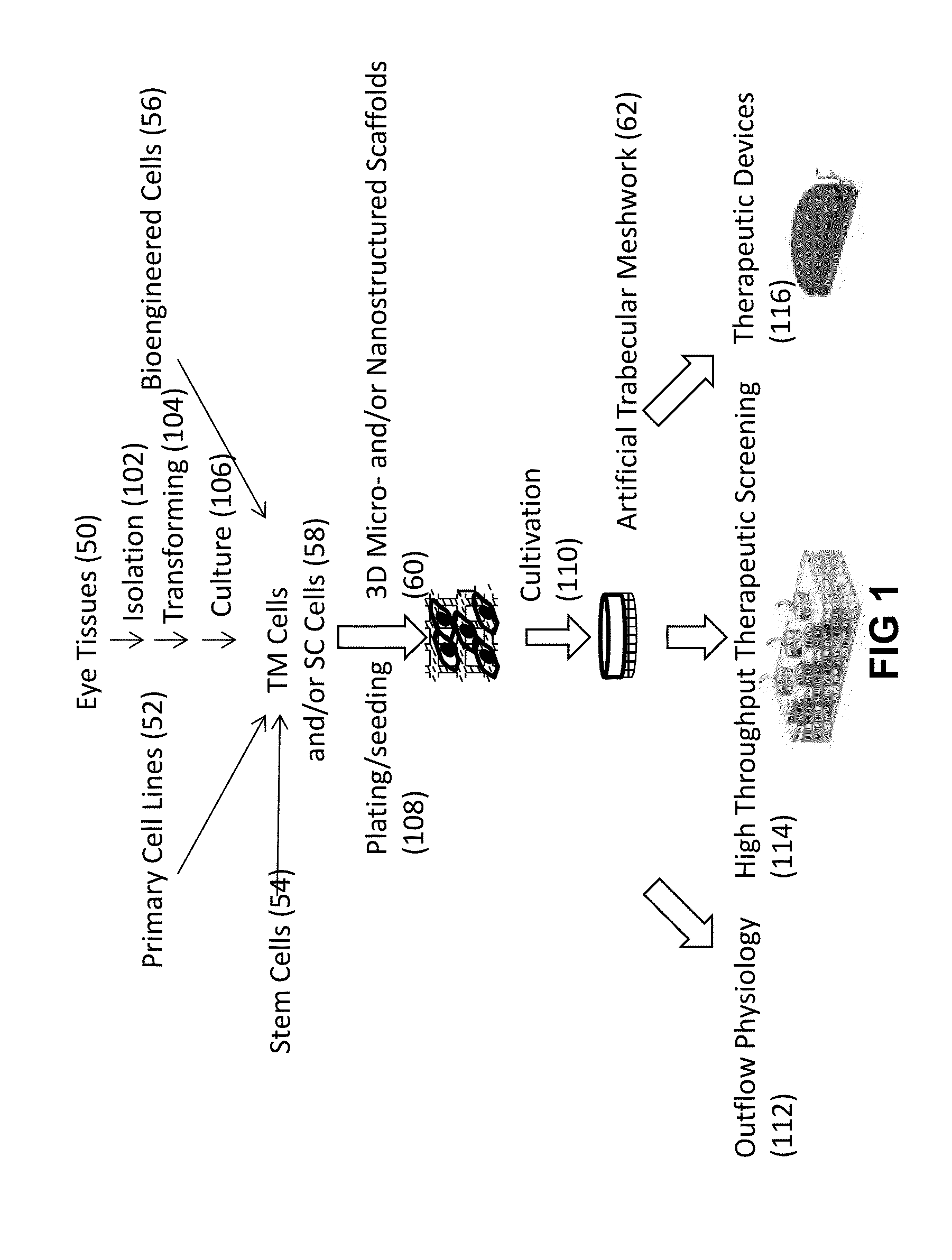
[0112]1. Primary Human Trabecular Meshwork (HTM) cells were purchased (from ScienCell Research Laboratories, Carlsbad, Calif.); or isolated from the juxtacanalicular and corneoscleral region of donor human eye tissue.

[0113]2. The HTM cells were plated in flasks, glass coverslips, and micropatterned 3-D micro- and nanostructured scaffolds coated with HTM biocompatible agents such as poly-L-lysine, gelatin etc., and cell proliferation compared; the HTM biocompatible coating which provided the optimal HTM cell proliferation was selected.

[0114]2.a. The HTM cells were plated in poly-L-lysine-coated 75 cm2 cell culture flasks (2 μg poly-L-lysine / cm2) and cultured in Improved MEM (Cellgro, Manassas, Va.) with 10% fetal bovine serum (ScienCell Research Laboratories, Carlsbad, Calif.).

or

[0115]2.b. The HTM cells were plated in gelatin-coated (75 cm2 cell culture flasks (1% sterile gelatin solution) and cultured in Improved MEM (Cellgro, Manassas, Va.) wit...

example 2

3-D Micro- and Nanostructured Scaffold Fabrication

[0123]To produce the scaffolds with varying dimensions of micro- and / or nanostructures, using standard photolithography techniques shown in FIG. 4, a 3-D micro- and nanostructured scaffold with an architecture (FIG. 3A; 3B; 3C; 9) very similar to the specific features of the TM observed in vivo. 1. A silicon wafer (FIG. 4; 2) was cleaned using Piranha (3:1 H2SO4:H2O2).

[0124]2. Rinsed with dionized water.

[0125]3. Then dried with nitrogen.

[0126]4. A release layer (FIG. 4; 1) was then spun on the wafer at 3000 rpm using a spin coater. In this example, Omnicoat™ treatment was used on previously cleaned silica wafer for the release layer (FIG. 4; a).

[0127]5. Baked on a hot plate at 200° C. for 1 min.

[0128]6. A substrate (in this example SU-8 2010) was applied by spin-coating to a final thickness of approximately 20 μm, (FIG. 4; b).

[0129]7. Then baked at 95° C. for 10 min.

[0130]8. Cooled to room temperature.

[0131]9. A resist is thus produc...

example 3

Culture of HTM Cells on SU-8 Scaffolds

[0140]1. SU-8 3-D micro- and nanostructured scaffolds were coated by soaking for 30 minutes in a HTM biocompatible coating agent (for example poly-L-lysine or gelatin) to promote HTM cell attachment.

[0141]2. Coated SU-8 3-D micro- and nanostructured scaffolds were removed and allowed to air-dry overnight in a sterile environment like a sterile tissue culture hood.

[0142]3. SU-8 3-D micro- and nanostructured scaffolds were allowed to rest at the bottom of a 24-well plate while preventing direct contact between 3-D micro- and nanostructured scaffold and the bottom of the 24-well plate. For the present invention, a structure was designed and constructed for preventing direct contact between 3-D micro- and nanostructured scaffold and the bottom of a cell culture plate. Aluminum tape rings were cut, autoclaved and placed around the borders of the previously sterilized scaffolds. These tape rings allowed the 3-D micro- and nanostructured scaffolds to r...


PUM


Abstract

The present invention relates to methods of manufacture and utility of an artificial trabecular meshwork [TM] that utilizes micro- and nanofabricated materials bioengineered to mimic the structure and function of native outflow system of the eye.

Description

CROSS REFERENCE TO RELATED APPLICATIONS[0001]This application claims priority to U.S. Provisional Patent Application Ser. No. 61 / 600,988 filed on Feb. 20, 2012, the entire contents of which are incorporated herein by reference.FIELD OF STUDY[0002]This invention is in the field of bioengineering. More particularly, the present invention is directed to bioengineering complex 3-dimensional (3-D) micro- and nanostructured scaffolds which after appropriate modifications are seeded with multiple live human, animal or bioengineered cells to produce functional units that closely resemble the physiology of the outflow pathways of the eye. Particular embodiments involve configurations that can be used with a 3-D cell culture chamber for high-throughput screening of medications for their effect on intraocular pressure (IOP) as well as configurations that can be used as therapeutic devices in glaucoma.BRIEF DESCRIPTION OF THE BACKGROUND OF THE INVENTION[0003]The field of bioengineering combines...

Claims


Application Information

Patent Timeline
IPC IPC(8): G01N33/483A61F9/007
CPCA61F9/00781G01N33/4833C12N5/0621C12N2503/02C12N2513/00C12N2533/30C12N2533/32C12N2533/54
Inventor BERGKVIST, MAGNUSBRENNER, SARA ANNDANIAS, IOANNISSHARFSTEIN, SUSAN TINAXIE, YUBING
Owner THE RES FOUND OF STATE UNIV OF NEW YORK
Learn More