
Computed-tomography system and method for determining volume information for a body

a computed tomography and body technology, applied in the field of computed tomography system, can solve problems such as inability, and achieve the effect of reducing memory requirements

Active Publication Date: 2013-05-23
SIEMENS HEALTHCARE GMBH
View PDF10 Cites 7 Cited by
  • Summary
  • Abstract
  • Description
  • Claims
  • Application Information

AI Technical Summary

Benefits of technology

The patent describes a method for producing tomograms that have significantly fewer image artifacts than traditional methods. The method uses a combination of gradient-based optimizing algorithms, such as the Newton and Gauss-Newton methods, to create tomograms with minimal distortion. Additionally, the method takes advantage of the fact that tomograms can be computed based on projected images of a body that was not significantly moved during the recording process. This allows for simultaneous computing of voxel data for different voxels, resulting in more efficient data processing.

Problems solved by technology

Hence, with that method it is not possible, for example, to show functionally, which is to say dynamically, how a contrast medium spreads along a patient's blood vessels.

Method used


Image

  • Computed-tomography system and method for determining volume information for a body
  • Computed-tomography system and method for determining volume information for a body
  • Computed-tomography system and method for determining volume information for a body

Examples


Embodiment Construction

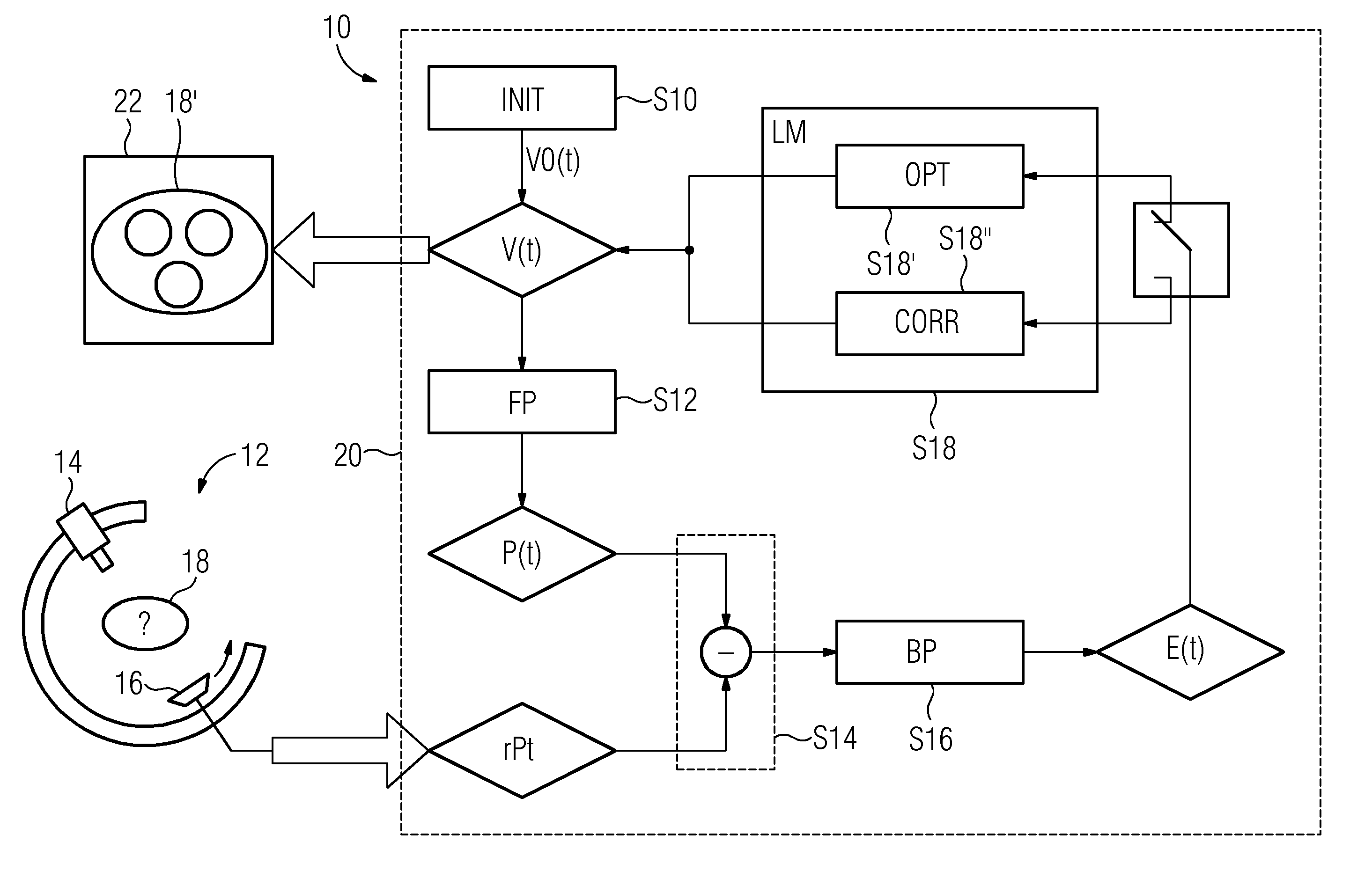
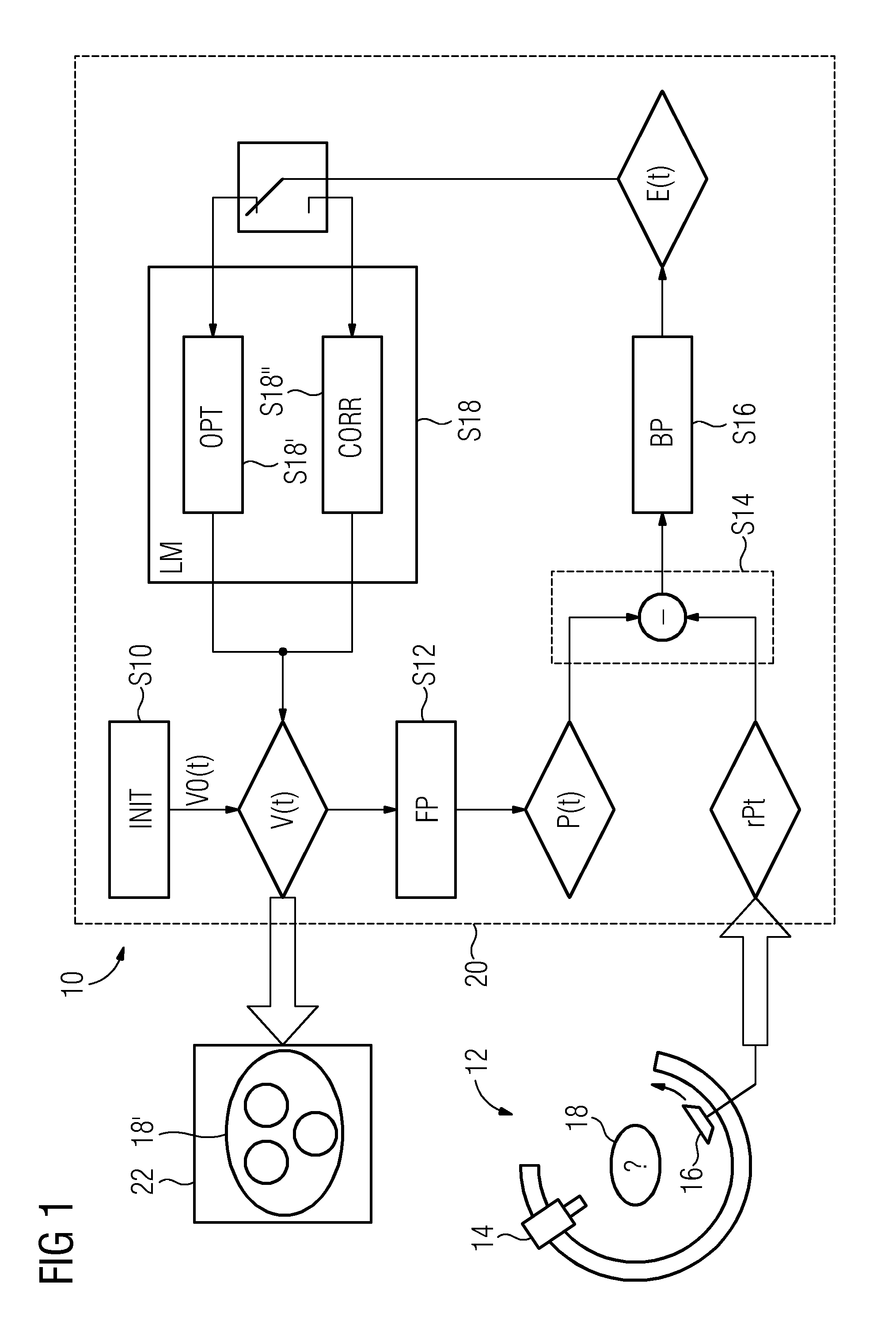
[0031]FIG. 1 is a flowchart relating to a method 10 for computing time-dependent voxel data V(t) from predefined real projection-image data rP(t). Real projection-image data rP(t) may have been obtained by, for example, a C-arm CAT scanner 12. An x-ray source 14 of a C-arm CAT scanner 12 may have been rotated along with an x-ray detector 16 around a human patient's body 18. The intensity values—registered by x-ray detector 16—of individual pixels of a sensor of x-ray detector 16 are combined in a numeric vector forming real projection-image data rP(t). A sequence of sectional images 18′, that will be displayed on a screen 22, of CAT scanner 12 is then computed for the patient's body 18 by a data-processing device 20 of CAT scanner 12.

[0032]Real projection-image data rP(t) may have been obtained, for example, in a recording cycle of eight seconds (8 sec) during which a C-arm was swiveled through an angular range of 200° to 210° with projection images being obtained having a uniform a...


PUM


Abstract

A tomogram of a body is provided. Projection-image data obtained by a radiation-based projection method is used for providing the tomogram. Initial voxel data are first specified for a plurality of voxels of the body. Synthetic projection-image data are generated based upon a projection rule modeling a course of the projection method. Projection-error data is determined by comparing the synthetic projection-image data with the real projection-image data. The projection-error data are imaged on the basis of a back-projection rule dependent on the projection rule so that voxel-error data are produced. Correction data is generated from the voxel-error data by a gradient-based optimizing algorithm, wherein corrected voxel data are generated using the correction data.

Description

CROSS REFERENCE TO RELATED APPLICATIONS[0001]This application claims priority of German Patent Application No. 10 2011 086 771.6 DE filed Nov. 22, 2011. All of the applications are incorporated by reference herein in their entirety.FIELD OF INVENTION[0002]A method for determining volume information for a body is provided, wherein projection-image data are used which were produced by a radiation-based projection method. Further, a computed-tomography system is provided.BACKGROUND OF INVENTION[0003]Image information about a body's internal structure may be determined non-invasively by radiation-based projection methods, for example computed tomography. The body may be the body of a patient, for example, whether human or animal. An inanimate object such as, for instance, a sample of material or a machine, may also be understood here as a body.[0004]To be able to mutually differentiate the body's individual constituents, differences in said constituents are used in regard of their absor...

Claims


Application Information

Patent Timeline
Patent Type & Authority Applications(United States)
IPC IPC(8): G06T7/00
CPCG06T11/003G06T7/0012
Inventor BOESE, JANDEUERLING-ZHENG, YUHEILAND, SABINEWAGNER, MARTIN
Owner SIEMENS HEALTHCARE GMBH
Learn More