
Method for quantifying drug delivery using contrast-enhanced ultrasound

a contrast-enhanced ultrasound and drug delivery technology, applied in the field of quantitative drug delivery using contrast-enhanced ultrasound, can solve the problems of inability to provide fix-quantity drug delivery, inferior sensibility of partial contrast agents, and additional challenges, and achieve high mechanical index, enhance signal intensity, and specific mechanical index of ultrasound

Inactive Publication Date: 2013-05-09
TAIPEI CITY HOSPITAL
View PDF7 Cites 0 Cited by
  • Summary
  • Abstract
  • Description
  • Claims
  • Application Information

AI Technical Summary

Benefits of technology

The present invention provides an instant and quantitative method for delivering drugs to target sites using ultrasound and microbubbles. The method measures the released and unreleased amount of drugs in the tested object with various time intervals and in a non-invasive way. Schedule setting for brain barrier treatment is also provided. The invention uses contrast agent to enhance the signal intensity of ultrasound and measures the variation in microbubble concentration to calculate the drug delivery at the target site. The system includes an ultrasound device, an injection device, and a signal processing device. The invention allows for the real-time monitoring of drug delivery and understanding of pharmacokinetics of drug-loaded microbubbles.

Problems solved by technology

However, in clinical applications, other factors pose additional challenge.
However, all of these have their limitations.
Despite the advantage of high tissue and spatial precision, none rigid pseudo signal and ionizing radiation of MRI, it doesn't allow to be used on individuals with implants such as pacemakers or defibrillators, the sensibility of partial contrast agent is inferior, and cannot provide fix-quantity drug delivery.
These make it difficult to measure vascular sonographic signal accurately, at or near the drug delivery zone.

Method used


Image

  • Method for quantifying drug delivery using contrast-enhanced ultrasound
  • Method for quantifying drug delivery using contrast-enhanced ultrasound
  • Method for quantifying drug delivery using contrast-enhanced ultrasound

Examples


embodiments

Embodiment 1

An Ultrasound Quantifying System for Monitoring Drug Delivery

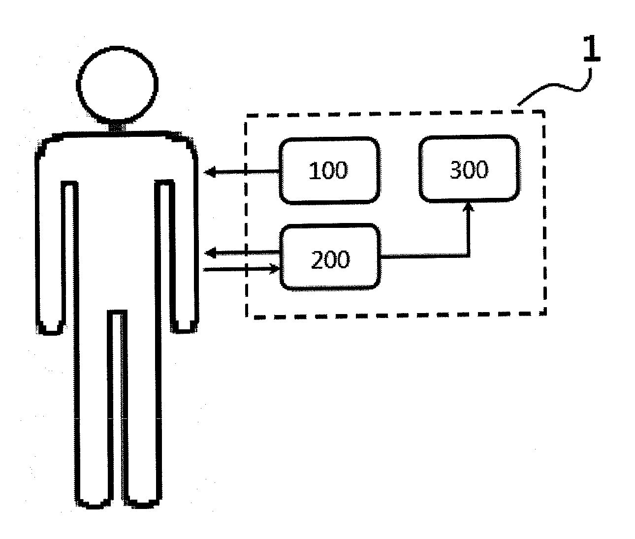
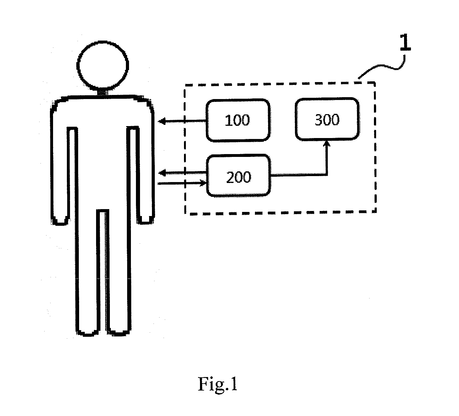
[0026]The ultrasound quantifying system (1) for monitoring drug delivery uses a contrast-enhanced ultrasound system to measure the signal intensity of peripheral vessels in order to quantify the drug delivery at target site. FIG. 1 shows the basic structure of the ultrasound quantifying system (1) of the presentation. Wherein the ultrasound quantifying system (1) includes: an injection device (100) that injects microbubble contrast agent into the tested object via a catheter or an intravenous injection; an ultrasound device (200) that records the variation of ultrasound signal intensity of the peripheral vessels of the tested object; and a signal processing device (300) that processes the ultrasound signal data obtained from the peripheral vessels of the tested object, converts the data into systemic microbubble concentration and thus calculates the drug delivery at the target site.

[0027]The ultrasound device (...

embodiment 2

Ultrasound Signal Saturation Experiment

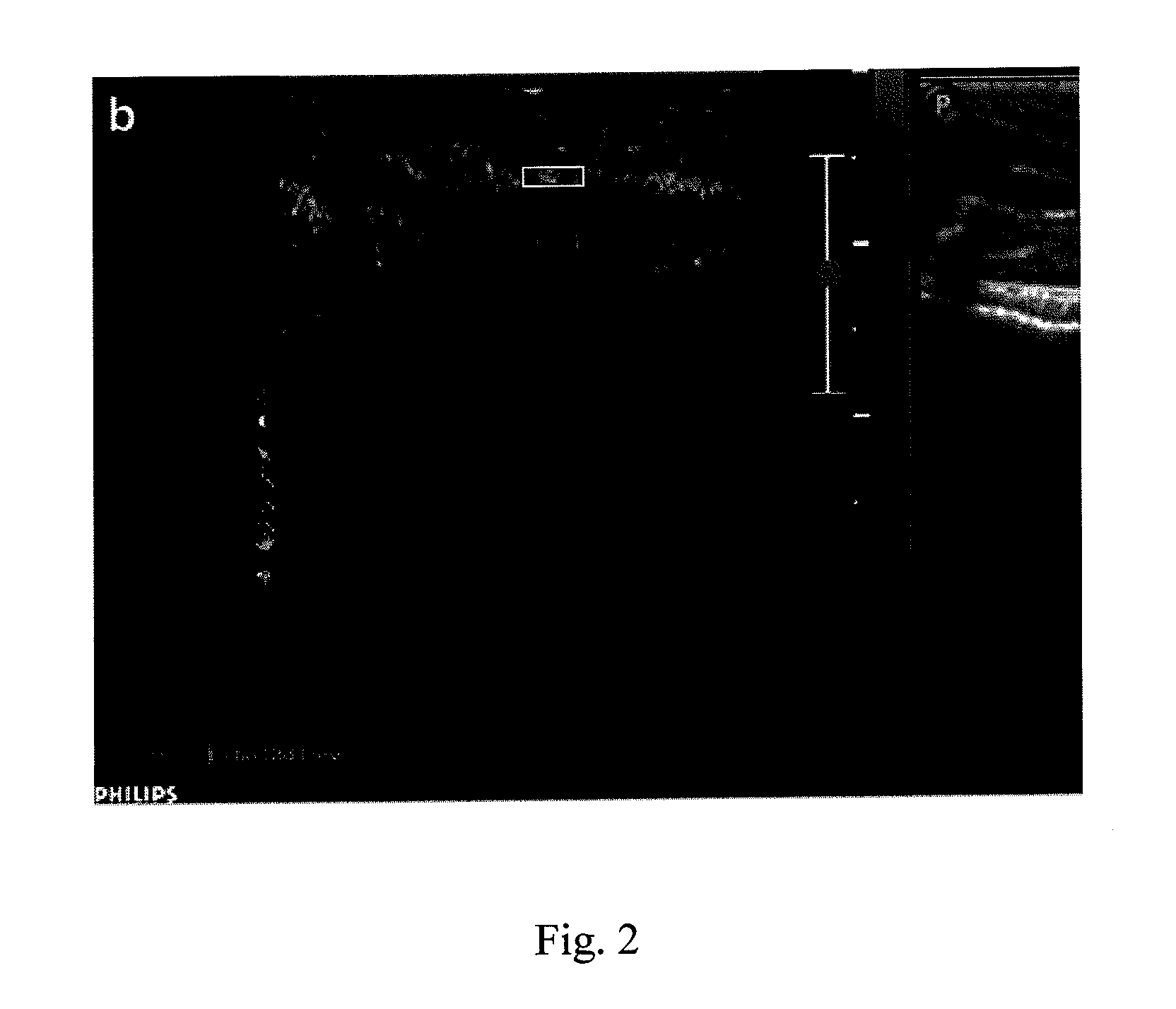
[0031]The rats averaging 330 g were anesthetized intraperitoneally with chloral hydrate of 450 mg / kg. Then the rats were placed in a supine position and the hairs of both things were shaved for ultrasound experiment. A 24-gauge catheter was placed in the tail vein and a three-way stopcock was attached to the end of the catheter with one branch used to administer microbubble contrast agents in the vein bolus and the second for flushing with physiologic saline. The microbubble contrast agents can be anyone of the following substances: ALBUNEX®, SONOZOID®, SONOVUE®, SONOVIST®, OPTISON®, LEVOVIST® or DEFINITY®. In this preferred embodiment, the powder contrast agent SonoVue (Bracco, Milan, Italy) was used in this study. 5 mL of physiologic saline was added to 25 mg of the powder in order to make a microbubble contrast agent suspension of sulfur hexafluoride with the average diameter of 2.5 μm which is considered to be of no difference with saline i...

embodiment 3

Microbubble Destruction Experiment

[0035]In this preferred embodiment, two sets of ultrasound equipments are used. Besides the above-mentioned Philips iU22 ultrasound system (210), another Philips iU22 ultrasound system equipped with a L17-5 linear array transducer (220, frequency range 5˜17 MHz) was used for microbubbles destruction. Wherein, for the first ultrasound system (210), the probe of L12-5 linear array transducer still held orthogonal to the skin and parallel to the long axis of the thigh and the right femoral artery is still chosen for signal measurement. And, for the second ultrasound system (220), the probe of L17-5 linear array transducer was held orthogonal to and in full contact with the skin of the left upper thigh and parallel to the long axis of the thigh. The ultrasound was focused on the adductor muscles of the left hind limb. The transducer position and contact area were the same for each experiment in this preferred embodiment. The experimental setup is shown ...


PUM


Abstract

The present invention relates to a method for quantifying drug delivery with the characteristic of measuring the signal intensity of peripheral vessels with a contrast-enhanced ultrasound to quantify the drug delivery at a target site. The present invention can be used for quantifying UTMD (ultrasound triggered microbubble destruction) targeted drug delivery, also for qualifying the unreleased amount of the long-acting drug in the tested object, and for helping setting the treatment schedule for ultrasound disruption of blood-brain barrier.

Description

BACKGROUND OF THE INVENTION[0001]1. Field of the Invention[0002]The present invention relates to a method for quantifying drug delivery. More specifically, it relates to the method and the application for quantifying microbubble drug delivery using contrast-enhanced ultrasound.[0003]2. Description of the Related Art[0004]Ultrasound-Triggered Microbubble Destruction (UTMD) is a promising strategy to increase local drug concentration at pathologic site while reducing systemic toxicity wherein drug is loaded in micron-sized or nanometer-sized bubbles whose destruction is then triggered by ultrasound at the treatment site in order to deliver drug to the target. However, in clinical applications, other factors pose additional challenge. First, the ultrasound is attenuated to different degrees before they reach the target due to different tissue thickness and composition and tumor structure. Second, the concentration of micro-bubbles in the target area can vary depending on vascular facto...

Claims


Application Information

Patent Timeline
Patent Type & Authority Applications(United States)
IPC IPC(8): A61B8/14
CPCA61B8/085A61B8/0891A61B8/0808A61B8/481A61B8/4477
Inventor HUNG, SHUO-HUICHEN, RAN-CHOUTSAI, TUNG-HU
Owner TAIPEI CITY HOSPITAL
Learn More