
Integrated fiber optic ophthalmic intraocular surgical device with camera

a fiber optic camera and intraocular surgery technology, applied in the field of ophthalmic surgical devices, can solve problems such as difficulty in viewing the internal parts hidden behind the iris

Inactive Publication Date: 2013-03-28
MIRLAY RAM SRIKANTH
View PDF2 Cites 44 Cited by
  • Summary
  • Abstract
  • Description
  • Claims
  • Application Information

AI Technical Summary

Benefits of technology

The patent describes a fiber optic camera assembly that allows a surgeon to see important parts of the eye during surgery. The camera is attached to surgical instruments and can capture still and live images of the eye. The images can be displayed on a TV or monitor and recorded using a VCR. The surgical instruments used in the eye surgery are slim and long, and can bend gently for easy maneuverability inside the eye. Overall, the fiber optic camera assembly provides a unique view of the eye and helps in improving the precision and safety of eye surgery.

Problems solved by technology

But it is difficult to view the internal parts hidden behind the iris, which are opaque in nature.

Method used


Image

  • Integrated fiber optic ophthalmic intraocular surgical device with camera
  • Integrated fiber optic ophthalmic intraocular surgical device with camera
  • Integrated fiber optic ophthalmic intraocular surgical device with camera

Examples


Embodiment Construction

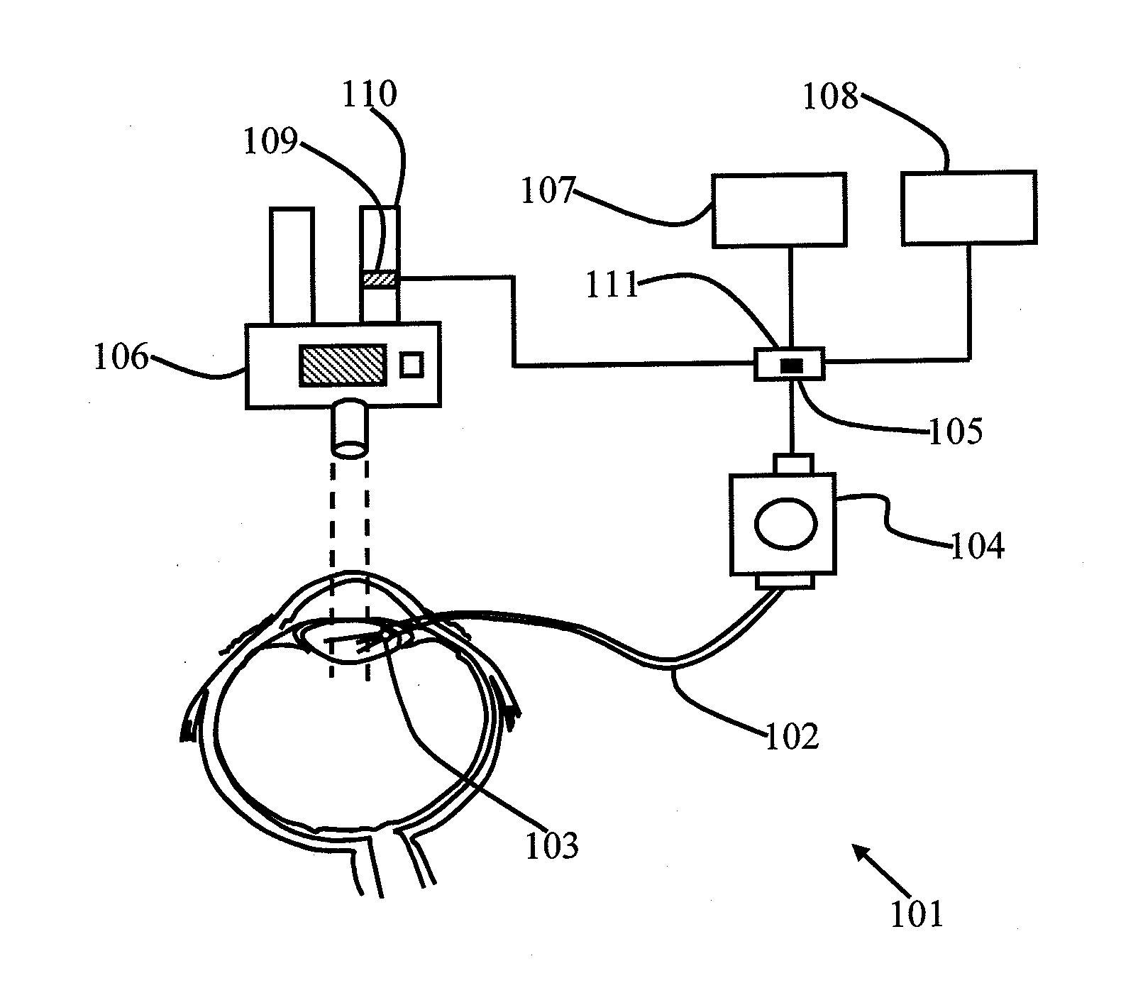
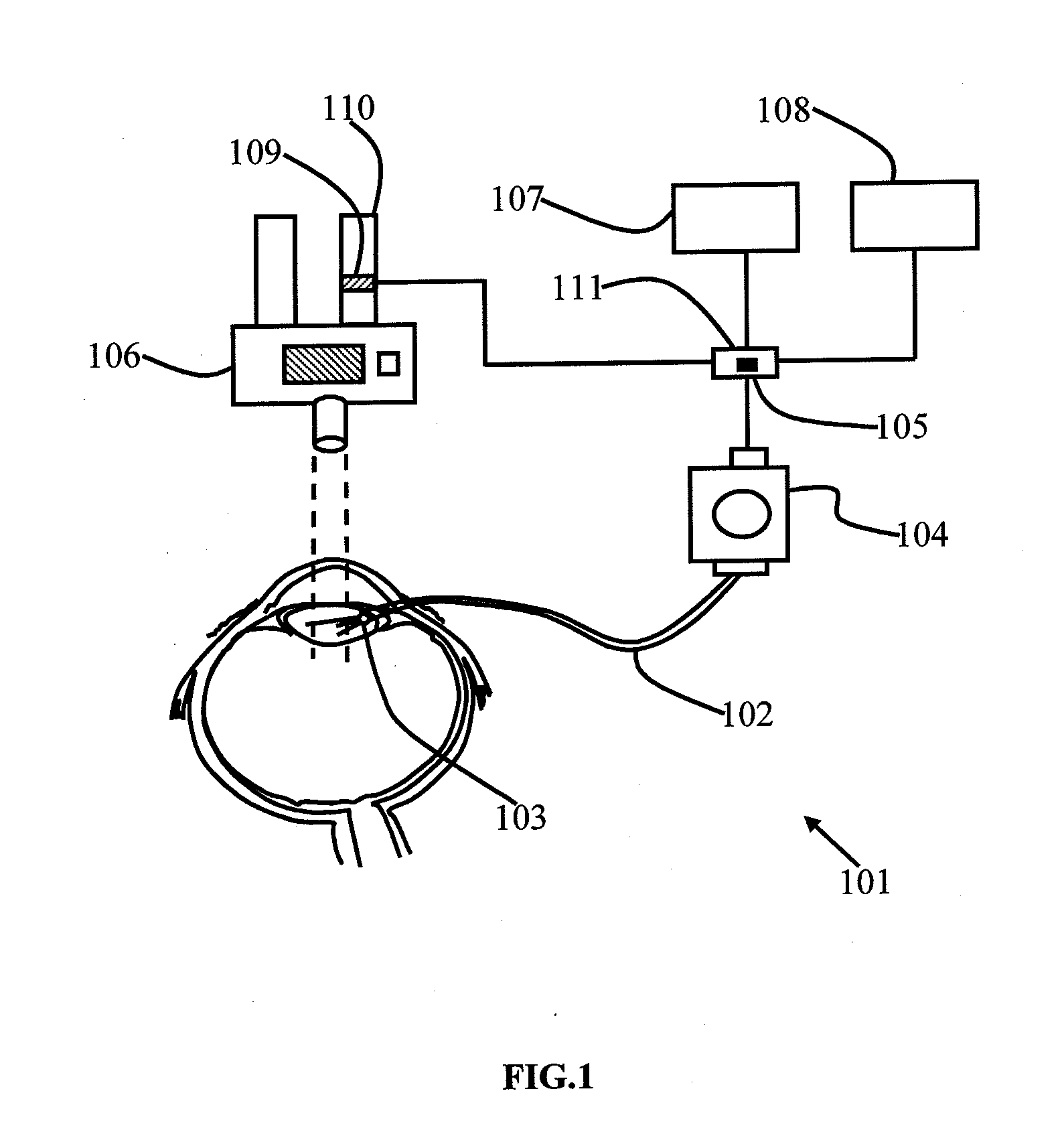
[0009]An objective of the embodiments herein is to provide an ophthalmic surgical microscope with a fiber optic camera to view the essential and vital internal structures of the eye during an eye surgery.

[0010]Another objective of the embodiments herein is to provide an ophthalmic surgical microscope with a fiber optic camera to view of the internal structures, normally hidden behind the iris, which are opaque in nature.

[0011]Yet another objective of the embodiments herein is to provide an ophthalmic surgical microscope with a fiber optic camera having a switch-over mechanism so that the surgeon switches from a fiber optic cable view to a direct microscopic view, or vice versa, while performing an eye surgery.

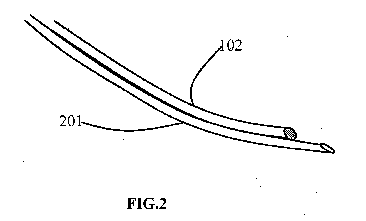
[0012]Yet another objective of the embodiments herein is to provide a fiber optic camera that is slim, long and can bend gently, and provide easy entry and maneuverability inside the eye during surgery.

[0013]Yet another objective of the embodiments herein is to provide a fiber ...


PUM


Abstract

A fiber optic ophthalmic surgical microscope with camera assembly comprises a fiber optic cable (102), a micro lens unit (103), a surgical instrument attached with the fiber optic cable (102), a signal splitter (105), a surgical operating microscope (106), a switch-over mechanism (111) and at least a device for viewing the images. The surgical instrument includes a chopper, a dialer, sinsky's hooks, a manipulator, micro forceps, a coaxial irrigation and aspiration (Infusion Aspiration) canula, bimanual Infusion Aspiration canula or combination thereof. The switch-over mechanism is a button. The button is provided on the signal splitter. The device for viewing the images is selected from a group comprising TV monitor and VCR.

Description

BACKGROUND[0001]1. Technical field[0002]The embodiments herein generally relate to an ophthalmic surgical device and particularly to an ophthalmic microscope with a camera for capturing and viewing still and live images or videos from inside the mammalian eye externally on a TV or monitor or during surgery. The embodiments herein more particularly relate to an ophthalmic surgical microscope with a fiber optic camera.[0003]2. Description of the Related Art[0004]A camera system provides a doctor / surgeon to view the various internal parts of the eye. The field of surgical microscopy encompasses a wide variety of applications, along with the various operating equipment used for these applications.[0005]A surgical operating microscope is a standard tool used in the operating room. The ophthalmic microscope is typically a binocular head that is angled. Video microscopy is also an added feature in a surgical operating microscope. Many of the models have optional beam splitters to split the...

Claims


Application Information

Patent Timeline
IPC IPC(8): A61B3/14A61B90/20
CPCA61B3/145A61B3/0008
Inventor MIRLAY, RAM SRIKANTH
Owner MIRLAY RAM SRIKANTH
Learn More