
Preparation method of antigen-immobilized immuno- fluorescence slide and immuno-fluoroscence slide prepared thereby

a fluorescence slide and immunofluorescence technology, applied in the direction of fluorescence/phosphorescence, instruments, peptides, etc., can solve the problems of high-cost assay instruments, high-sensitivity increase, and variability of evaluation protocols, and achieve easy and highly-sensitive effects

Inactive Publication Date: 2013-01-31
KOREA FOOD RES INST
View PDF1 Cites 29 Cited by
  • Summary
  • Abstract
  • Description
  • Claims
  • Application Information

AI Technical Summary

Benefits of technology

The present invention provides an immunosensor slide that can easily and highly-sensitively measure a biomarker, such as C-reactive protein, which is associated with various health conditions. The slide is coated with C-reactive protein and reacted with an antibody that binds to a fluorescent nanoprobe. This technology can also be used to simultaneously measure other biomarkers, such as low density lipoprotein, fibrinogen, or angiotensin II, which are present in blood at different concentrations. The technical effect of this invention is to provide a reliable and efficient tool for evaluating food function and health.

Problems solved by technology

However, this method causes non-uniform results, and also, requires variability of evaluation protocols, skilled experts, and high-cost assay instruments.
When an in vivo cardiovascular functionality of a food is evaluated by measuring a C-reactive protein as a biomarker present in blood of a lab animal by using a biosensor, an increase in sensor sensitivity is still a critical issue and also, ease of measurement is endlessly pursued.

Method used


Image

  • Preparation method of antigen-immobilized immuno- fluorescence slide and immuno-fluoroscence slide prepared thereby
  • Preparation method of antigen-immobilized immuno- fluorescence slide and immuno-fluoroscence slide prepared thereby
  • Preparation method of antigen-immobilized immuno- fluorescence slide and immuno-fluoroscence slide prepared thereby

Examples


example 1

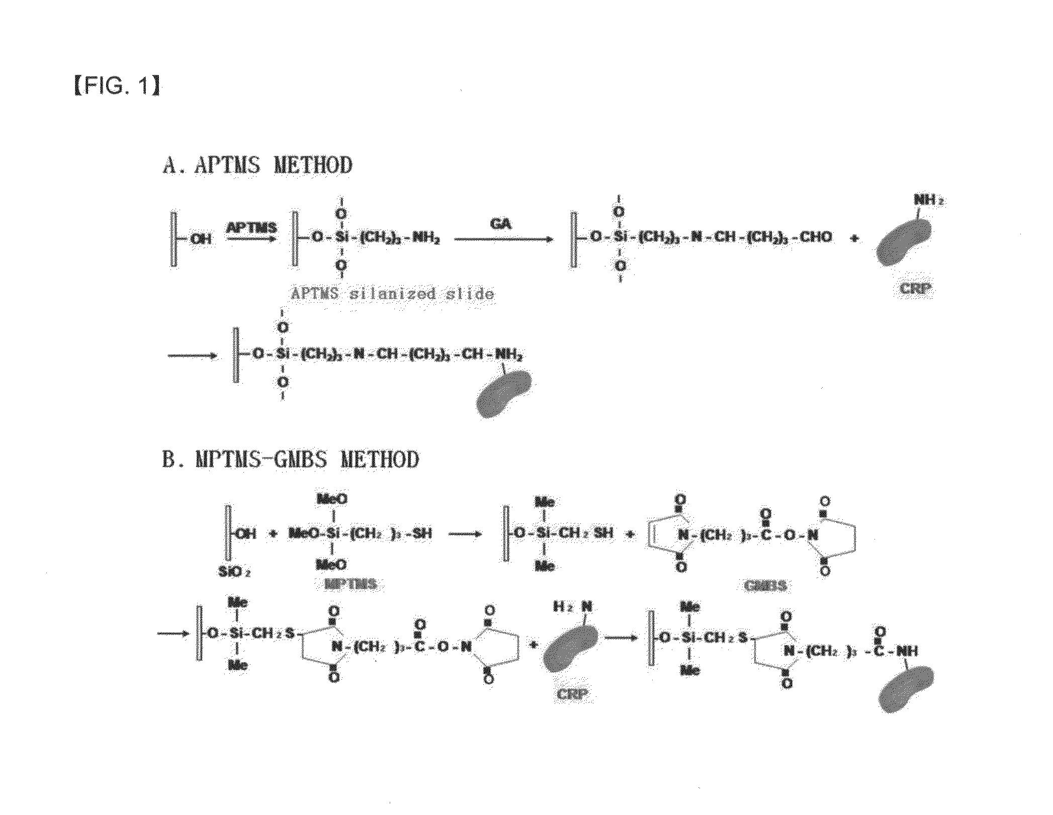
Immobilization of Antigen on Slide Treated with APTMS-GA

[0066]An antigen was immobilized on a slide as follows. That is, for surface cleaning, a slide was immersed in a piranha solution (H2SO4:H2O2=3:1, volumetric ratio) for 10 minutes, washed with distilled water, sonicated in distilled water for 5 minutes, washed by hybridization with hot water having a temperature of 90° C. for 1 hour, placed on kimwipes, and then, dried for 30 minutes at room temperature.

[0067]To perform silanization to introduce an amino group (—NH2), which is needed to immobilize an antigen, to the surface of the slide, the surface-cleaned slide was placed in a petri dish containing an APTMS solution in which 10% APTMS was dissolved in acetone and a reaction was performed at room temperature for 1 hour. Front and rear sides of the slides were sequentially washed with a 50 mM phosphate buffer solution (pH 7.4) and distilled water, three times for each, and then, immersed in a beaker containing distilled water f...

example 2

Immobilization of Antigen on Slide Treated with MTS-GMBS

[0070]Immobilizing of an antigen on a slide was performed through the following processes. That is, for surface cleaning, a slide was immersed in a mixed solution including a strong hydrochloric acid (35%) and methanol (70%) at a volumetric ratio of 1:1 for 30 minutes, and then, the slide was washed three times with distilled water. After 30 minutes of immersion in a strong sulfuric acid, front and rear surfaces of the slide were washed three times with distilled water, and washed with boiled distilled water and placed on kimwipes and dried at room temperature for 30 minutes.

[0071]To perform silanization to introduce a thiol group (—SH), which is a reaction group needed to immobilize an antigen, to the surface of a slide, 2 ml of (3-mercaptopropyl)trimethoxy silane (MPTMS) was added to 98 ml of toluene to prepare a 2% MPTMS solution. The slide was placed on a petri dish containing the solution and a reaction was performed at ro...

example 3

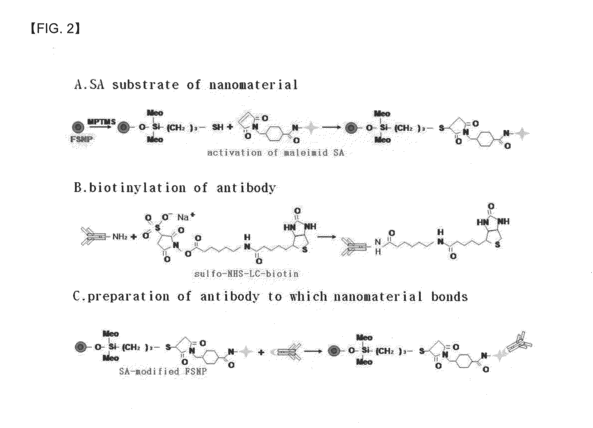
Preparation of FSNP Modified with SA

[0075]2 mg of FSNP and 10 ml of ethanol were added to a cap tube having a capacity of 25 ml and the mixture was shaken well, and then, sonicated to completely dissolve FSNP in ethanol for 15 minutes. Thereafter, 100 ml of MPTMS was added to the cap tube and a reaction was performed at room temperature for 4 hours while stirring at a low sped of 100 rpm. The reaction mixture was centrifuged at a temperature of 4° C. for 30 minutes at a rate of 13000 rpm and the obtained precipitate was washed three times with ethanol, and after each washing, centrifuging was performed under the same conditions as described above to obtain precipitates. The cap of the tube was separated and the tube was lightly covered with an aluminum foil and dried at room temperature. Each of the precipitates was dissolved in 4 ml of 50 mM phosphate buffer solution (pH 7.4) to prepare a thiol-modified FSNP solution.

[0076]0.25 mg of SA-msaleimide and 5 ml of 50 mM phosphate buffer...


PUM

PropertyMeasurementUnit
temperatureaaaaaaaaaa
sizeaaaaaaaaaa
temperatureaaaaaaaaaa

Abstract

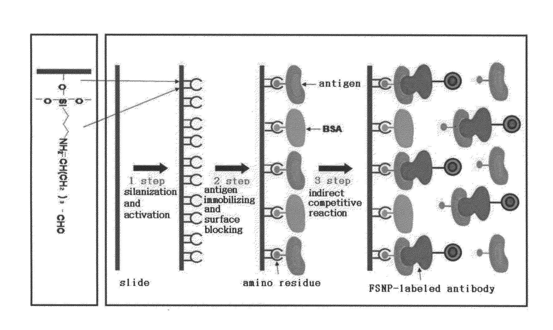
A method of preparing an antigen-immobilized immuno-fluorescence slide, the method comprising: immobilizing a C-reactive protein on a slide to prepare a protein chip; mixing an antibody that specifically binds to a target protein, with streptavidin to label the antibody with a fluorescent nanoparticle; immuno-reacting the antibody by competitive mixing, assaying with a fluorescence camera, wherein the immobilizing of the C-reactive protein on the slide comprises: modifying the slide with 3-aminopropyltrimethoxysilane to prepare a modified slide; hydrating the slide modified with 3-aminopropyltrimethoxysilane; activating the modified slide by using a glutaraldehyde solution; dissolving a C-reactive protein at a concentration of 0.01-0.5 mg / ml in a 30-70 mM phosphate buffer solution (pH 6.5-7.8) to prepare an antigen solution for immobilization; placing a petri dish comprising the slide on a spotting guide and spotting 1-100 μl of the antigen solution on spotting points; and performing a reaction on the slide prepared as described above for 1-6 hours to immobilize the antigen, and an immune-fluorescence slide prepared by using the method.

Description

TECHNICAL FIELD[0001]The present invention relates to a method of preparing an antigen-immobilized immuno-fluorescence slide including: immobilizing a C-reactive protein on a slide to prepare a protein chip, mixing an antibody that specifically binds to a target protein, with streptavidin, to label the antibody with a fluorescent nanoparticle, immune-reacting the antibody by competitive mixing, assaying with a fluorescence camera, and an immuno-fluorescence slide made by using the method.BACKGROUND ART[0002]Biochips include various kinds of probes immobilized on a unit area of a surface of a solid support, and when such biochips are used, only with a small amount of sample, disease diagnosis, high-throughput screening (HTS), and enzyme activity measuring may be easily performed in a large scale.[0003]Most of methods of immobilizing a probe on a slide include immobilizing a probe on a glass slide pre-treated with a coating material. For these methods, various surface chemical materia...

Claims


Application Information

Patent Timeline
Patent Type & Authority Applications(United States)
IPC IPC(8): G01N21/64C07K17/02B82Y15/00
CPCG01N33/54366G01N2333/4737
Inventor KIM, NAM SOOCHO, YONG JINKIM, CHONG TAI
Owner KOREA FOOD RES INST
Learn More