
Systems and methods for measuring translation of target proteins in cells

a target protein and cell technology, applied in the field of systems and methods for measuring the rate of translation of a target protein in cells, can solve the problems of not being suitable for living cells analysis, not being able to provide real-time or dynamic measurements of translation activity, and not being able to correlate well with actual synthesis of the protein

Active Publication Date: 2012-07-19
ANIMA CELL METROLOGY
View PDF2 Cites 6 Cited by
  • Summary
  • Abstract
  • Description
  • Claims
  • Application Information

AI Technical Summary

Benefits of technology

[0027]The present invention advantageously provides readouts of the rate of synthesis of a selected protein of interest in a cell, including that localized to a subcellular compartment of the cell. The present invention can be carried out in any type of cell, including primary cells and cell lines, with relatively mild intervention in the cellular machinery and without requiring single ribosome analysis. The invention can be utilized for numerous applications, including, but not limited to, diagnostic assays, macroscopic assays, microscopic assays, high-throughput screening, cell enrichment and cell sorting, applications in biomanufacturing, and in numerous protein synthesis-related diseases. Measurements of rates of protein translation can be obtained in real time, which is crucial for instantly monitoring responses to various stimuli, particularly changes in environmental conditions, such as temperature, or exposure to specific compounds, such as small molecule drug candidates, biotherapeutic agents, or any other substances which potentially affect protein synthesis. The present invention is thus advantageous for studying the effects of drugs and drug candidates on cells in disease states characterized by aberrations in protein translation, and for studying rates of synthesis of target therapeutic proteins in stem cell and recombinant cell culture systems.
[0101]According to other embodiments, a method of the present invention further comprises the steps of (a) administering to the biological sample a drug candidate; (b) detecting the FRET signals emitted by the biological sample, as described herein; and (c) comparing the FRET signals detected in the presence of the drug candidate, to that detected in the absence of the drug candidate, thereby evaluating the effect of the drug candidate on protein translation.

Problems solved by technology

Currently, collagen is measured with DNA microarrays, which indicate the existence of collagen transcripts, but do not correlate well with actual synthesis of the protein.
Cell assays that quantify production of a single specific protein typically require cell lysis and / or permeabilization, making then unsuitable for analysis of living cells.
Those methods which are suitable for intact cells and organelles, such as metabolic labeling with radioactive tracers, are unable to isolate a signal from a particular transcript or protein of interest, and thus do not provide real-time or dynamic measurements of translation activity.

Method used


Image

  • Systems and methods for measuring translation of target proteins in cells
  • Systems and methods for measuring translation of target proteins in cells
  • Systems and methods for measuring translation of target proteins in cells

Examples


example 1

Labeling Two Parts of the Translational Machinery as a Fret Pair

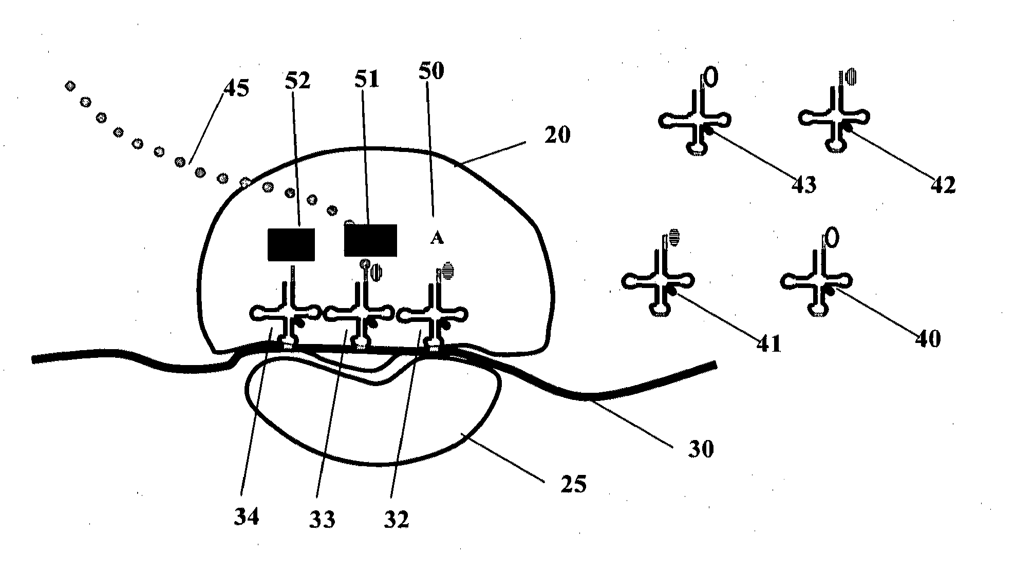
[0393]Two different tRNA molecules are labeled as a FRET pair. For example, some tRNAs (FIG. 1) can be labeled with a donor fluorophore, and others with a corresponding acceptor fluorophore such as Cy3 (excitation and emission peaks are 550 and 570 nm, respectively) and Cy5 (excitation and emission peaks are 650 and 670 nm, respectively); when translation is active, such tRNAs are immobilized in two adjacent sites (A and P or P and E) of the ribosome, thereby producing a FRET pair which produces measurable FRET signals. These signals indicate that the A and P sites are populated with labeled tRNAs. A small signal indicates that a low percentage of the A and P sites are populated, and therefore that the translation apparatus is in a state of low production rates. Additional exemplary, non-limiting FRET combinations are listed in Table 3.

TABLE 3Exemplary FRET combinations.Donor fluorophoreAcceptor fluorophoreCy3Cy5Rhoadmi...

example 2

Introduction of the Labeled tRNAs into CHO Cells

[0398]Labeled tRNAs are transfected into CHO cells using TransMessenger transfection reagent (Qiagen, Hilden, Germany) according to the manufacturer's protocol. Transfected cells are placed under a microscope equipped for single molecule detection (Zeiss, Oberkochen, Germany) with an image acquisition device operable at a sufficient rate (10-100 frames per second), and computational units that can acquire and analyze the resulting images and data (FIG. 2). Translation of the tRNA of interest is measured.

[0399]For high-throughput screening, transfected cells are cultured in a 96-well plate format, compatible with automated screening. A robot administers one compound out of the library being screened into each well and translation detection is performed. A suitable sampling regime is adopted. The effect of the compound on translation activity is detected in comparison with negative control signal.

example 3

Diagnostic Applications

[0400]A selected yeast tRNA is labeled with donor fluorophore and stored. Another selected yeast tRNA is labeled with acceptor fluorophore and stored. Prior to the assay, two aliquots of donor- and acceptor-labeled tRNA are mixed. The mixture is transfected into the cells to be diagnosed, for example, by using the transfection kit RNAiFect™ (Qiagen, Hilden, Germany). The cells may be human cells, for example, human cells obtained from a tissue removed by biopsy. The transfected cells are introduced into a 96 well-plate. Signals are collected from the plates using a fluorescent plate reader and are subjected to computerized analysis / es. Typically, the parameters derived for the analysis are: average signal strength, average signal deviation, or percentage of labeled tRNA of a given species in each well. These parameters are monitored over time and in response to treatment. Values before and / or after treatment are compared to known standards to infer clinical pa...


PUM

PropertyMeasurementUnit
pressureaaaaaaaaaa
wavelengthsaaaaaaaaaa

Abstract

The present invention relates to systems and methods for measuring the rate of translation of a target protein in cells, which are based on the detection of translation of one or more predetermined codon pairs during synthesis of the target protein. The detection is provided by a FRET signal emitted from labeled tRNA molecules which are juxtaposed during synthesis of the protein.

Description

FIELD OF THE INVENTION[0001]The present invention relates to systems and methods for measuring the rate of translation of a target protein in cells, which are based on the detection of translation of one or more predetermined codon pairs during synthesis of the target protein. The detection is provided by a FRET signal emitted from labeled tRNA molecules which are juxtaposed during synthesis of the protein.BACKGROUND OF THE INVENTIONThe Process of Protein Synthesis[0002]Protein synthesis is one of the most central life processes. A protein is formed by the linkage of multiple amino acids via peptide bonds, according to a sequence defined by the template messenger RNA (mRNA). Protein synthesis occurs in the ribosomes, the protein manufacturing plants of every organism and nearly every cell type.[0003]Ribosomes are ribonucleoprotein particles consisting of a small and large subunit. In bacteria these subunits have sedimentation coefficients of 30 and 50, and thus are referred to as “3...

Claims


Application Information

Patent Timeline
Patent Type & Authority Applications(United States)
IPC IPC(8): G01N21/64
CPCG01N33/5023G01N33/542G01N21/6428G01N2201/061
Inventor SMILANSKY, ZEEVCOOPERMAN, BARRY S.GOODMAN, YALE E.
Owner ANIMA CELL METROLOGY
Learn More