
Markers for endometrial cancer

Active Publication Date: 2012-05-17
GEADIC BIOTEC AIE
View PDF3 Cites 3 Cited by
  • Summary
  • Abstract
  • Description
  • Claims
  • Application Information

AI Technical Summary

Benefits of technology

[0064]The methods of the present invention may further comprise providing a uterine fluid sample obtained from a patient with a pipelle device or syringe wherein the patient has a risk factor or symptom of endometrial cancer; contacting said sample with an agent capable of preserving, preventing, or lessening the degradation of RNA in said uterine fluid sample; determining in said sample the expression level of mRNA corresponding to from 1 to 20 herein described markers (preferably 2 to 8 markers) and one or more endogenous genes using quantitative PCR; normalizing the expression level of from 1 to 20 (preferably 2 to 8 markers) herein described biomarkers with the one or more endogenous genes; comparing the normalized level of the from 1 to 20 (preferably 2 to 8 markers) biomarkers to a control value wherein differential expression of from 1 to 20 (preferably 2 to 8 markers) of the biomarkers indicates endometrial cancer or an increased likelihood of endometrial cancer.
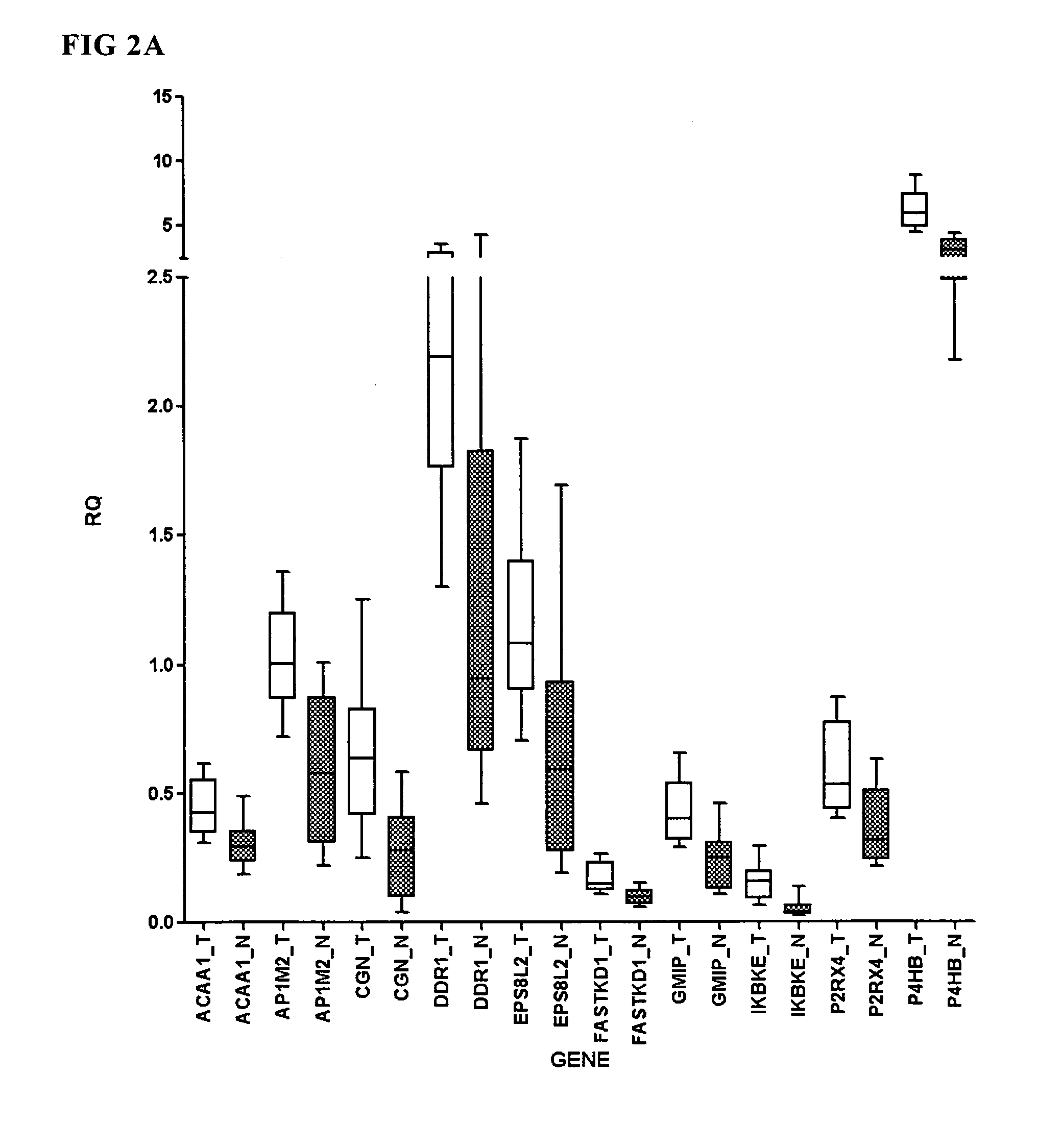
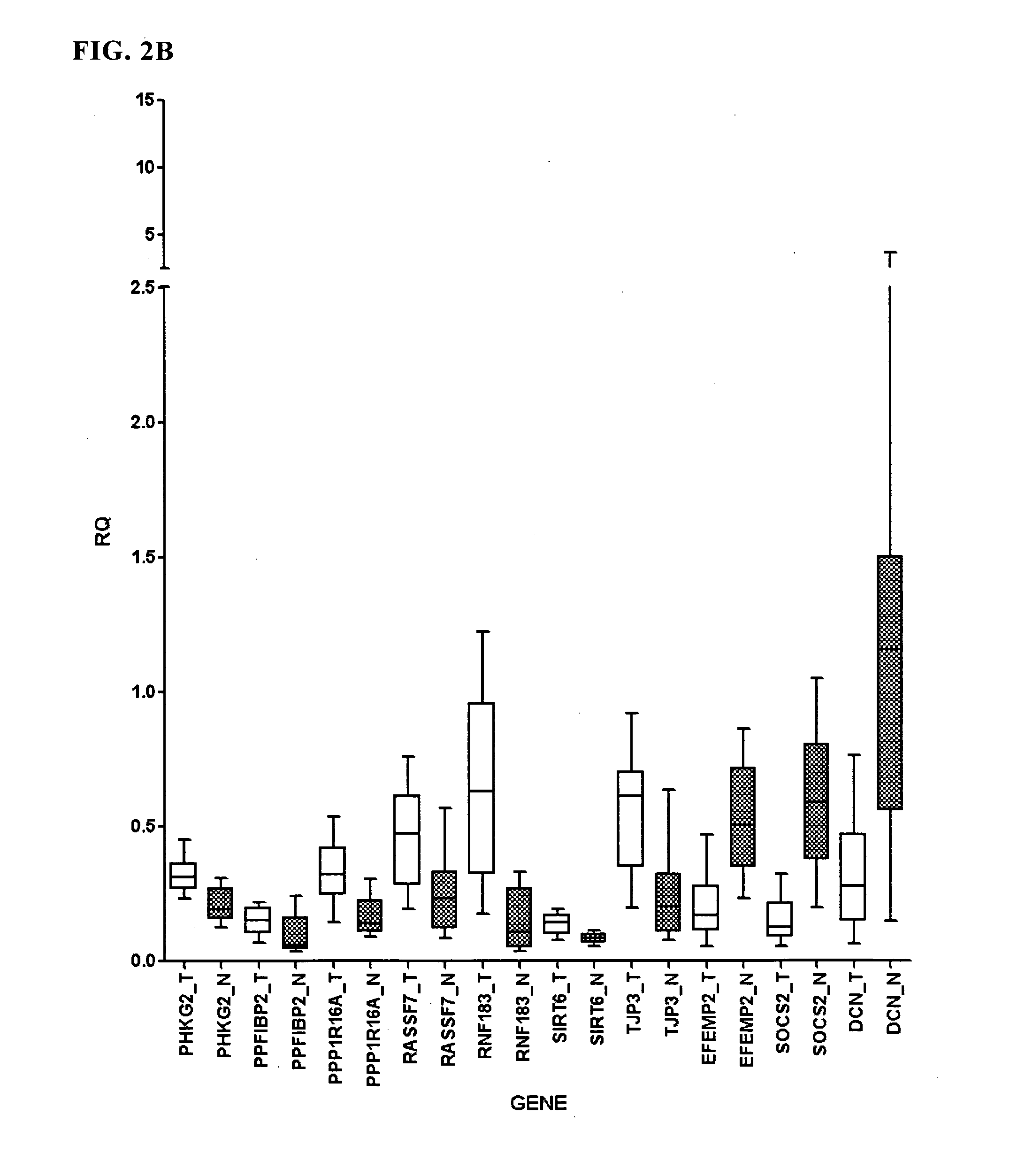
[0076]In one embodiment, the invention provides a method for diagnosing endometrial cancer comprising obtaining a sample from an individual and determining the level of from 2 to 7 biomarkers chosen from GMIP, IKBKE, P4HB, SOCS2, EPS8L2, PPP1R16A, and TJP3 wherein if said markers are differentially expressed compared to a control value, then the individual is diagnosed with endometrial cancer and / or an increased likelihood of endometrial cancer. As result of the studies disclosed herein, it was surprisingly found that combinations (e.g., profiles and / or fingerprint patterns) of biomarkers chosen from GMIP, IKBKE, P4HB, SOCS2, EPS8L2, PPP1R16A, and TJP3 have excellent sensitivity and specificity for endometrial cancer and the AUROC values for various combinations of these markers are indicative of the ability of these markers to separate patients having endometrial cancer from those not having endometrial cancer.

Problems solved by technology

Unfortunately, 20% of patients present with myometrial invasion and / or lymph node affectation, which are main indicators related to poor prognosis, decrease in survival rate, and more advanced disease.
Another important issue for clinicians for diagnosis of endometrial cancer relates to synchronous cancers.
Current methods of diagnosing endometrial cancer often create discomfort to the patient and sometimes rely on subjective interpretation of visual images.
Current methods for detecting endometrial cancer include the dilation and curettage method which is considered the gold standard, but this method is invasive, can cause significant discomfort, and may require a trained pathologist for interpretation, and therefore is not suitable as a general screening tool.
TVS has a high rate of false positives since other conditions besides endometrial cancer can produce a thicker endometrium.
One potential problem with the use of TVS in pre- and peri-menopausal women is that the thickness of the endometrium varies as a function of the phase of the menstrual cycle.

Method used


Image

  • Markers for endometrial cancer
  • Markers for endometrial cancer
  • Markers for endometrial cancer

Examples


example 1

Identification of Endometrial Cancer Biomarkers

[0380]In order to identify biomarkers for predicting and / or diagnosing endometrial cancer, gene expression levels from fifty-six endometrial primary tumors in several differentiation stages were compared with 10 normal (i.e., not having endometrial cancer) endometrial tissues by DNA microarray technique. This technique allows us to check the expression of the whole genome in a particular type of cell, tissue, organ, or in this case, check the differential gene expression between endometrial cancer and healthy endometrial tissue. A microarray chip contains small DNA sequences arranged in a regular pattern with specific addresses for probes for typically thousands of genes.

[0381]The amount of specific mRNAs in a sample can be estimated by it hybridization signal on the array.

Sample Description

[0382]Tumor samples were obtained from patients who underwent surgery and control tissue was obtained from non affected regions of endometrial tissu...

example 2

Uterine Fluid Sample Preparation

[0398]Endometrial aspirates were collected with the help of a Cornier pipelle, after complete informed consent was obtained from all patients. The aspirate (uterine fluid) was immediately transferred to an eppendorf tube containing 500 microliters of a RNA preserving solution (RNA later, Ambion). The sample was centrifuged and the pellet containing a representative population of cells from the uterine cavity was further processed for RNA extraction (Qiagen). Quality tests (Bioanalyzer) were performed before the analysis of gene expression by Taqman technology for the selected markers of endometrial carcinoma.

example 3

Correlation of Biomarkers in Primary Tumor and in Uterine Fluid

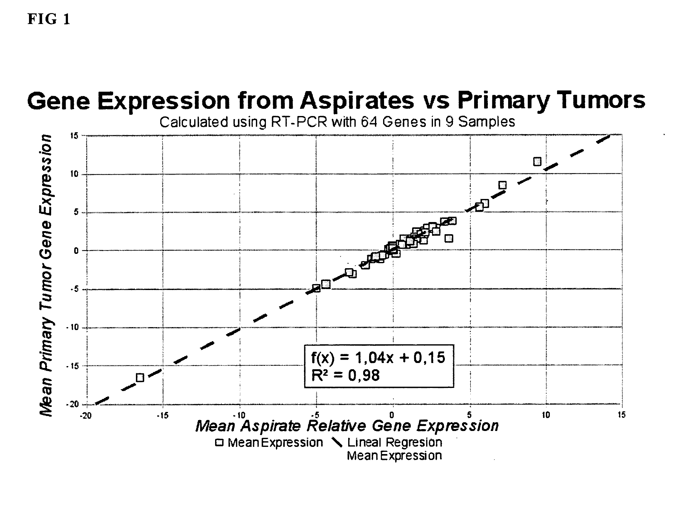
[0399]The levels of biomarkers from primary tumor sample and uterine fluid sample obtained by the procedure of Example 2 were compared as by RT-PCR following the general RT-PCR protocol as described in Example 4. The biomarkers in this study included ACAA1, AP1M2, CGN, DDR1, EPS8L2, FASTKD1, GMIP, IKBKE, P2RX4, P4HB, PHKG2, PPFIBP2, PPP1R16A, RASSF7, RNF183, SIRT6, TJP3, EFEMP2, SOCS2, and DCN whose expression level was found to be surprisingly correlated between the primary tumor and endometrial aspirates (uterine fluid). See FIG. 1. As can be seen in FIG. 1, the expression level of a number of biomarkers of endometrial cancer are correlated in uterine fluid and primary tumor. In particular, it was found that there was a high level of correlation of expression of biomarkers corresponding to ACAA1, AP1M2, CGN, DDR1, EPS8L2, FASTKD1, GMIP, IKBKE, P2RX4, P4HB, PHKG2, PPFIBP2, PPP1R16A, RASSF7, RNF183, SIRT6, TJP3, EFEMP2, ...


PUM

PropertyMeasurementUnit
Fractionaaaaaaaaaa
Fractionaaaaaaaaaa
Fractionaaaaaaaaaa

Abstract

The invention relates to the surprising finding that biomarkers corresponding to ACAA1, AP1M2, CGN, DDR1, EPS8L2, FASTKD1, GMIP, IKBKE, P2RX4, P4HB, PHKG2, PPFIBP2, PPP1 R16A, RASSF7, RNF183, SIRT6, TJP3, EFEMP2, S0CS2, and DCN are differentially expressed in control samples as compared to samples from patients having endometrial cancer and are therefore useful for detecting endometrial cancer. In particular these biomarkers having excellent sensitivity, specificity, and / or the ability to separate affected from non affected individuals. Furthermore, the inventors found that the differential expression of these biomarkers in primary endometrial cancer tumor tissue is correlated to their expression level in uterine fluid samples as compared to control values. Thus these biomarkers are robust in that they are found to be differentially expressed in several different types of samples from affected individuals.

Description

FIELD OF THE INVENTION[0001]The invention relates to the detection diagnosis, and prognosis of uterine cancer. The invention relates to the surprising finding that biomarkers corresponding to ACAA1, AP1M2, CGN, DDR1, EPS8L2, FASTKD1, GMIP, IKBKE, P2RX4, P4HB, PHKG2, PPFIBP2, PPP1R16A, RASSF7, RNF183, SIRT6, TJP3, EFEMP2, SOCS2, and DCN are differentially expressed in control samples as compared to samples from patients having endometrial cancer and are therefore useful for detecting endometrial cancer. In particular these biomarkers having excellent sensitivity, specificity, and / or the ability to separate affected from non affected individuals. Furthermore, the inventors found that the differential expression of these biomarkers in primary endometrial cancer tumor tissue is correlated to their expression level in uterine fluid samples as compared to control values. Thus these biomarkers are robust in that they are found to be differentially expressed in several different types of sa...

Claims


Application Information

Patent Timeline
IPC IPC(8): C40B30/04C12Q1/68C40B40/10C07H21/02C07K16/18C40B40/06G01N33/566C07H21/04
CPCC07K16/3069C12Q1/6886C12Q2600/112G01N2800/60C12Q2600/16G01N33/57442C12Q2600/156C07K2317/34C12Q1/6837C12Q2600/158
Inventor POSADA, MIGUEL ABALDOLL, ANDREASMORENO, ANTONIO GILMAES, TAMARAPEREZ, CRISTINAPUIGJANER, JAUME REVENTOSROSSELL, ELISABET
Owner GEADIC BIOTEC AIE
Learn More