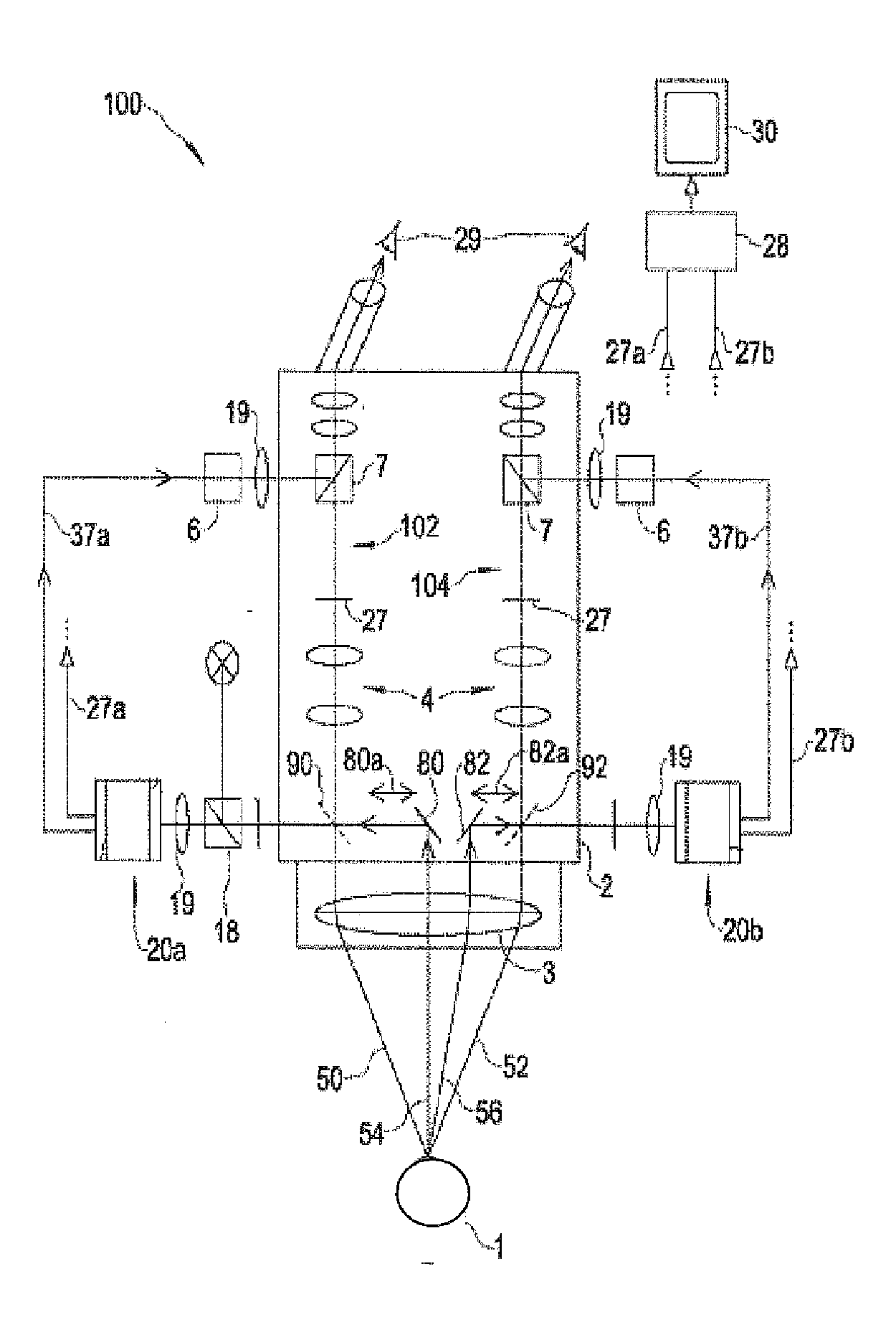
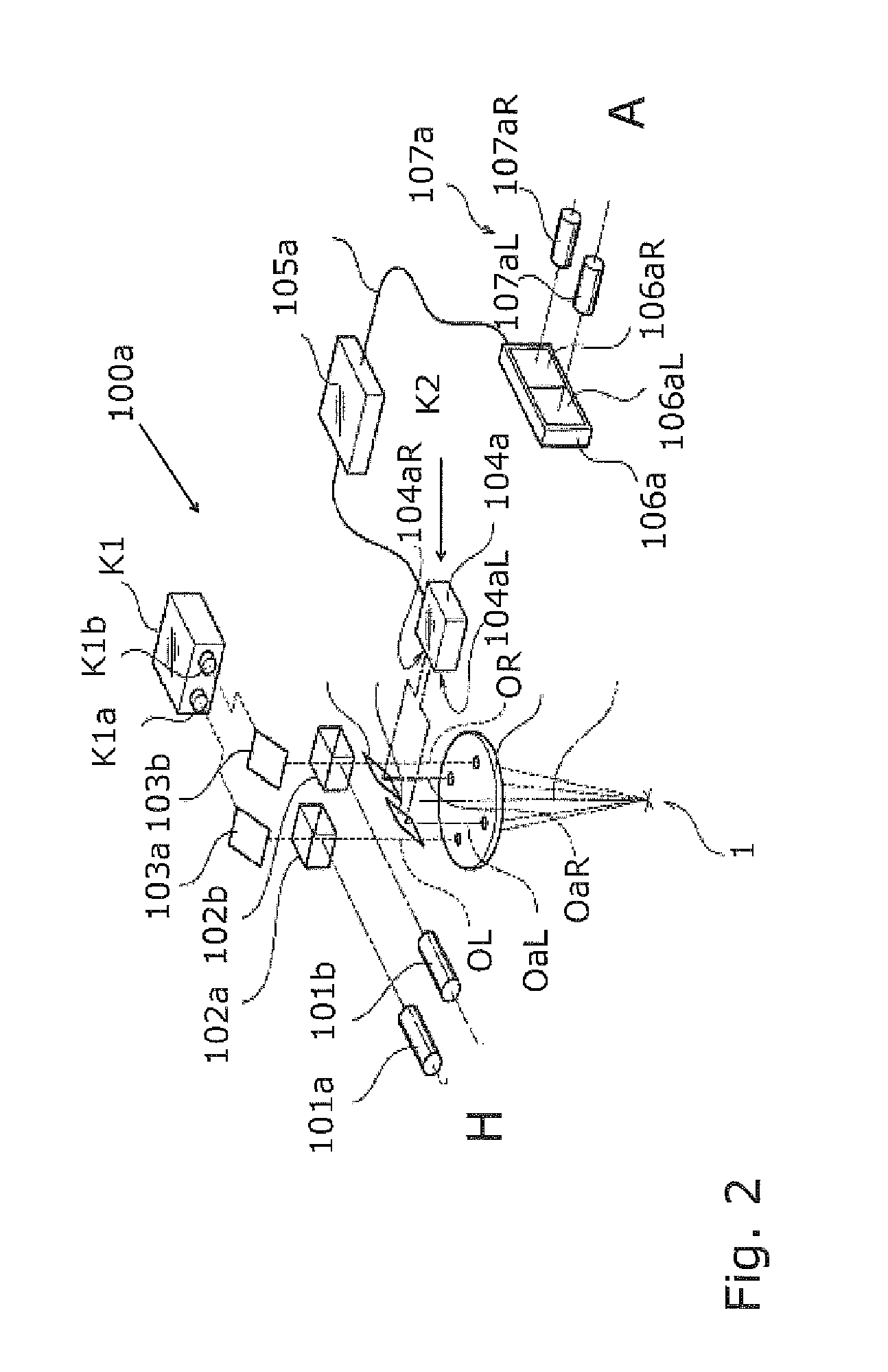
[0031]On the other hand, the video
chip in the right partial video observation beam path could exhibit, for example, particularly high sensitivity to red emitted light, so that even weak fluorescence emission light yields a strong video
signal and the weak fluorescence phenomenon appears bright. The result would be that an observer for whom the images of the video
chip or chips were reproduced on a display would perceive, in one stereoscopic observation beam path, both good background illumination and a strong emitted light, both of which would moreover contrast well with one another. Similarly advantageous effects could be achieved with the invention using a wide variety of light
wavelength regions, for example for IR or UV observation of object fields.
[0032]A refinement of the invention can, however, also be embodied in such a way that different spectral sensitivities are provided pixel-by-pixel within a partial image. The pixels could, for example, be subdivided in
checkerboard fashion, such that on the straight sides, respectively adjacent pixels each have a different sensitivity. In electronic terms, in accordance with this refinement of the invention, particularly good light efficiency could thus be achieved with a single acquired image (although then with reduced resolution) both in one light wavelength region and in a region different therefrom. In terms of depiction on a display, not only could a distinction therefore be made between a right and a left partial image, but different spectral sensitivities within one partial image could also be emphasized. With a configuration of this kind it would be possible to acquire and depict stereoscopic images of high
light intensity and sensitivity in different spectral regions using one and the same apparatus. A
checkerboard configuration of this kind having different spectral sensitivities can also be used advantageously in other applications, independently of the other features disclosed. The disclosure in this regard thus offers priority protection for a novel video
chip of this kind. The conventional configuration (RGB structure) of color
video imaging units of video chips, with the corresponding different color sensitivities of the individual RGB pixels, is not encompassed by this novel video chip for lack of novelty. What is at issue according to the present invention is the aforementioned different spectral sensitivities in the special-illumination region and in the emitted light region, and not those that serve merely for conventional
color imaging.
[0033]Preferred
image reproduction can be achieved by the fact that the special-illumination surgical video stereomicroscope is set up, by means of a unitary display, to inject an image acquired by the first region (e.g. left region) of the video imaging unit, and an image acquired by the second region (e.g. right region) of the video imaging unit, simultaneously into at least one
visual observation output, overlaid either stereoscopically or monoscopically.
[0034]According to an embodiment of the invention, a three-dimensional depiction of the acquired fluorescence images can be achieved in simple fashion in that the video imaging unit comprises a
video camera having a unitary video chip, to each of whose two halves a partial video observation beam path is allocated; and that a likewise unitary display, to whose left and right image halves a respective left and right observation beam path is respectively allocated, is used for
reproduction; or that a conventional stereo
video camera having video chips modified according to the present invention serves as an imaging unit, the stereoscopic image of which is depicted (as is known per se) by being injected into a stereoscopic observation beam path, or on a 3D display.
[0035]The present invention may be embodied as a special-illumination surgical video stereomicroscope for detecting and assisting the treatment of areas of a specimen in an
object field, preferably having a first white-light
surgical microscope illumination device having a
light source that irradiates the
object field with
white light at least during operational service, and selectably in a special light wavelength region during observational service. The stereomicroscope may have an observation beam path for guiding the light radiating from the specimen in the object field, and at least one
stereoscopic video observation beam path having at least one first observation filter device in the video observation beam path, which device is, in the utilization state, principally entirely transparent in the emitted light wavelength region (a
fluorescent light wavelength region, in the case of a fluorescence technique), and may optionally be partly transparent in the special light wavelength region (an excitation light wavelength region, in the case of a fluorescence technique). The
stereoscopic video observation beam path may have a photoelectronic video imaging unit that is coupled to a video
reproduction unit which is capable of presenting video images from the video imaging unit in the observation beam path to an observer by means of data injection.


