
Tumor treatment using ultrasound cavitation

a tumor and cavitation technology, applied in the field of tumor treatment using ultrasound cavitation, can solve the problems of insufficient vascular structure, inability to properly organize new vessels, and inability to effectively treat tumors, and achieve the effect of improving the interaction of the ultrasound field

Inactive Publication Date: 2012-02-02
KONINKLIJKE PHILIPS ELECTRONICS NV +1
View PDF5 Cites 1 Cited by
  • Summary
  • Abstract
  • Description
  • Claims
  • Application Information

AI Technical Summary

Benefits of technology

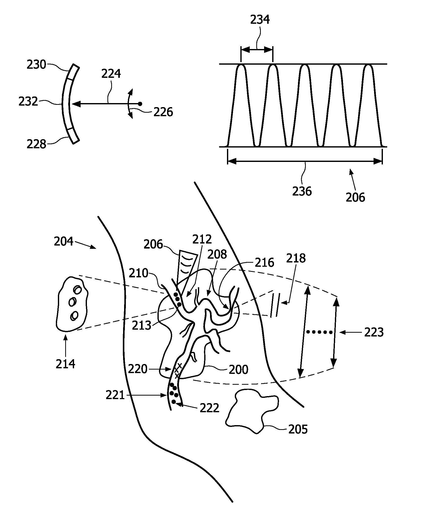
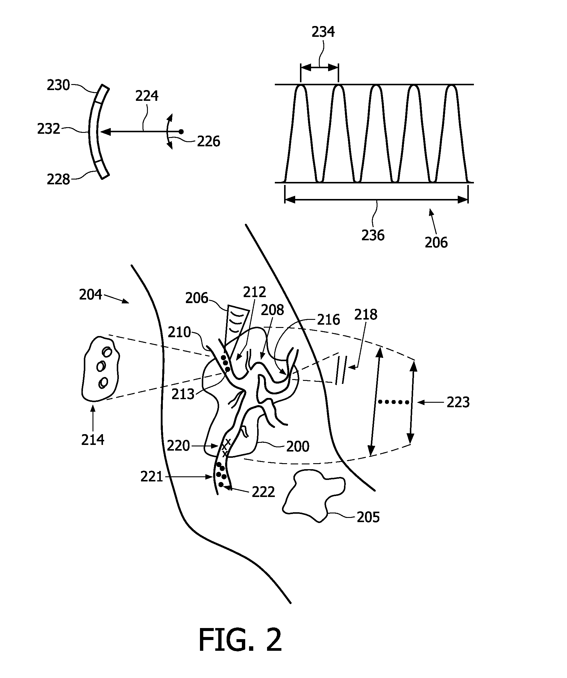
[0010]The inventors of the present application have discovered that ultrasound cavitation effects, potentiated by microbubbles, can induce changes in the tumor physiology for extended periods of time and thereby can cause restriction of tumor growth and even regression, with reduction of tumor size.
[0011]In accordance with the present invention, acute localized disruption of blood flow is induced to treat neoplasia. This vasospasm effect is sensitized in tumor vasculature and, therefore, normal tissue in the treated zone is spared differentially. In addition, the critical state of tumor perfusion allows a greater probability and severity of ischemia subsequent to flow disruption.
[0013]Test results of the present inventors show the vasospasm effect to be triggered within seconds after the ultrasound treatment, offering capability of direct monitoring in real-time.
[0014]As proposed herein, use is made of microbubbles which respond in an ultrasound field by oscillating with stimulation from the sound wave. This oscillation can be controlled to be stable (continuous) and unstable (rapid implosion). These oscillations allow for enhanced interaction of the ultrasound field with tissue through the intermediate effects of the movement of the surface of the microbubble.
[0036]As a further variation, the sequencing selectively causes in real time, in conjunction with an outcome of the monitoring, the administering of the agent for facilitating the cavitation.

Problems solved by technology

However, these methods are still in the development phase, and one of the unresolved issues is that the drug or genetic molecules released by such methods may be washed away from the treatment site by the blood flow, so that localized release may not result in the ideal distribution of drug molecules in the body.
These new vessels are poorly organized and display numerous deficiencies or abnormalities not found in normal tissues.

Method used


Image

  • Tumor treatment using ultrasound cavitation
  • Tumor treatment using ultrasound cavitation
  • Tumor treatment using ultrasound cavitation

Examples


example

[0078]Reduction of blood flow in the tumor vasculature after microbubble destruction by ultrasound.

[0079]Evaluate tumor blood flow by real-time ultrasound contrast imaging after high-amplitude focused ultrasound treatment in a murine model.

[0080]Microbubbles were prepared from perfluorobutane gas and stabilized with a phosphatidylcholine / PEG stearate shell. MC38 mouse colon adenocarcinoma cells (J. Schlom, NIH) were subcutaneously administered in the hind leg of C57BL / 6 mice. After the tumor reached >5-6 mm size, anesthetized mice were placed under the focused ultrasound transducer. Intravenous administration of 0.05-0.1 ml microbubbles was performed, immediately followed by 1.2 MHz 5 MPa insonation, delivered to the tumor as ten 1 Hz PRF 100K-cycle pulses (TIPS™ system, Philips). Insonation was repeatedly performed, i.e., on essentially a daily basis, to achieve reduction in tumor size.

[0081]Ultrasound contrast imaging during and after insonation was performed with CL15 transducer ...


PUM


Abstract

Growth in body tissue is slowed, arrested or reversed. In one aspect, this is accomplished by providing bubbles (S315), and delivering, to cause temporary change in physiology that at least one of retards, arrests and reverses said growth, a series of one or more pulses (206) of energy to respective focal points (213) at the site of the growth. In another aspect, temporary change in physiology, such as transient vasospasm (216) in vasculature of a neoplasm, is induced via the mechanical, non-thermal effects of fluid cavitation caused by the pulses. The bubbles, for facilitating the cavitation, in some embodiments, are afforded by administration of a microbubble agent to the host.

Description

FIELD OF THE INVENTION[0001]The present invention relates to growth control of body tissue, and more particularly to applying energy to retard, arrest or reverse growth.BACKGROUND OF THE INVENTION[0002]Cancers of all forms remain a major killer worldwide. A significant limitation of existing and prospective cancer therapies has been and continues to be toxic side effects.[0003]The heating effect of continuous-wave (CW) focused ultrasound is used to kill cells in the treatment of uterine fibroid. Unfortunately, the extent of the treated area is difficult to control, because blood circulation has a strong local effect on the temperature field that develops in the tissue.[0004]Non-heating approaches involving ultrasonic cavitation are known to cause a range of bioeffects including hemolysis, hemorrhage in the microvasculature, sonoporation, transient opening of the blood brain barrier, apoptosis and cell death. The type and severity of the bioeffects depend on many factors, including u...

Claims


Application Information

Patent Timeline
Patent Type & Authority Applications(United States)
IPC IPC(8): A61N7/00
CPCA61B8/481A61N2007/0039A61N7/00A61B2017/22008A61B17/22A61B18/00
Inventor CHIN, CHIEN TINGHALL, CHRISTOPHER STEPHENKLIBANOV, ALEXANDER L.
Owner KONINKLIJKE PHILIPS ELECTRONICS NV
Learn More