
Use of Stem Cells to Prevent Neuronal Dieback

a stem cell and neuronal technology, applied in the field of neuronal injury treatment, can solve the problems of high concentration of inhibitory myelin breakdown products, high concentrations of ed-1+ cells, and the role of neuroinflammation in regeneration and regeneration failure, so as to reduce neuronal injury, reduce neuronal injury, and reduce the adhesion of ed-1+ cells

Pending Publication Date: 2011-12-01
ABT HOLDING COMPANY +1
View PDF10 Cites 11 Cited by
  • Summary
  • Abstract
  • Description
  • Claims
  • Application Information

AI Technical Summary

Benefits of technology

[0008]The inventors observed that ED-1+ cells, such as activated macrophages and microglia, adhered to dystrophic axons and that this was necessary for retraction. They found that application of the cells, or conditioned medium from the cells, to the dystrophic axons reduced or prevented adhesion. They further found that application of conditioned medium from culturing the cells had neurostimulatory effects and significantly increased neurite outgrowth / regeneration.
[0016]These results are achieved by administering cells in sufficient proximity to the lesion, for a time sufficient, and in sufficient amount to reduce axonal retraction in neuronal injury and reduce neuronal injury that is associated with axonal retraction.

Problems solved by technology

The environment of a spinal cord lesion is extremely complex.
Oligodendrocytes within the lesion die, leading to demyelination, which results in high concentrations of inhibitory myelin breakdown products (Yiu and He, 2006; Xie and Zheng, 2008).
While the inhibitory effects of proteoglycans and myelin on axonal growth were well-established, the role of neuroinflammation in regeneration and regeneration failure remained highly controversial (Popovich and Longbrake, 2008).
However, studies have indicated that macrophage infiltration results in increased lesion size, decreased growth of regenerating fibers, and increased death of neurons spared by the initial lesion (Fitch et al., 1999; McPhail et al., 2004; Donnelly and Popovich, 2007).

Method used


Image

  • Use of Stem Cells to Prevent Neuronal Dieback
  • Use of Stem Cells to Prevent Neuronal Dieback
  • Use of Stem Cells to Prevent Neuronal Dieback

Examples


example i

[0155]“Glial Scar Model” Aggrecan-laminin opposing spot gradients (Tom et al., 2004; Steinmetz et al., 2005). These references are incorporated by reference for teaching the glial scar model. This model provides an assay for the effectiveness of cells, proteins, medium, etc., in reducing adhesion / retraction in vitro.

[0156]PGs can induce the so-called dystrophic state in axons if the inhibitory matrix is presented in a spatial organization that more closely resembles that which develops after lesions in vivo. To do this, spots of a solution of the PG aggrecan and the growth-promoting molecule laminin were placed on nitrocellulose coverslips and air dried.

[0157]A consistent artifact of drying produced a crude gradient in which the rim of the spot contained an increasingly higher concentration of aggrecan than in the center. The very outermost part of the rim contained a lower concentration of laminin than any more central region. The optimal ECM concentrations (0.7 mg / ml aggrecan and ...

example ii

Axonal Retraction and Macrophages

Summary

[0158]In vivo, a close correlation was found between dystrophic retraction clubs at the ends of severed axons and ED-1+ cells following a dorsal column crush spinal cord injury (FIG. 3). The in vitro model of the glial scar (Tom et al., 2004; Steinmetz et al., 2005) was applied to examine the interactions between axons and ED-1+ cells in real time. Direct cell-cell contact between dystrophic growth cones and ED-1+ macrophages induced a long distance axonal retraction (FIGS. 4, 5). The result of using clodronate liposomes (Popovich et al., 1999) for macrophage depletion in vivo was significant reduction in axonal retraction in the clodronate-treated animals compared to controls. These data indicate that ED-1+ cells are directly responsible for retraction of injured spinal cord axons through physical cell-cell interactions.

Results

1. Ascending Dorsal Column Sensory Axons Retract Extensively Following Spinal Cord Injury

[0159]It was considered by t...

example iii

Stem Cells can Prevent Adhesion of Activated Macrophages to DRGs

Results

1. Model

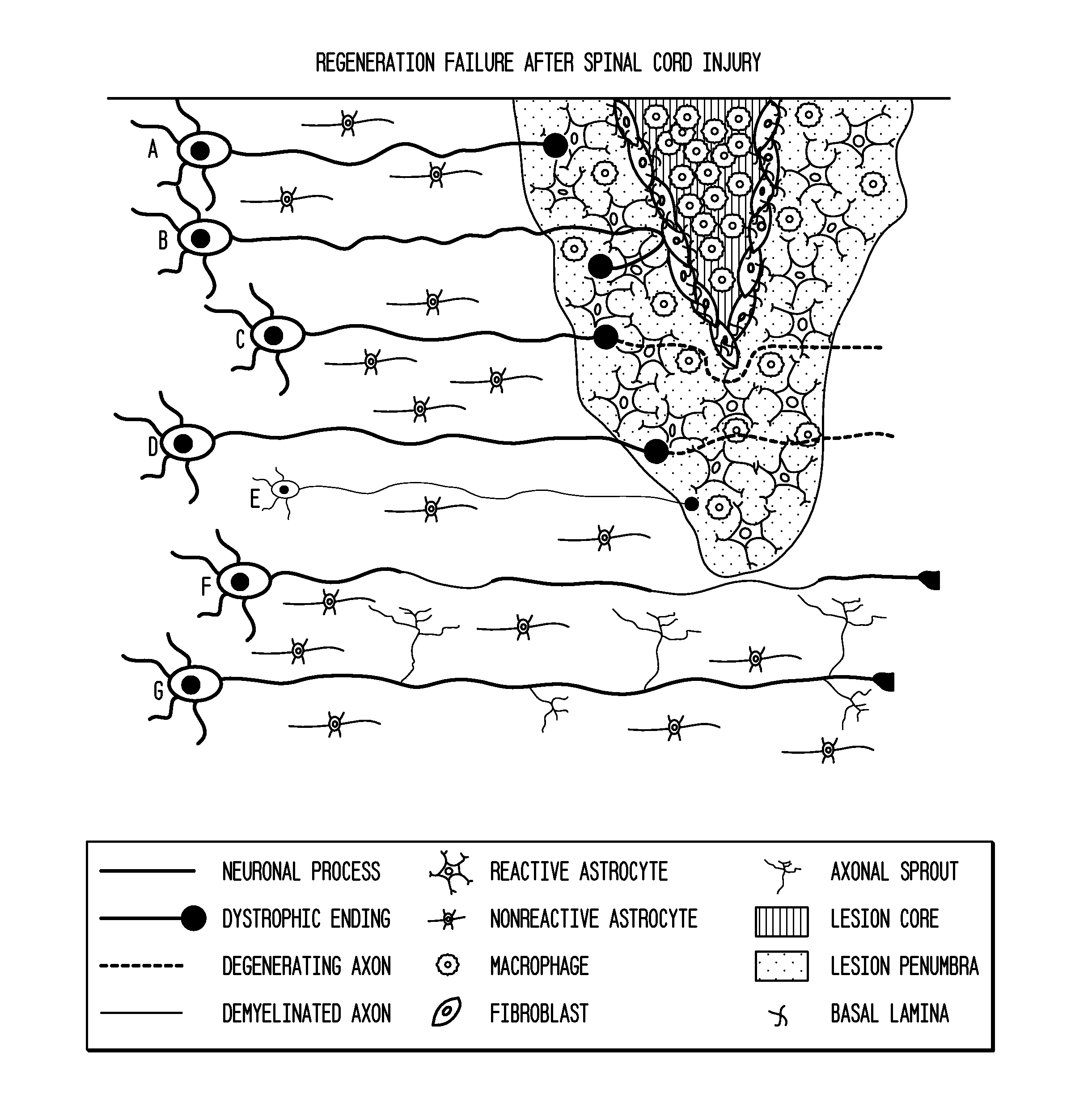
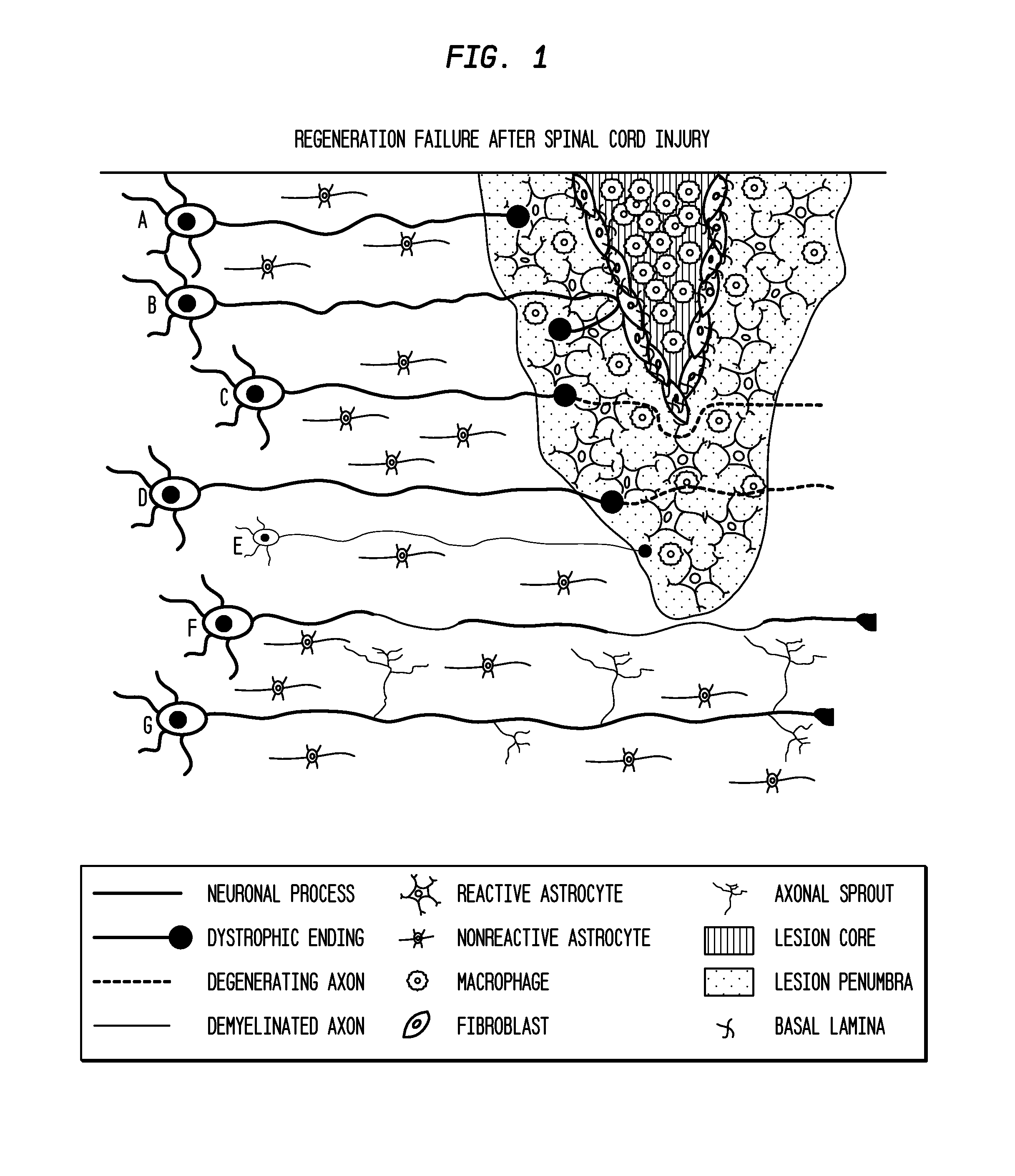
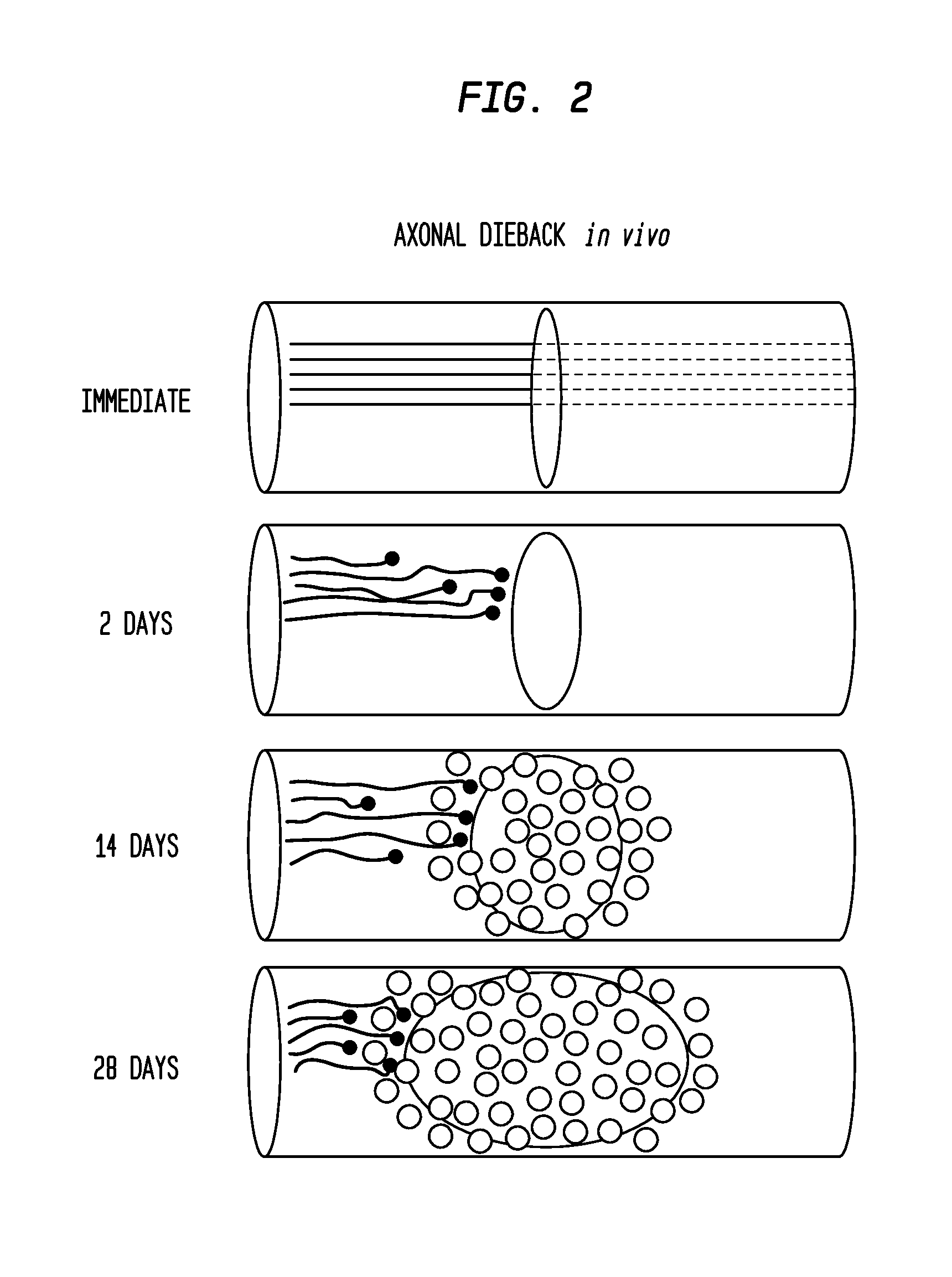
[0188]Following dorsal column crush injury, regenerating axons encounter macrophages and microglia and form dystrophic endings. This is shown schematically in FIG. 1. Previous work from the inventors' laboratory has shown that macrophage infiltration is correlated with axonal dieback following dorsal column crush injury (FIGS. 2 and 3). After characterizing the extent of axonal dieback of the ascending dorsal column sensory axons following injury, the inventors established an in vitro model of dieback, which can be used to evaluate various treatment strategies. The in vitro assay consists of cultured adult DRG neurons on a substrate of opposing gradients of the growth-promoting protein laminin and the potently inhibitory chondroitin sulfate proteoglycan aggrecan (Tom et al., 2004). This spot gradient is sufficient to stall axonal growth and induce the formation of dystrophic growth cones like those observ...


PUM

PropertyMeasurementUnit
thickaaaaaaaaaa
timeaaaaaaaaaa
concentrationsaaaaaaaaaa

Abstract

The invention is generally directed to treatment of neuronal injury. In particular, the invention is directed to reducing axonal retraction (“dieback”) that occurs as a result of the interaction of activated macrophages with dystrophic axons that are produced during nervous system acute or chronic injury. The invention is also directed to promoting axonal growth / regeneration. The invention is specifically directed to using stem cells or their secreted cellular factors, such as would be produced in conditioned cell culture medium, to ameliorate or prevent axonal dieback and / or promote growth / regeneration of axons.

Description

FIELD OF THE INVENTION[0001]The invention is generally directed to treatment of neuronal injury. In particular, the invention is directed to reducing axonal retraction (“dieback”) that occurs as a result of the interaction of activated macrophages with dystrophic axons that are produced during nervous system acute or chronic injury. The invention is also directed to promoting axonal growth / regeneration. The invention is specifically directed to using stem cells or their secreted cellular factors, such as would be produced in conditioned cell culture medium, to ameliorate or prevent axonal dieback and / or promote growth / regeneration of axons.BACKGROUND OF THE INVENTIONAxonal Retraction[0002]After spinal cord injury, a glial scar forms that poses a major impediment to CNS regeneration (Silver and Miller, 2004). In the region of forming scar tissue, the ends of the regenerating axons cease extending and become swollen and distorted into various bizarrely shaped “growth cones” that can r...

Claims


Application Information

Patent Timeline
Patent Type & Authority Applications(United States)
IPC IPC(8): A61K35/12C12N5/079A61P25/16A61P25/00A61P25/28A61P25/08A61K38/18C12N5/0786A61K35/28A61K35/30A61K35/545
CPCA61K2035/124C12N5/0607A61K35/12A61K35/30A61K35/545A61K35/28A61K2300/00A61P21/00A61P21/02A61P25/00A61P25/08A61P25/16A61P25/28A61P43/00A61P9/00A61P9/10
Inventor BUSCH, SARAH A.HORN, KEVIN P.MAYS, ROBERT W.SILVER, JERRY
Owner ABT HOLDING COMPANY
Learn More